A new cembranolide diterpene, sarcophytonolide V (1), along with 6 known compounds, isosarcophytonolide D (2), (4Z,8S*,9R*,12E,14E)-9-hydroxy-1-(prop-1-en-2-yl)-8,12-dimethyl-oxabicyclo[9.3.2]-hexadeca-4,12,14-trien-18-one (3), (7E,11E)-3,4-epoxy-7,11,15-cembratriene (4), (1S*,3S*,4S*,7E,11E)-3,4-epoxy-13-oxo-7,11,15-cembratriene (5), (-)-eunicenone (6), and 2-[(E,E,E)-7′,8′-epoxy-4′,8′,12′-trimethylcyclotetradeca-1′,3′,11′-trienyl]propan-2-ol (7) were isolated from the Bornean soft coral Sarcophyton sp. Their structures were elucidated based on spectroscopic data, such as nuclear magnetic resonance (NMR) and high resolution electron spray ionization mass spectroscopy (HRESIMS). These compounds were evaluated for their biological activity against marine pathogenic fungi.
Soft corals genus Sarcophyton (Alcyoniidae) are known to be a rich source of diterpenes, where cembrane represent the most commonly encountered structural type.1,2 In addition, some of them have exhibited very interesting biological activities such as anticancer and antibacterial.2,3 In the course of our recent investigation, one population of Sarcophyton sp. was collected from the coastal waters in Sepanggar Bay, Sabah, North Borneo and has led to the isolation of a new cembranolide, sarcophytonolide V (1), along with 6 known compounds (2-7), as shown in Figure 1. Here, we report the isolation, structure elucidation, and antifungal activities of these secondary metabolites.
Structures of compounds 1 to 7.
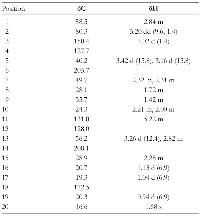
Sarcophytonolide V (1) was obtained as a colorless oil with −60.8 (c 0.32, CHCl3). Its molecular formula, C20H28O4, was deduced based on high resolution electron spray ionization mass spectroscopy (HRESIMS) [M + H]+ ion at m/z 333.2067 (calculated for C20H29O4, 333.2060). Thus, seven degrees of unsaturation were determined for compound 1. Infrared spectroscopy (IR) absorptions at νmax 1762 and 1703 cm−1, suggested the presence of carbonyl functionality. The presence of α, β-unsaturated γ-lactone system was obvious with the presence of δC 172.5, 150.4, 127.7, and 80.3; δH 7.02 and 5.20, 2 ketone carbonyls at δC 208.1 and 205.7, 2 trisubstituted double bonds at δC 150.4, 131.0, 128.0, and 127.7; δH 7.02 and 5.22, and isopropyl group at δC 28.9, 20.3, and 19.3; δH 2.28, 1.13, and 1.04 (Table 1). The two ketone carbonyls, ester carbonyl, and two pairs of double bonds accounted for five degrees of unsaturation. The remaining two degrees of unsaturation were attributed to the bicyclic system in compound 1.
1H-/13C-NMR Data (600 MHz/150 MHz, CDCl3) for Compound 1 (δ in ppm, J in Hz).
Position
δC
δH
1
58.5
2.84 m
2
80.3
5.20 dd (9.6, 1.4)
3
150.4
7.02 d (1.4)
4
127.7
5
40.2
3.42 d (15.8), 3.16 d (15.8)
6
205.7
7
49.7
2.32 m, 2.31 m
8
28.1
1.72 m
9
35.7
1.42 m
10
24.3
2.21 m, 2.00 m
11
131.0
5.22 m
12
128.0
13
56.2
3.26 d (12.4), 2.82 m
14
208.1
15
28.9
2.28 m
16
20.7
1.13 d (6.9)
17
19.3
1.04 d (6.9)
18
172.5
19
20.3
0.94 d (6.9)
20
16.6
1.68 s
Two 1H–1H correlation spectroscopy (COSY) spin systems were present; H-15/H-1/H-2 and H2-7/H-8/H2-9/H2-10/H-11. Key heteronuclear multiple bond correlation spectroscopy (HMBC) were H3-20 to C-11, C-12, and C-13; H3-19 to C-7, C-8, and C-9; H3-17 to C-1, C-15, and C-16; H3-16 to C-1, C-15, and C-17; H2-13 to C-14; H2-7 to C-6; H2-5 to C-3, C-6, and C-18; H-15 to C-14; H-2 to C-4, these correlations lead to the establishment of the cembranolide-type skeleton of compound 1. Therefore, planar structure of compound 1 was established as shown in Figure 2. Position of α, β-unsaturated γ-lactone at C-4 (α), C-3 (β), C-2 (γ), and C-18 (carbonyl carbon) was deduced from HMBC between H-3 and C-2, C-4, C-5, and C-18 and between H-2 and C-1. Vinyl methyl group attached at C-12 was observed by HMBC between H3-20 and C-11, C-12, and C-13 and between H2-10 and C-11 and C-12. The last methyl group was assigned at C-8 based on HMBC between H3-19 and C-7, C-8, and C-9, biogenetic considerations were also taken into account in this placement. Geometry of carbon–carbon for H3-20 was defined as E based on its chemical shift that was <20 ppm. The 1H and 13C NMR data (Table 1) of compound 1 were near identical to sarcophytonolide C, where methylene at C-14 in sarcophytonolide C was replaced by a carbonyl in compound 1.4 Presence of carbonyl at C-14 resulted in a downfield shifted in chemical shifts at C-1 and C-13 in compound 1 when compared to sarcophytonolide C. The relative stereochemistry of compound 1 was assigned on the basis of a 2D NOESY experiment (Figure 2). Correlations were observed between H-3, H-8 and H2-13, and between H-2, H3-16 and H3-17. These correlations suggested H-2 and H-8 are on the same face, while H-1 is on the opposite face. These relative configurations were identical to those closely related analogs, sarcophytonolides C, E and J.4
1H–1H COSY (bold), and key HMBC (single-headed arrows) and NOE (double-headed arrows) correlations of compound 1.
Known compounds were identified as isosarcophytonolide D (2), (4Z,8S*,9R*,12E,14E)-9-hydroxy-1-(prop-1-en-2-yl)-8,12-dimethyl-oxabicyclo[9.3.2]-hexadeca-4,12,14-trien-18-one (3), (7E,11E)-3,4-epoxy-7,11,15-cembratriene (4), (1S,3S,4S,7E,11E)-3,4-epoxy-13-oxo-7,11,15-cembratriene (5), (-)-emicenone (6), and 2-[(E,E,E)-7′,8′-epoxy-4′,8′,12′-trimethylcyclotetradeca-1′,3′,11′-trienyl]propan-2-ol (7) by comparing their spectroscopic data with those reported in the literature.5-9 These compounds were tested for their antifungal activities against 8 strains of marine fungi (Exophiala sp. NJM 1551, Ochroconis humicola NJM 1503, Haliphthoros milfordensis IPMB 1603, Lagenidium thermophilum IPMB 1401, Fusarium moniliforme NJM 8995, Fusarium oxysporum NJM 0179, Haliphthoros sabahensis IPMB 1402, and Fusarium solani NJM 8996). Compound 1 exhibited inhibition against hyphal growth of O. humicola and H. milfordensis at minimum inhibitory concentration (MIC) 6.25 µg/mL.
Experimental
General
NMR (ECA 600 MHz, Jeol, Japan); liquid chromatography electrospray ionisation ion trap time of flight mass spectrometry (LC-ESI-IT-TOF-MS) (Shimadzu, Japan); fourier transform infrared spectroscopy (Nicolet, Thermo Fisher Scientific, America); AUTOPOL IV automatic polarimeter (Rudolph Research Analytical, America). All instruments are located at the Laboratory of Natural Products Chemistry, Institute for Tropical Biology and Conservation, Universiti Malaysia Sabah.
Biological Material
A specimen of Sarcophyton sp. was collected from Sepanggar Bay, North Borneo, (6o4.683′N, 116o4.710′E) on July 2016. The voucher specimen (BORMI0054) was deposited in the BORNEENSIS Collection of the Institute for Tropical Biology and Conservation, Universiti Malaysia Sabah, Malaysia.
Extraction and Isolation
Fresh soft coral (0.6 kg wet weight) was extracted in methanol (MeOH) at room temperature for 3 days. The resulting raw crude extract was concentrated in vacuo and partitioned between ethyl acetate (EtOAc)/distilled water (H2O), which EtOAc fraction was further partitioned with n-hexane/90 % MeOH. The resulting crude extracts were subjected to column chromatography (CC) eluting with a gradient of n-hexane and EtOAc with increasing polarity. Fraction 1 obtained in n-hexane-EtOAc (9:1) gave compound 4 (5.7 mg; 2.7%) after purification by preparative thin layer chromatography (TLC) using n-hexane. The fraction 2, obtained in n-hexane-EtOAc (8:2), was subjected to preparative TLC with n-hexane-EtOAc (9:1) and toluene-EtOAc (9:1) to yield compounds 5 (27.0 mg; 12.9%) and 6 (12.3 mg; 5.9%). The fraction 3 obtained in n-hexane-EtOAc (7:3) gave compounds 1 (9.7 mg; 4.6%), 2 (9.0 mg; 4.3%), and 7 (21.3 mg; 10.1%) after purification by preparative TLC using n-hexane-EtOAc (8:2), toluene-EtOAc (8:2), and CHCl3. The fraction 4, obtained in n-hexane-EtOAc (5:5), was subjected to preparative TLC with n-hexane-EtOAc (7.5:2.5) to yield compound 3 (22.1 mg; 10.5%). Percentages of compounds were the average of the respective compounds in 90% MeOH crude.
HRESIMS: m/z 333.2067 [M + H]+ (calculated for C20H29O4, 333.2060).
Antifungal Activity
The MIC against 8 strains of marine fungi was performed by incorporating the compound solutions (100, 50, 25, 12.5, and 6.25 µg/mL) onto peptone yeast glucose sea water agar in petri dish.10 The MIC was determined visually as the lowest concentration showing no hyphal growth when they were incubated at 25°C for 7 days.
Footnotes
Acknowledgments
The authors would like to thank the Sabah Biodiversity Centre for Access Permit to conduct research as part of research program supported by SaBC Biotechnology enhancement grant (GL00070).
Declaration of Conflicting Interests
The author(s) declared no potential conflicts of interest with respect to the research, authorship, and/or publication of this article.
Funding
The author(s) disclosed receipt of the following financial support for the research, authorship, and/or publication of this article: The authors received financial support to carry out this research from Sabah Biodiversity Center via GL00070 Research Grant. However, no financial support was given for authorship, and/or publication of this article.
References
1.
BowdenBF.CollJC.MitchellSJ. Studies of Australian soft corals. XVIII. Further cembranoid diterpenes from soft corals of the genus Sarcophyton. Aust J Chem. 1980;33(4):879-884.doi:10.1071/CH9800879
2.
LiuX.ZhangJ.LiuQet al. Bioactive cembranoids from the South China Sea soft coral Sarcophyton elegans. Molecules. 2015;20(7):13324-13335.doi:10.3390/molecules200713324
3.
LiangL-F.KurtánT.MándiAet al. Structural, stereochemical, and bioactive studies of cembranoids from Chinese soft coral Sarcophyton trocheliophorum. Tetrahedron. 2018;74(15):1933-1941.doi:10.1016/j.tet.2018.02.059
4.
TakamuraH.KikuchiT.IwamotoKet al. Unified total synthesis, stereostructural elucidation, and biological evaluation of sarcophytonolides. J Org Chem. 2018;83(18):11028-11056.doi:10.1021/acs.joc.8b01634
5.
YanX-H.GavagninM.CiminoG.GuoY-W. Two new biscembranes with unprecedented carbon skeleton and their probable biogenetic precursor from the Hainan soft coral Sarcophyton latum. Tetrahedron Lett. 2007;48(30):5313-5316.doi:10.1016/j.tetlet.2007.05.096
6.
GrkovicT.WhitsonEL.RabeDCet al. Identification and evaluation of soft coral diterpenes as inhibitors of HIF-2α induced gene expression. Bioorg Med Chem Lett. 2011;21(7):2113-2115.doi:10.1016/j.bmcl.2011.01.127
7.
RaviBN.FaulknerDJ. Cembranoid diterpenes from a South Pacific soft coral. J Org Chem. 1978;43(11):2127-2131.doi:10.1021/jo00405a009
8.
RodríguezAD.LiY.DhasmanaH.BarnesCL. New marine cembrane diterpenoids isolated from the Caribbean gorgonian Eunicea mammosa. J Nat Prod. 1993;56(7):1101-1113.doi:10.1021/np50097a014
9.
CollJC.HawesGB.LiyanageN.OberhansliW.WellsRJ. Studies of Australian soft corals. I. A new cembrenoid diterpene from a Sarcophyton species. Aust J Chem. 1977;30(6):1305-1309.doi:10.1071/CH9771305
10.
KamadaT.PhanC-S.HamadaT.HataiK.VairappanCS. Cytotoxic and antifungal terpenoids from Bornean soft coral, Sinularia flexibilis. Nat Prod Commun. 2018;13(1):17-19.doi:10.1177/1934578X1801300106