



Abstract
Introduction
Caesalpinia sappan L. is a leguminous plant, also known as Su Mu or Chi mu. Sappan is widely distributed in China and East Asian countries and has been used as a medicinal plant for several years.1,2 Several chemical components have been isolated from sappan, which can be divided into 5 categories based on their skeleton types including hematoxylin, proto hematoxylin, hyperisoflavones, hematoxylin chalcone, and phenylpropanoid.3-6 Previous studies report that sappan promotes blood circulation and alleviates blood stasis, exhibits detumescence and pain relief, and is used for treatment of injuries, fractures and tendons, stasis, swelling, and pain from falls. 1
Pharmacological studies report that sappan induces apoptosis and growth of several cancer cells, such as U266 cells, oral cancer cells, head and neck cancer cells.7,8 A previous study reported that ethyl acetate extract of the dried heartwood of sappan can inhibit acute myeloid leukemia cells by inducing mitochondrial apoptosis and promoting differentiation, which are implicated in reactive oxygen species activation. 9 However, studies have not elucidated the exact components in sappan that exhibit antitumor effects and the pathways and targets involved.
Advances in bioinformatics, systems biology and multipharmacology, have resulted in the development of more cost-effective drugs. 10 Network pharmacology has changed the research paradigm from the current “one target, one drug” model to a new “network target, multi-component” model. 11 Network pharmacology allows the rationalization and compatibility of traditional Chinese medicine by providing detailed compound target and target pathway networks.12,13
The current study explored the potential antitumor mechanism of sappan using network pharmacology methods. Potential molecular targets of sappan were identified and pathways and networks of sappan compounds were predicted. Molecular docking studies were then conducted to predict the interactions between the sappan compounds and its predicted target. The findings show that sappan can effectively inhibit the proliferation of tumor cells and provide a basis for development of novel drugs.
Materials and Methods
Network Pharmacology-Based Analysis
Phytochemicals extracted from sappan were retrieved from Traditional Chinese Medicine on Immuno-Oncology (TCMIO) database (http://tcmio.xielab.net/). 14 Protein targets and pathway enrichment analyses were conducted using sappan active substances retrieved from TCMIO.
Expression Analysis of AKT from Public Datasets
Expression analysis of PI3K and AKT in cancer cells was performed following previously described methods. 15 The z value of AKT mRNA expression for each sample was determined by comparing the mRNA expression level with the expression level of the gene in a reference population. The query “AKT1” or “PIK3CG” was used in the Cancer Genome Atlas and cancer cell lines encyclopedia (CCLE) (Broad Institute, Novartis Institutes for Biomedical Research). 16 RNA-Seq expression data were grouped based on the clinical characteristics (tumor type and histology) of the selected samples.
Preparation of Sappan Ethanol Extract Stock Solution
Extraction was performed using 50 g of sappan, and10 times 55% ethanol–water solution was added to the sample. The mixture was soaked overnight and reflux extraction was performed 3 times at 80°C, with each time performed for 2 h. The extracts from the 3 extractions were then combined. The extract was concentrated under low pressure and transferred to a clean vial. The solvent was evaporated and the extract was retained. Some amount of the dry ethanol extract was weighed and dimethyl sulfoxide was added to completely dissolve the extract. This portion was used as an ethanol extract reserve solution of sappan (SE) at a concentration of 100 mg/mL.
Cell Culture and Treatment
HepG2 cells were cultured in Dulbecco’s modified eagle medium containing 10% fetal bovine serum at 37°C and 5% CO2. Cells in the logarithmic growth phase were inoculated in a 96-well plate with 1 × 10⁵ cells/mL, and 100 μL per well. After 12 h of culture, set a final concentration of 25, 50, 100, and 200 μg/mL was added and cultured in a cell incubator for 24/36/48/72 h. 3-(4,5-Dimethylthiazol-2-Yl)-2,5-Diphenyltetrazolium Bromide solution was added, and cultured in the cell culture box for 4 h. The supernatant was then obtained and the OD value was determined at 490 nm. The inhibition rate was calculated as follows: IR (inhibitor ratio) % = OD (sample)/OD (blank control) × 100%.
Western Blot Analysis
Cell lysis and western blot analysis were performed following a method described previously. 17 12% sodium dodecyl sulfate–polyacrylamide gel electrophoresis was used to separate protein samples. The primary antibody was Akt (CST, 1:1000) and p-Akt (CST, 1:1000). Cells were incubated overnight with primary antibodies at 4°C. Samples were then washed thrice with TBST and then incubated with a secondary antibody for 1 h at room temperature. β-actin (Wanleibio, Cat: wl01114, c 1:15000) protein was used as the internal control for western blot analysis. Image analysis software (ImageJ) was used to quantify proteins.
Statistical Analysis
Statistical differences between multiple treatments and control were analyzed using a one-way analysis of variance with Turkey's post-hoc test. Statistical analyses were performed using GraphPad Prism 5 (GraphPad Software, Inc). P < .05 was considered statistically significant.
Results
Pathway Enrichment and Networks Analysis of Sappan
A total of 30 sappan-derived compounds were retrieved from TCMIO (Table S1). Immuno-oncology (IO) network analysis was performed using the TCMIO database. Enrichment fractions of 4 hub targets including PI3CG, PTEN2, TP53, and EGFR were significant (Figure 1A). Further, pathway enrichment analysis showed that the top 10 enriched pathways were implicated in cancer. Pathways implicated in cancer were significantly enriched for 18 targets, (P = 4.984 × 10⁻¹², Figure 1B). A PI3K-Akt signaling pathway is an important signal transduction pathway in cells and is a major target for the development of antitumor drugs. Subsequent analyses were focused on the PI3K-Akt signaling pathway which showed 13 targets. (P = 1.739 × 10⁻⁷, Supplemental Table S2).

Network pharmacology analysis.
PIK3CG and AKT Expression Analysis
Expression of PIK3CG and AKT1 mRNA was explored in cancer cells (n = 2041 samples). PIK3CG showed the highest expression level in lymphoma, whereas its expression level was lower in solid tumors (Figure 2A). AKT1 was highly expressed in solid tumors, with the highest expression observed in breast cancer (Figure 2B).

PIK3CG (A) and AKT1 (B) expression in cancer cell line encyclopedia.
Further, the effect of sappan compounds on solid tumors was explored by conducting bioinformatics analysis on different subtypes of AKT. The findings showed that the expression level of AKT1 and AKT2 was similar in different tumor tissues, however, the distribution of AKT1 and AKT3 was significantly different across tumor tissues (Figure 3A to C). Moreover, expression of AKT1 in breast cancer cells was explored and the finding showed that the mean expression level of AKT1 was 5.29 (P = 2.18722 × 10⁻⁸, Figure 3D).
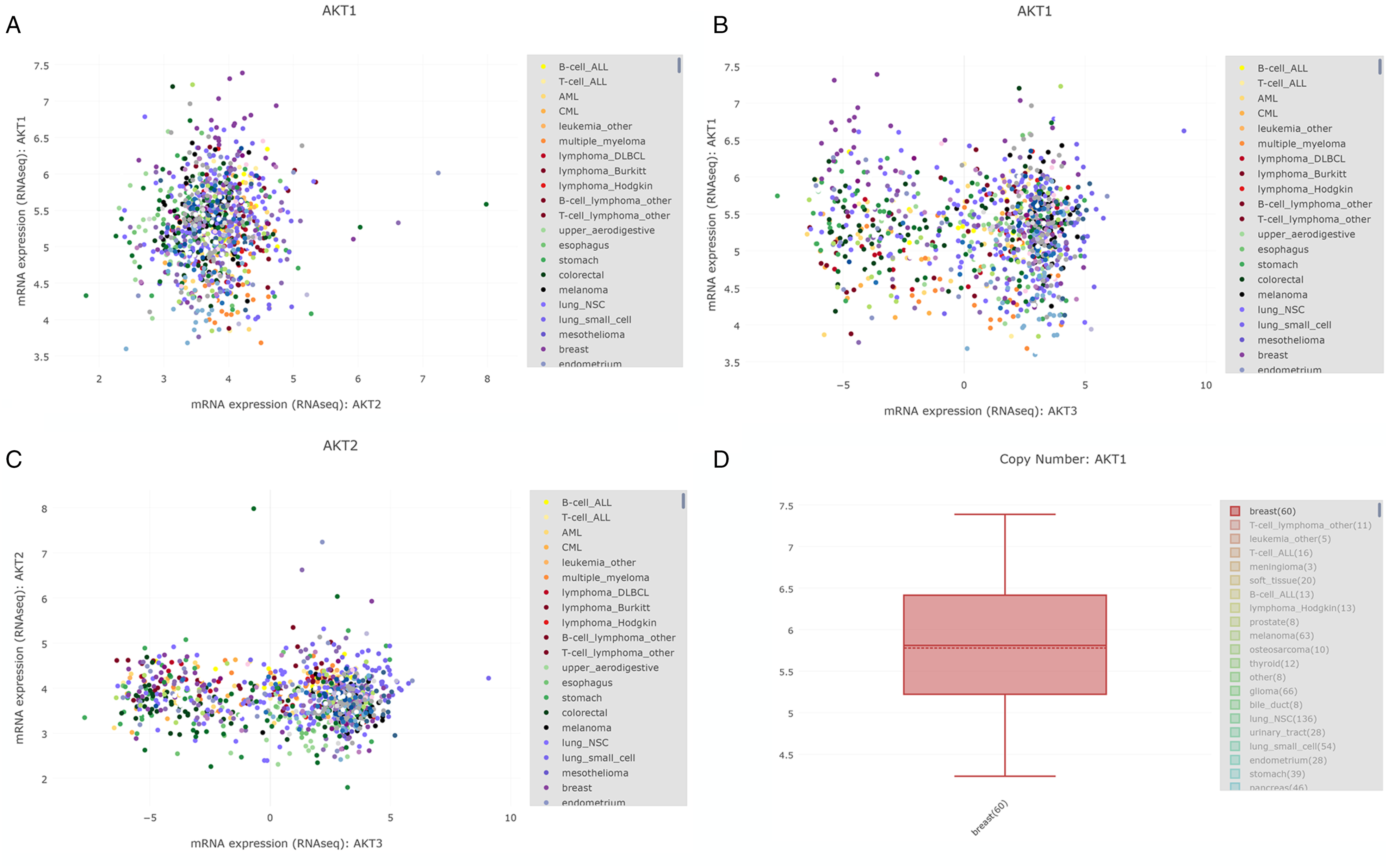
Akt mRNA expression in cancer cell line encyclopedia (CCLE).
Molecular Docking
Molecular docking was performed to explore the chemical composition of sappan and the mode of action of sappan compounds in modulating Akt. The top 8 compounds with a score higher (−14.665) than that of co-crystallized ligand (−12.186) are listed in Table 1. (3R,5R)-1,3,4,5-tetrakis(((E)-3-(3,4-dihydroxyphenyl)acryloyl)oxy)cyclohexane-1-carboxylic acid showed several hydrogen bond donors and receptors, has and formed hydrogen bonds with several key amino acids including Glu193, Met282, and Asp293 (Figure 4A). Binding poses of compounds 2 to 6 at Akt active site showed that the compounds interacted with Met282 and Asp293 through hydrogen bonds (Figure 4C to F).
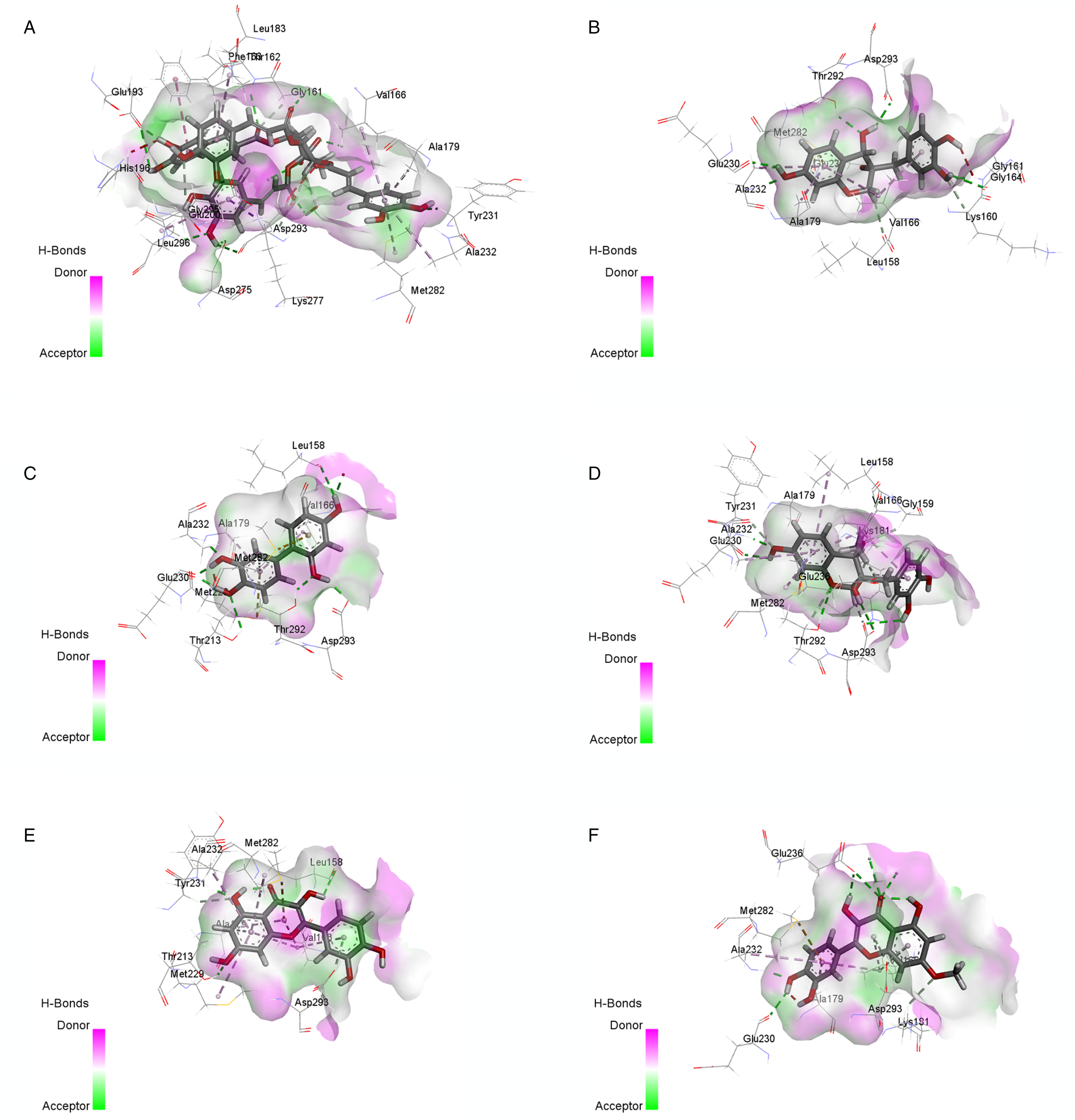
Molecular docking predicted the binding mode of the compounds from Caesalpinia sappan with the top 6 scores to Akt protein.
The Docking Score of the Compounds from Caesalpinia Sappan.

Sappan Ethanol Extract (SE) Induced HepG2 Cells Apoptosis
Different concentrations of SE were incubated with HepG2 cells for 24, 36, 48, and 72 h to explore its inhibitory effect on HepG2 cell proliferation. The findings showed that HepG2 cell viability was inhibited in a dose-dependent and time-dependent manner (Figure 5A).

Sappan extract (SE) can induce apoptosis of HepG2 cells.
Further, HepG2 cells were treated with 25, 50, and 1000 μg/mL of SE for 48 h. Proteins were extracted and expression levels of Akt and p-Akt (ser473) were determined by western blot analysis. The expression level of p-Akt after SE treatment was significantly lower compared with the level of the control group (Figure 5B). These findings showed that SE can inhibit the activity of Akt protein by down-regulating the phosphorylation level of p-Akt.
Discussion
In the present study, data from public databases were used to predict the interaction between sappan and its potential protein targets and its anti-tumor pharmacological effects. In addition, similarities and differences in mRNA expression of different subtypes of Akt in various tumor cells were explored. Moreover, the mode of interaction of sappan compounds with Akt was explored.
Network pharmacology uses network theory and systems biology, therefore, it is an important method in drug discovery.18,19 Network pharmacology has been widely used to explore the “complex protein/gene-disease” relationship, determine its effect on biological systems from a wholistic perspective, and is used to explore the complex relationship between drugs and diseases.20,21 Therefore, the method can be used for predicting interactions between traditional Chinese medicines and their targets. In the current study, TCMIO online database was used to analyze and predict the network pharmacology of sappan compounds. The findings showed that the PI3K-Akt pathway is significantly correlated with the antitumor activity of sappan compounds. 9
Akt is a serine/threonine-protein kinase with a molecular weight of 56 KD. The name was derived from high homology with the retroviral oncogene v-Akt in T-cell lymphoma and similar structure with protein kinase A and protein kinase C. Akt is an anti-apoptotic protein including Akt1/PKBα, Akt2/PKBβ, and Akt3/PKBγ.22,23 Sale et al 24 used subtype-specific antisense oligonucleotide probe strategies to explore the function of Akt and its 3 subtypes and the findings showed that 3 Akt subtypes are involved in the activation of downstream substrates. In the current study, a public database was used to compare mRNA expression levels of the 3 subunits of Akt in different tumor cells for the first time. The findings showed that expression levels of AKT1 and AKT2 were similar in different tumor tissues.
The molecular docking technique was used to explore interactions between Akt using the crystalline structure (PDB: 2 × 39) and the chemical components in sappan. The findings showed that the binding energy of compound 1 to the protein target was higher compared with that of the native ligand molecule (4-amino-1-(7H-pyrrolo [2,3-d] pyrimidin-4-yl) piperidine-4-carboxamide), 25 and formed several stable hydrogen bonds.
Furthermore, SE significantly induced apoptosis of HepG2 cells. Western blot assay was used to explore the expression level of phosphorylated Akt protein. These findings show that sappan extract can inhibit signal transduction by modulating the expression of Akt and related proteins in HepG2 cells, therefore, it exhibits a potential anticancer effect.
Conclusion
In summary, the current study established a network of antitumor molecular targets targeted by sappan compounds. In addition, public databases were used to explore the expression levels of the targets in different tumor tissues. The chemical components in sappan were docked to the crystal structure of Akt to explore interactions between the target and ligands. The findings from the current study provide a basis for further studies on the antitumor effects of sappan compounds.
Supplemental Material
sj-docx-1-npx-10.1177_1934578X211039922 - Supplemental material for Integrative Analysis to Uncover the Molecular Mechanisms of Caesalpinia sappan L. for Anti-Cancer Activity
Supplemental material, sj-docx-1-npx-10.1177_1934578X211039922 for Integrative Analysis to Uncover the Molecular Mechanisms of Caesalpinia sappan L. for Anti-Cancer Activity by Bing Liu and Hao Lian in Natural Product Communications
Supplemental Material
sj-docx-2-npx-10.1177_1934578X211039922 - Supplemental material for Integrative Analysis to Uncover the Molecular Mechanisms of Caesalpinia sappan L. for Anti-Cancer Activity
Supplemental material, sj-docx-2-npx-10.1177_1934578X211039922 for Integrative Analysis to Uncover the Molecular Mechanisms of Caesalpinia sappan L. for Anti-Cancer Activity by Bing Liu and Hao Lian in Natural Product Communications
Footnotes
Acknowledgments

Declaration of Conflicting Interests
The authors declared no potential conflicts of interest with respect to the research, authorship, and/or publication of this article.
Funding
The authors received no financial support for the research, authorship and/or publication of this article.
Supplemental Material
Supplemental material for this article is available online.
References
Supplementary Material
Please find the following supplemental material available below.
For Open Access articles published under a Creative Commons License, all supplemental material carries the same license as the article it is associated with.
For non-Open Access articles published, all supplemental material carries a non-exclusive license, and permission requests for re-use of supplemental material or any part of supplemental material shall be sent directly to the copyright owner as specified in the copyright notice associated with the article.
