One new oleanane-type triterpene glycoside, oleanolic acid-[28-O-β-d-glucopyranosyl]-3-O-[β-d-glucopyranosyl(1→6)-β-d-glucopyranosyl](1→3)[α-l-arabinofuranosyl(1→4)]-β-d-glucuronopyranoside (1), and 3 known ones {oleanolic acid-[28-O-β-d-glucopyranosyl]-3-O-[β-d-galactopyranosyl(1→3)]-[β-d-glucopyranosyl(1→2)]-β-d-glucuronopyranoside (2) chikusetsusaponin IVa methyl ester (3), and chikusetsusaponin IV (4)} were isolated from the leaves of Aralia armata. Their chemical structures were elucidated using a combination of high-resolution electrospray ionization mass spectrometry, 1-dimensional and 2-dimensional nuclear magnetic resonance (NMR) spectral data, as well as comparison with data in the previous literature. This is the first report of full NMR spectroscopic data of 2. Compounds 1-4 displayed weak cytotoxic activity toward KB and HepG2 cell lines, with half-maximal inhibitory concentration50 values ranging from 24.2 ± 0.3 to 32.6 ± 0.8 µM in in vitro assay.
The genus Aralia (family Araliaceae) contains more than 70 species and many of which are used medicinally in Asia and the Americas. The main chemical constituents of Aralia species include triterpenoid glycosides, sterols, diterpenoids, and acetylenic lipids,1 and many of which have been evaluated for their potential as lead compounds for drug discovery. Several species of Aralia have been used in folk medicine for treating diabetes, hepatitis, stomach ulcer, and other diseases.1-3 Phytochemical study of A. armata leads to the isolation of oleane-type triterpene glycosides as the main components.3,4 In Vietnam, 8 Aralia species are found: A. armata, A. chinensis, A. cordata, A. dasyphylla, A. decaisneana, A. planchoniana, A. thomsonii, and A. vietnamensis.5Aralia armata is used in traditional medicine to treat hepatitis, arthritis, stomach ache, and malaria.5 Three oleanane-type triterpene saponins, narcissiflorin, stipuleanosid R1, and stipuleanosid R2, have been isolated from A. armata growing in Vietnam.6 As part of our continuing research program to find bioactive components of Aralia species,7 we report herein the identification of 1 new (1) and 3 known oleanane-type triterpene saponins (2-4) from the leaves of A. armata. The cytotoxic activity of the isolated compounds on some cell lines was also evaluated.
Results and Discussion
Compounds 1-4 were obtained as white amorphous powders from MeOH extract of the leaves of A. armata. Compounds 3 and 4 were identified as chikusetsusaponin IVa methyl ester (3),6 and chikusetsusaponin IV (4)6 (Figure 1) by analyzing their high-resolution electrospray ionization mass spectrometry, 1-dimensional (1D) and 2D nuclear magnetic resonance (NMR) spectral data, as well as by comparison with the previous literature (Supplemental Table S1, Figures S15-S26).
Chemical structures of compounds 1-4.
The molecular formula of 1 was determined to be C59H94O28 from its HR-ESI-MS ion peak at m/z 1249.5860 [M − H]− (calcd. for C59H93O28: 1249.5853) (Supplemental Figure S1). Analyzing the NMR spectra of 1 (Supplemental Figures S2-S8) found that this compound had oleanolic acid as its aglycone, and 5 sugars, 1 glucuronopyranosyl, 1 arabinofuranosyl, and 3 glucopyranosyls, which matched with the molecular formula of C59H93O28. The double bond at C-12/C-13, the carboxylate C-28, and oxygenated methine group at C-3 were identified by signals at δH 5.27/δC 123.8 (CH)/144.8(C), δC 178.1, and δH 3.15/δC 90.7, respectively. The 5 sugar units were identified by their anomeric signals at δH 5.37/δC 95.7, δH 4.36/δC 106.3, δH 4.87/δC 104.4, δH 5.20/δC 108.3, and δH 4.37/δC 104.6, determined from the heteronuclear single quantum correlation (HSQC) spectrum. One glucopyranosyl was attached to C-28 by an ester linkage, and the moiety arabinofuranosyl-(1→3)-glucuronopyranoside was suggested to link to C-3 by an ether linkage by comparison of their NMR data with those of 3-O-α-l-arabinofuranosyl-(1→4)-β-d-glucuronopyranosylhederagenin 28-O-β-d-glucopyranosyl ester8; further confirmation was obtained from the HSQC, H-H correlation spectroscopy (COSY), and heteronuclear multiple bond correlation (HMBC) spectra. A glucosyl unit at C-28 was verified by the set of signals from C-1′ to C-6′ as δC 95.7, 73.8, 78.1, 71.0, 77.9, and 62.2, which had HSQC cross-peaks with signals at δH 5.37 (d, J = 8.0 Hz), 3.35, 3.43, 3.44, 3.31, and 3.70/3.82, respectively, and confirmed by HMBC from glc H-1′ (δH 5.37) to C-28 (δC 178.1). The glucopyranosyl(-(1→6)-glucopyranosyl moiety was confirmed to be attached to C-3′′ of the glucuronopyranosyl sugar by the observation of HMBC from glc H1′′′′ (δH 4.37) to glc H-6′′′ (δC 69.5), and from glc H-1′′′ (δH 4.87) to gluA C-3′′ (δC 82.1) (Figure 2). Furthermore, the coupling constants (J = 7.5-8.0 Hz) observed for the gluA and glc anomeric protons in the 1H NMR spectrum of compound 1 indicated that these sugar linkages must be in the β-form; ara H-1′′′ appeared as a broad singlet confirming the α-form of this sugar linkage. The monosaccharides in the sugar residue were further confirmed to be d-glucuronic acid, d-glucose, and l-arabinose by hydrolysis, conversion to thiazolidine derivatives, high-performance liquid chromatography (HPLC) analysis and comparison of their retention times with those of standard monosaccharide derivatives prepared using the same procedure.9 Based on the biosynthesis of the oleanane skeleton, the 2 protons H-5 and H-9, and the methyl group at C-27 all had α-orientations. The H-3 proton had an axial orientation, elucidated from the observation of nuclear Overhauser effect (NOE) cross-peaks between Hα-5 (δH 0.79) and Hα-3 (3.15), as well as by the larger coupling constant of H-3 (JH-2/H-3 = 12.0 Hz). Consequently, compound 1 was determined to be oleanolic acid-[28-O-β-d-glucopyranosyl]-3-O-[β-d-glucopyranosyl(1→6)-β-d-glucopyranosyl](1→3)[α-l-arabinofuranosyl(1→4)]-β-d-glucuronopyranoside, a new compound and named as araliaarmoside (Figure 1).
The 1H-1H correlation spectroscopy (COSY) and key heteronuclear multiple bond correlation (HMBC) of compound 1.
The molecular formula of 2 was determined to be C54H86O24 from its HR-ESI-MS ions at m/z 1153.5208 [M + 35Cl]− (calcd. for C54H86O2435Cl: 1153.5198) and 1155.5154 [M + 37Cl]− (calcd. for C54H86O2437Cl: 1155.5168)] (Supplemental Figure S9). The NMR spectra of 2 (Supplemental Figures S10-S14) showed that its aglycone was oleanolic acid, and the sugar moieties were similar to those of compounds 3 and 4. The NMR data of the aglycone and the 28-O-β-d-glucopyranoside unit of 2 were in accordance with those of compounds 3 and 4 suggesting that compounds 2-4 have the same oleanolic acid skeleton with 1 glycosylic unit attached to C-28 by an ester linkage. The other sugar moiety of 2 was identified as β-d-galactopyranosyl(1→3)-[β-d-glucopyranosyl(1→2)]-β-d-glucuronopyranoside, which was attached to C-3 of the aglycone, similar to the sugar moieties of calendasaponin C (Supplemental Table S1)10 and glycoside A;11 this was further confirmed from the HSQC and HMBC spectra. The HMBC correlations from gal H-1′′′′ (δH 4.72) to gluA C-3′′ (δC 88.8), from glc H-1′′′ (δH 4.99) to gluA C-2′′ (δC 78.9), from gluA H-1′′ (δH 4.49) to C-3 (δC 92.0), as well as from gal H-5′′′′ (δH 3.59) to gal C-1′′′′ (δC 104.4)/C3′′′′ (δC 75.2) and from glc H-2′′′ (δH 3.18) to glc C-1′′′ (δC 103.2)/C-3′′′ (δC 78.1) confirmed the sugar moiety as 3-O-β-d-galactopyranosyl(1→3)]-[β-d-glucopyranosyl(1→2)]-β-d-glucuronopyranoside. The HMBC from glc H-1′ (δH 5.40) to C-28 (δC 178.1) confirmed that the other glucose was linked to C-28. Consequently, compound 2 was identified as oleanolic acid-[28-O-β-d-glucopyranosyl]−3-O-[β-d-galactopyranosyl(1→3)]-[β-d-glucopyranosyl(1→2)]-β-d-glucuronopyranoside. This compound had been isolated from Calendula officinalis and named as glycoside A, but only the carbon chemical shifts of the sugar moieties and mass spectral data had been reported.6 To the best of our knowledge, this is the first time that the full NMR data of this compound have been reported (Table S1, Supplemental Figures S9-S14).
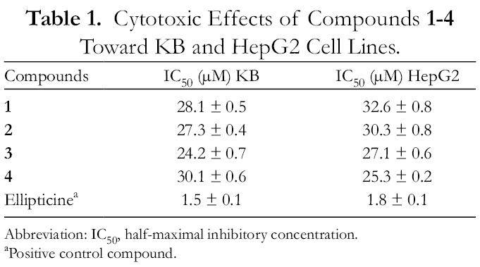
Compounds 1-4 were evaluated in vitro for their cytotoxic activity by using the 3-[4,5-dimethylthiazol-2-yl]-2,5-diphenyltetrazolium bromide (MTT) assay against 2 human cancer cell lines (KB and HepG2). Compounds 1-4 displayed weak cytotoxic activity toward both cell lines with half-maximal inhibitory concentration (IC50) values ranging from 24.2 ± 0.3 to 32.6 ± 0.8 µM (Table 1).
Cytotoxic Effects of Compounds 1-4 Toward KB and HepG2 Cell Lines.
HR-ESI-MS was carried out on an Agilent 6530 Accurate Mass Quadrupole Time-of-Flight (QTOF) liquid chromatography (LC)/MS. The QTOF instrument was set at 2 GHz extended dynamic range resolution mode, negative ESI capillary voltage of 3500 V, fragmentor voltage of 175 V, MS scan ranging from m/z 100-1700, and an MS acquisition rate of 1.0 spectra/second. The NMR spectra were recorded on a Bruker 500 MHz spectrometer and optical rotation on a Jasco P2000 polarimeter. Column chromatography was performed using silica gel, reverse phase C-18, and diaion HP-20 resins as the stationary phase. HPLC was carried out using an AGILENT 1100 HPLC system. For thin-layer chromatography, precoated silica gel 60 F254 and RP-18 F254S plates were used. The compounds were visualized by spraying with a 5% solution of sulfuric acid in ethanol, followed by heating with a heat gun.
Plant Material
Aralia armata (Wall.) Seem leaves, collected in Vinh Phuc province, Vietnam, in December 2017, were identified by Dr Nguyen, The Cuong at the Institute of Ecology and Biological Resources, VAST. A voucher specimen (coded: NCCT-P71) was deposited at the Institute of Marine Biochemistry, VAST.
Extraction and Isolation
The dried leaves of A. armata (6 kg) were powdered and then ultrasonically extracted with methanol (MeOH), 3 times (each with 15 L of MeOH for 30 minutes). After filtration, the solvent was removed in vacuo to give 240 g of dry extract, which was suspended in water and successively partitioned with dichloromethane and ethyl acetate to give organic soluble fractions and a water layer. The water layer was chromatographed on a diaion (HP-20) column washing with water to remove salts and oligosaccharides. Saponins were stepwise eluted by MeOH/water (25%, 50%, 75%, and 100% vol of MeOH) to give 4 fractions AAW1-AAW4. Fraction AAW2 (15.5 g) was separated by silica gel column chromatography, eluting with dichloromethane/MeOH (1/0-0/1, v/v) to give 5 fractions AAW2A- AAW2E. Fraction AAW2C was further fractionated on a reverse phase C18 column, eluting with MeOH/water (2/2.5, v/v) to give 4 fractions AAW2C1- AAW2C4. Fraction AAW2C3 was chromatographed on a silica gel column, eluting with dichloromethane/acetone/water (1/4/0.5, v/v/v) to yield compounds 1 (12.0 mg) and 2 (11.0 mg), respectively. Fraction AAW2C4 was chromatographed on a reverse-phase C18 column, eluting with acetone/water (2/5, v/v) to give compound 4 (11 mg). Fraction AAC2 (40 g) was chromatographed on a silica gel column eluting with dichloromethane/MeOH/water (1.5/5/0.1, v/v/v) to give compound 3 (9 mg).
The assignments were made from 1-dimensional NMR (1H, 13C, distortionless enhancement by polarization transfer) and 2-dimensional NMR (heteronuclear single quantum correlation, heteronuclear multiple bond correlation, H-H correlation spectroscopy, nuclear Overhauser effect spectroscopy) spectra.
Research on the chemical constituents of A. armata leaves led to the isolation of 4 oleanane-type triterpene glycosides including 1 new one, oleanolic acid-[28-O-β-d-glucopyranosyl]-3-O-[β-d-glucopyranosyl(1→6)-β-d-glucopyranosyl](1→3)[α-l-arabinofuranosyl(1→4)]-β-d-glucuronopyranoside (1) and 3 known ones, oleanolic acid-[28-O-β-d-glucopyranosyl]-3-O-[β-d-galactopyranosyl(1→3)]-[β-d-glucopyranosyl(1→2)]-β-d-glucuronopyranoside (2) chikusetsusaponin IVa methyl ester (3), and chikusetsusaponin IV (4). Their chemical structures were elucidated by using a combination of HR-ESI-MS, 1D and 2D NMR spectral data, as well as by comparison with the previous literature. This is the first report of the full NMR spectroscopic data of 2. Compounds 1-4 displayed weak cytotoxic activity toward KB and HepG2 cell lines with IC50 values ranging from 24.2 ± 0.3 to 32.6 ± 0.8 µM in in vitro assay. The above results further confirmed that oleanane-type triterpene glycosides are typically present in Aralia species.
Supplemental Material
online supplementary file 1 - Supplemental material for Araliaarmoside: A New Triterpene Glycoside Isolated From the Leaves of Aralia armata
Supplemental material, online supplementary file 1, for Araliaarmoside: A New Triterpene Glycoside Isolated From the Leaves of Aralia armata by Pham Hai Yen, Nguyen Thi Cuc, Phan Thi Thanh Huong, Nguyen Xuan Nhiem, Nguyen Thị Hong Chuong, Giang Thi Kim Lien, Bui Huu Tai, Nguyen Van Tuyen, Chau Van Minh and Phan Van Kiem in Natural Product Communications
Footnotes
Declaration of Conflicting Interests
The author(s) declared no potential conflicts of interest with respect to the research, authorship, and/or publication of this article.
Funding
The author(s) disclosed receipt of the following financial support for the research, authorship, and/or publication of this article: This research is funded by Vietnam National Foundation for Science and Technology Development (NAFOSTED) under grant number 104.01-2017.08.
ORCID iD
Phan Van Kiem
Supplemental Material
Supplemental material for this article is available online.
References
1.
JasonAC.EllaSHC. The medicinal chemistry of genus Aralia. Curr Top Med Chem. 2014;14(24):2783-2801.
2.
MiaoH.SunY.YuanYet al. Herbicidal and cytotoxic constituents from Aralia armata (wall.) seem. Chem Biodivers. 2016;13(4):437-444.doi:10.1002/cbdv.201500130http://www.ncbi.nlm.nih.gov/pubmed/26948515
3.
BiL.TianX.DouF.HongL.TangH.WangS. New antioxidant and antiglycation active triterpenoid saponins from the root bark of Aralia taibaiensis. Fitoterapia. 2012;83(1):234-240.doi:10.1016/j.fitote.2011.11.002http://www.ncbi.nlm.nih.gov/pubmed/22088497
4.
HuM.OgawaK.SashidaY.Pei-GenX.XiaoPG. Triterpenoid glucuronide saponins from root bark of Aralia armata. Phytochemistry. 1995;39(1):179-184.doi:10.1016/0031-9422(94)00902-6
5.
ChiVV. Dictionary of Vietnamese medicinal plants. Medicine Publishing House; 2012:956-957.
6.
ShaoCJ.KasaiR.XuJ-D.TanakaO. Saponins from roots of Kalopanax septemlobus (Thunb.) Koidz., Ciqiu: Structures of Kalopanax-saponins C, D, E and F. Chem Pharm Bull. 1989;37(2):311-314.doi:10.1248/cpb.37.311
7.
TrangDT.NhiemNX.DTHet al. Oleanane saponins from the leaves of Aralia armata (Wall) Seem. Vietnamese J Med Material. 2015;1:12-17.
8.
Rengifo CarrilloM.Mitaine-OfferA-C.MiyamotoTet al. Oleanane-type glycosides from Pittosporum tenuifolium “variegatum” and P. tenuifolium “gold star”. Phytochemistry. 2017;140:166-173.doi:10.1016/j.phytochem.2017.04.013http://www.ncbi.nlm.nih.gov/pubmed/28500929
9.
TanakaT.NakashimaT.UedaT.TomiiK.KounoI. Facile discrimination of aldose enantiomers by reversed-phase HPLC. Chem Pharm Bull. 2007;55(6):899-901.doi:10.1248/cpb.55.899http://www.ncbi.nlm.nih.gov/pubmed/17541189
10.
YoshikawaM.MurakamiT.KishiA.KageuraT.MatsudaH. Medicinal flowers. III. Marigold (1): hypoglycemic, gastric emptying inhibitory, and gastroprotective principles and new oleanane-type triterpene oligoglycosides, calendasaponins A, B, C, and D, from Egyptian Calendula officinalis. Chem Pharm Bull. 2001;49(7):863-870.doi:10.1248/cpb.49.863http://www.ncbi.nlm.nih.gov/pubmed/11456093
11.
Vidal-OllivierE.BalansardG.FaureR.BabadjamianA. Revised structures of triterpenoid saponins from the flowers of Calendula officinalis. J Nat Prod. 1989;52(5):1156-1159.doi:10.1021/np50065a042
Supplementary Material
Please find the following supplemental material available below.
For Open Access articles published under a Creative Commons License, all supplemental material carries the same license as the article it is associated with.
For non-Open Access articles published, all supplemental material carries a non-exclusive license, and permission requests for re-use of supplemental material or any part of supplemental material shall be sent directly to the copyright owner as specified in the copyright notice associated with the article.