A new macrocyclic glycoside named helilobatoside A (1) and 5 known phenyl glycosides as 3,5-dimethoxy-4-hydroxyphenyl-1-O-β-d-glucopyranoside (2), tachioside (3), isotachioside (4), 1-(4-hydroxy-3-methoxyphenyl)-1-propanone-3-O-β-d-glucopyranoside (5), and 1-(4-hydroxy-3,5-dimethoxyphenyl)-1-propanone-3-O-β-d-glucopyranoside (6), were isolated from the wood of Heliciopsis lobata (Merr.) Sleumer. Their chemical structures were elucidated using a combination of high-resolution electrospray ionization mass spectrometry, 1-dimensional (1D) and 2D nuclear magnetic resonance (NMR) spectral data as well as by comparison with data in the previous literature. This is the first time the 13C NMR data of compounds 5 and 6 were reported and also were assigned by heteronuclear single quantum correlation and heteronuclear multiple bond correlation spectra. Compounds 2-6 were first isolated from Heliciopsis genus. The isolated compounds were evaluated for their antioxidant and hepatoprotective activities in vitro. Compound 2 showed potential as an antioxidant in a 2,2-diphenyl-1-picryl-hydrazyl-hydrate assay (half-maximal inhibitory concentration [IC50] = 6.07 ± 0.17 µg/mL) and in thio-barbituric acid reactive substances assay (IC50 = 89.55 ± 8.26 µg/mL). This compound could also reduce the toxic effects of carbon tetrachloride on HepG2 survival and significantly protect the viability of cells up to 52.25 ± 4.36% at the 100 µg/mL treatment (P < 0.05). Thus, with obtained results, the hepatoprotective activity of compound 2 could be related to radical scavenging and limited the lipid peroxidative activities.
Heliciopsis lobata (Merr.) Sleumer belongs to Proteaceae family, which was used for the treatment of several diseases as a traditional medicine in China and Vietnam.1-4 Several researches focused on investigating chemical constituents from leaves of this plant and indicated that phenolic glycosides and arbutin derivatives were the major groups2-5 together with grevillic acid, grevillone, 4-hydroxy-trans-cinnamic acid, and daucosterol.6 In Vietnam, H. lobata grows scattered from northern to southern areas. The dried wood of this species was used as an herbal medicine independently or in combination with the other materials for hepatoprotection or treating liver diseases.1 This article reports the isolation, structural determination of 1 new macrocyclic glycoside and 5 known phenyl glycosides from the wood of H. lobata, and their antioxidant and hepatoprotective activities also were further screened.
Results and Discussion
Compound 1 was obtained as a white amorphous powder from methanol extract of the wood of H. lobata. The infrared (IR) spectrum of 1 showed a broad absorption band of hydroxyl, carbonyl, aromatic ring, and ether groups at 3401, 1711, 1450, 1073 cm−1, respectively (Supplemental Figure S33). The 1H nuclear magnetic resonance (NMR) spectrum of compound 1 (Supplemental Figure S2) showed 5 signals of 2 aromatic rings from 7.19 to 7.96 ppm. Two signals at δH 7.97 and 7.18 (each, 2H) suggested for the presence of a para-substituted aromatic ring, and 3 inter-related signals in the ABX system at δH 7.81 (1H, dd, J = 8.5, 2.0 Hz), 7.39 (1H, d, J = 8.5 Hz), and 7.42 (1H, d, J = 2.0 Hz) demonstrated the presence of a 1,3,4-trisubstituted aromatic ring. The 2 sugar units were identified by 2 anomeric proton signals at δH 5.36 (d, J = 8.0 Hz) and 5.25 (d, J = 7.5 Hz). The remaining signals of the 2 sugars appeared in the region of the chemical shift from 3.20 to 4.50 ppm, and 1 methoxy group was identified by a singlet signal at δH 3.79 (3H). The 13C NMR spectrum (Supplemental Figure S3) together with the heteronuclear single quantum correlation (HSQC) spectrum (Supplemental Figure S5) of compound 1 confirmed the presence of 25 signals typical for 27 carbon atoms. Four signals of the para-substituted ring were identified at δC 160.7, 115.9, 131.2, and 122.6; 6 signals of the 1,3,4-trisubstituted aromatic ring were determined at δC 149.9, 148.5, 122.5, 123.0, 114.4, and 112.3; 2 carbonyl ester groups were at δC 165.3 and 165.9, and a methoxy group was at δC 55.6. The remaining 12 carbon signals belong to 2 sugar molecules including 2 anomeric carbons at δC 98.3 and 96.9 and 2 oxygenated methylene carbons at δC 68.2 and 65.1. The formations of the 2 ester linkages were also demonstrated by the heteronuclear multiple bond correlation (HMBC) interactions between the H-6′″ protons (δH 4.36/4.14) of the first sugar and carbon at δC 165.3, and between H-6″ protons (δH 4.06/4.42) of the second sugar and carbon at δC 165.0. There were also the HMBC correlation between the anomeric proton at δH 5.36 with carbon C-4 (δC 160.7) of the para-aromatic ring, and between the second anomeric proton at δH 5.25 with carbon C-4′ (δC 149.9) of the other aromatic ring. On the HSQC spectrum (Supplemental Figure S5), the proton signals at δH 7.81, 7.39, and 7.42 interacted with the corresponding carbon at δC 123.0, 114.4, and 112.3. Simultaneously HMBC interaction between H-6′ and C-4′, C-2′, C-7′, between H-2′ and C-4′, C-7′, and between methoxy protons at δH 3.79 with C-3′ (δC 148.5) confirmed the methoxy group attached to C-3′ of the 1,3,4-trisubstituted aromatic ring. Similarly, the chemical shift values of the remaining protons and carbon were determined precisely by 2-dimensional analysis of HSQC and HMBC spectra (Supplemental Figures S5, S6) and confirmed the linkages between the 2 sugars and the 2 aromatic rings as shown in Figure 1. With the analytical results mentioned above, the chemical structure of 1 was suggested to be similar to the chemical structure of clemahexapetoside B (1a),7 in which compound 1 lacked a methoxy group at C-3 position compared with 1a. Careful analysis and comparison of the NMR data of compounds 1 and 1a showed a complete coincidence of the NMR spectral values of 2 sugar molecules, including an allose and a glucose (Table 1), as well as the corresponding match between the NMR spectral values of the vanillic acid unit. The differences occurred in the para-substituted aromatic ring of 1 compared with the vanillic acid unit of 1a. This evidence further confirmed the loss of a methoxy group at C-3 of 1 as shown in Supplemental Table S1. The large proton coupling constants of the 2 anomeric protons at δH 5.25 (J = 7.5 Hz) and δH 5.36 (J = 8.0 Hz) proved that the 2 sugar linkages must be in the β-form. In addition, the 1H-1H correlation spectroscopy (COSY) cross-peaks between H-1″/H-2″/H-3″/H-4″/H-5″/H-6″ protons of the glucose and between H-1′″/H-2′″/H-3′″/H-4′″/H-5′″/H-6′″ protons of the allose were observed (Supplemental Figure S4) confirming the NMR assignments of these sugars. Proton H-3′″ appeared as a triplet (J = 3.0 Hz) confirming H-3′″ must be equatorial orientation as in clemahexapetoside B.7 Thus, the expected molecular formula of 1 is C27H30O15. This was further confirmed by the high-resolution electrospray ionization mass spectrometry (HR-ESI-MS) with the appearance of an ion peak at m/z 593.1499 [M − H]− (calcd. for C27H29O15: 593.1506) (Supplemental Figure S1). Furthermore, monosaccharides in the sugar residue were confirmed to be d-glucose and d-allose by hydrolysis (Supplemental Figure S32).7 From the spectral data and analysis above, the chemical structure of compound 1 was determined as shown in Figure 2, a new macrocyclic glycoside and named helilobatoside A.
The 1H-1H correlation spectroscopy and key heteronuclear multiple bond correlations of compound 1.
The 1H-NMR and 13C NMR Data for Compound 1 in DMSO-d6.
Abbreviations: NMR, nuclear magnetic resonance; HMBC, heteronuclear multiple bond correlation; HSQC, heteronuclear single quantum correlation; COSY, correlationspectroscopy; DMSO, dimethyl sulfoxide; Glc, β-D-glucopyranosyl;All, β-D-allopyranosyl.
The assignments done by 1-dimensional NMR (1H, 13C, DEPT), 2D NMR (HSQC, HMBC, H-H COSY) spectra.
a Measured in 125 MHz.
bMeasured in 500 MHz.
cOverlapped signals.
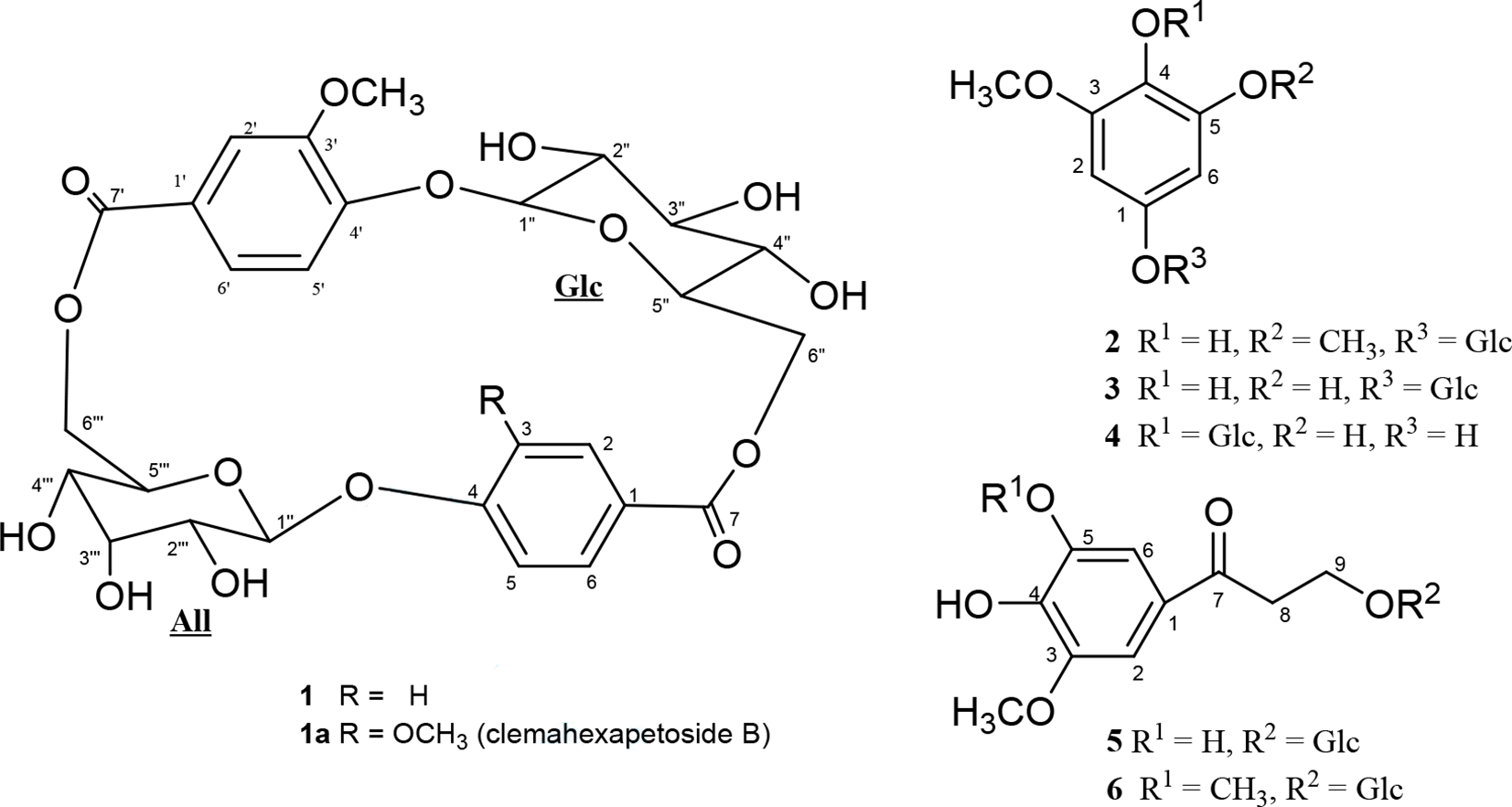
Chemical structures of compounds 1-6 and 1a. COSY, correlation spectroscopy; HMBC, heteronuclear multiple bond correlations.
By analyzing the HR-ESI-MS, 1D and 2D NMR spectra (Table 2), compounds 5 (Supplemental Figures S22–S26) and 6 (Supplemental Figures S27–S31) were identified as 1-(4-hydroxy-3-methoxyphenyl)-1-propanone-3-O-β-d-glucopyranoside (5),8,9 and 1-(4-hydroxy-3,5-dimethoxyphenyl)-1-propanone-3-O-β-d-glucopyranoside (6). However, to the best of our knowledge, this is the first time the 13C NMR data of compounds 5 and 6 were reported and also were assigned by HSQC and HMBC spectra. The other known compounds were identified as 3,5-dimethoxy-4-hydroxyphenyl-1-O-β-d-glucopyranoside (2),10 tachioside (3),11 isotachioside (4),11 by analyzing their HR-ESI-MS, 1D and 2D NMR spectral data as well as by comparison with the previous literature and found to match well (Supplemental Table S2, Figures S7–S21). This is the first report of compounds 2-6 from genus Heliciopsis.
Abbreviations: NMR, nuclear magnetic resonance; CD3OD, deuterated methanol.
NMR data were assigned by 1D and 2D NMR spectra.
aMeasured in CD3OD.
bMeasured in 125 MHz.
cMeasured in 500 MHz.
Compounds 1-6 were evaluated in vitro for their antioxidant and hepatoprotective activities. The results of their antioxidant activity are shown in Table 3. As reported, oxidative stress often relates to liver pathogenesis.12 Therefore, the antioxidant activity of compounds 1-6 was examined by 2,2-diphenyl-1-picryl-hydrazyl-hydrate assay (DPPH) radical scavenging and thio-barbituric acid reactive substances (TBARS) lipid peroxidation inhibition assays (Table 3). As a result, compound 2 showed the strongest potential antioxidant activity in DPPH assay (half-maximal inhibitory concentration [IC50] = 6.07 ± 0.17 µg/mL), while the others presented weaker activities with the IC50 values ranging from 43.34 ± 5.14 µg/mL to higher than 500 µg/mL. Again, compound 2 also presented much stronger inhibition of lipid peroxidation (IC50 = 89.55 ± 8.26 µg/mL) compared with that of other tested compounds (IC50 = 462.93 ± 28.86 µg/mL or higher than 500 µg/mL). Trolox, a reference compound, exhibited strong activity with the IC50 of 8.05 ± 0.62 µg/mL. Due to the very limited activity in TBARS assay, the compounds 1, 4-6 were not selected for further hepatoprotective assessment.
Antioxdant Activities of the Studied Compounds in DPPH and TBARS Assays
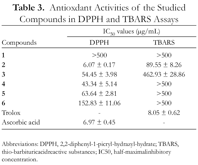
As reported, HepG2 cells are accepted for the in vitro study of polarized human hepatocytes.13 Therefore, carbon tetrachloride (CCl4) induced toxicity in the HepG2 cell line was used to determine the hepatoprotective effect of compound 2. The viability of HepG2 cells was strongly decreased when treated with 40 mM CCl4 for 2 hours (Table 4). CCl4 is commonly used as a chemical inducer of hepatocellular injury since this toxic agent causes lipid peroxidation, and these metabolic products would covalently bind to cellular lipids and proteins.14 In accordance with lipid peroxidative inhibition, compound 2 could reduce the toxic effect CCl4 on HepG2 cells (Table 3). At the highest tested concentration, 2 exhibited significantly hepatocellular protective activity against CCl4 toxic induction as the viability of cells maintained up to 52.25% ± 4.36% (P < 0.05). It is well recognized that radicals play a pivotal role in the cellular toxic effect of CCl4 of which could be prevented by using the antioxidant.14 Consequently, the hepatoprotective activity of 2 might be associated with radical scavenging and inhibition of lipid peroxidation.
Hepatoprotective Activities of Tested Compounds Against CCl4 Toxic Induction
The NMR spectra were recorded on a Bruker 500 MHz spectrometer. Optical rotation was measured on a Jasco P2000 polarimeter. HR-ESI-MS was carried out on an Agilent 6530 Accurate Mass Q-TOF LC/MS. The QTOF instrument was set at 2 GHz extended dynamic range resolution mode, negative ESI capillary voltage of 3500 V, fragmentor voltage of 175 V, MS scan ranging at m/z 100-1700, and MS acquisition rate of 1.0 spectra per second. Column chromatography was performed using silica gel, reverse phase C-18, and Diaion HP-20 resins as a stationary phase. Thin-layer chromatography was carried out using precoated silica gel 60 F254 and RP-18 F254S plates. The spots were visualized by spraying with a solution of sulfuric acid 5% in ethanol followed by heating with a heat gun.
Plant Material
The plant sample was collected at Bach Thong, Bac Kan province, Vietnam in December 2019. Its scientific name was identified as Heliciopsis lobata (Merr.) Sleumer by MSc. Nghiem Duc Trong at the Hanoi University of Pharmacy, Vietnam. A voucher specimen (coded: NCCT-P69B) was deposited at the Institute of Marine Biochemistry, VAST.
Extraction and Isolation
The dried wood of H. lobata (10 kg) was powdered and then ultrasonically extracted with methanol (MeOH) three times (each 20 L of MeOH in 30 minutes). After filtration, the solvent was removed in vacuo to give 150 g of methanol extract. This extract was suspended in water and successively partitioned with hexane and ethyl acetate to give organic soluble fractions and water layer. The water layer was chromatographed on a Diaion (HP-20) column washing with water to remove salts and oligosaccharides. Saponin compounds were stepwise eluted by methanol/water (25%, 50%, 75%, and 100% vol of methanol) to give4 fractions HLN1-HLN4. Fraction HLN3 (11.0 g) was chromatographed on a reverse-phase C18 column, eluting with acetone/water (1/3, v/v) to give 3 fractions HLN3A (5.6 g), HLN3B (5.5 g), and HLN3C (1.5 g). Compound 1 (7.2 mg) was obtained from fraction HLN3B (5.5 g) after running a chromatographic column with silica gel as a stationary phase and eluting with dichloromethane/acetone/water (1/3/0.5, v/v/v). The fraction HLN3A (5.6 g) was chromatographically separated on a silica gel column, eluted with dichloromethane/acetone/water (1/5/0.5, v/v/v) to yield compounds 2 (370.0 mg), 3 (23.0 mg), and 4 (25 mg). Finally, fraction HLN3C (1.5 g) was chromatographed on a silica gel column, eluted with dichloromethane/acetone/water (1/2.5/0.3, v/v/v) to yield compounds 5 (21.0 mg) and 6 (33 mg).
Helilobatoside A (1)
White amorphous powder, : +113.0° (c 0.1, MeOH); IR (potassium bromide [KBr]) ν (cm−1): 3401 (OH), 2934 (CH), 1711 (C = O), 1450 (Ar), 1073 (C-O-C) (see Figure S33); HR-ESI-MS m/z 593.1499 [M − H]− (calcd. for C27H29O15: 593.1506). 1 H NMR (imethyl sulfoxide [DMSO]-d6, 500 MHz) and 13C NMR (DMSO-d6, 125 MHz) data, see Table 1.
White amorphous powder, : −18.0° (c 0.1, MeOH); HR-ESI-MS m/z 387.1283 [M − H]− (calcd. for C17H23O10: 387.1291); 1 H NMR (CD3OD, 500 MHz) and 13C NMR (CD3OD, 125 MHz) data, see Table 2.
DPPH Scavenging Assay
Free radical-scavenging activity was investigated according to the method described by Pyrzynska et al.15 Prepared samples at different concentrations were examined by their reactivity with a methanolic DPPH solution. The decrease in the absorbance was measured at 517 nm. The calibration curve of % DPPH scavenging activity versus concentration was plotted to calculate IC50 values.
Antioxidant TBARS Assay
The TBARS assay is a well-recognized, established method for quantifying lipid peroxides. TBARS assay values are usually reported in malonaldehyde equivalents, a compound that results from the decomposition of polyunsaturated fatty acid lipid peroxides. The inhibition of lipid peroxidation by compounds was determined by following Zhu et al.16
In Vitro Hepatoprotective Activity Against CCl4 Induced Toxicity on HepG2 Cells
The HepG2 cells (ATCC HB-8065) were obtained from the American Type Culture Collection (ATCC, Rockville, MD, USA) and grown in Dulbecco’s modified Eagle medium supplemented with 10% fetal bovine serum and 1% penicillin/streptomycin (Invitrogen, Carlsbad, CA, USA) in a humidified incubator at 37 °C, 5% carbon dioxide. The cells at log phase were preseeded in 96-well plate at the density of 3 × 10⁴ cells/well and incubated overnight. Subsequently, cells were treated with different concentrations of samples with or without 40 mM CCl4 for 2 hours. The viability of cells was then determined by 3-(4,5-dimethylthiazol-2-yl)-2,5-diphenyltetrazolium bromide assay.17,18
Statistical Analysis
All experiments in this study were run in triplicate. The results are shown as the mean ± SD. Data were analyzed using GraphPad Prism 5.0 (GraphPad Software, Inc., San Diego, CA, USA). Statistically significant differences were determined at the P < 0.05 level.
Acid Hydrolysis and Confirmation of Monosaccharide
Compound 1 (1 mg) was heated at 105 °C in a sealed ampule with DMSO 100 µg/mL and 2 M trifluoro-acetic acid (TFA) 10 µL for 2 hours. After removing the TFA and DMSO, the reaction mixture was dissolved in water (10 mL) and extracted with ethyl acetate (2 × 5 mL). The water (H2O) layer was concentrated and the residue dissolved with acetonitrile (MeCN)-H2O (8:2); then sugars were detected by HPLC Hitachi Chromaster 5210. Chromatographic conditions: column, Asahipak NH2P-50 (4.6 mm × 25 cm); solvent, 80% MeCN in water; flow rate, 1.0 mL/min. Detections: An SEDEX 85 ELSD detector was used. The carrier gas was nitrogen; the drift tube temperature was set at 40 °C; pressure was set at 3.5 bar, and gain was set at 6. Peaks at a retention time of 11.2 and 13.7 were confirmed to be d-allose and d-glucose, respectively, by comparison of retention time with that of authentic d-allose and d-glucose, prepared in the same manner (Supplemental Figure S32).7
Conclusions
A new phenyl glycoside named helilobatoside A (1) and 5 known phenyl glycosides, 3,5-dimethoxy-4-hydroxyphenyl-1-O-β-d-glucopyranoside (2), tachioside (3), isotachioside (4), 1-(4-hydroxy-3-methoxyphenyl)-1-propanone-3-O-β-d-glucopyranoside (5), and 1-(4-hydroxy-3,5-dimethoxyphenyl)-1-propanone-3-O-β-d-glucopyranoside (6), were isolated from the wood of H. lobata (Merr.) Sleumer. Their chemical structures were elucidated using a combination of HR-ESI-MS, 1D and 2D NMR spectral data as well as by comparison with data from the previous literature. This is the first time the 13C NMR data of compounds 5 and 6 were reported and also were assigned by HSQC and HMBC spectra. Compounds 2-6 were first isolated from the Heliciopsis genus. The isolated compounds were tested for their in vitro antioxidant and hepatoprotective activities. The compound 2 exhibited as the most potential antioxidant activity among them in DPPH assay (IC50 = 6.07 ± 0.17 µg/mL) and in TBARS assay (IC50 = 89.55 ± 8.26 µg/mL) as well. In the hepatocellular protective assay, compound 2 could significantly protect HepG2 cells against CCL4 induced toxicity (P < 0.05). Summarizing altogether, compound 2 presents as a promising antioxidant and hepatoprotective agent.
Supplemental Material
Figure S1 - Supplemental material for Antioxidant and Hepatoprotective Activity of Phenyl Glycosides Isolated From Heliciopsis lobata
Supplemental material, Figure S1, for Antioxidant and Hepatoprotective Activity of Phenyl Glycosides Isolated From Heliciopsis lobata by Bui Van Trung, Do Thi Thao, Duong Hong Anh, Phan Van Kiem and Pham Hung Viet in Natural Product Communications
Footnotes
Declaration of Conflicting Interests
The author(s) declared no potential conflicts of interest with respect to the research, authorship, and/or publication of this article.
Funding
The author(s) disclosed receipt of the following financial support for the research, authorship, and/or publication of this article: The authors are indebted to the Vietnamese Governmental North-western Research Program for the valuable financial support (Grant No. KHCN-TB.11C/13-18).
ORCID iD
Phan Van Kiem
References
1.
ChiVV. Dictionary of Vietnamese Medicinal Plants. Medicine Publishing House; 2012:973-974.
2.
HeQ-Q.LiuM-S.JinD-J.KongL-Y. Phenolic glycosides from leaves of Hopiciopsis lobata. J Asian Nat Prod Res. 2006;8(4):373-377.doi:10.1080/10286020500172251http://www.ncbi.nlm.nih.gov/pubmed/16864450
3.
LiuM.KongL.FongWF.HeQ.JinD.ShenX. A new phenolic glucoside from the leaves of Heliciopsis lobata. Fitoterapia. 2008;79(5):398-399.doi:10.1016/j.fitote.2008.03.006http://www.ncbi.nlm.nih.gov/pubmed/18534782
4.
LiuM.KangS.ZhangJ.ZhangX. A new arbutin derivative from the leaves of Heliciopsis lobata. Nat Prod Res. 2010;24(19):1861-1864.doi:10.1080/14786419.2010.482938http://www.ncbi.nlm.nih.gov/pubmed/21104533
5.
QiW-Y.OuN.WuX-D.XuH-M. New arbutin derivatives from the leaves of Heliciopsis lobata with cytotoxicity. Chin J Nat Med. 2016;14(10):789-793.doi:10.1016/S1875-5364(16)30094-2http://www.ncbi.nlm.nih.gov/pubmed/28236409
6.
LiuM.HeQ.JinD.KongL. Studies on the chemical constituents of Heliciopsis lobata. Zhongguo Yaoxue Zazhi. 2005;40(12):893-894.
RolfA.LennartNL. Monoaryl and cyclohexenone glycosides from needles of Pinus sylvestris. Phytochemistry. 1988;27(2):559-562.
9.
KuoY-H.HuangS-L.ChangC-I. A phenolic and an aliphatic lactone from Diospyros maritima. Phytochemistry. 1998;49(8):2505-2507.doi:10.1016/S0031-9422(98)00360-4
10.
ChungM-I.LaiM-H.YenMH.WuRR.LinCN. Phenolics from Hypericum geminiflorum. Phytochemistry. 1997;44(5):943-947.doi:10.1016/S0031-9422(96)00644-9
11.
ShogoI.ManamiS.HiroshiK.HideakiO.KazuoY. Aromatic glycosides from Berchemia racemosa. Phytochemistry. 1987;26(10):1814-2811.
12.
LiS.TanH-Y.WangNet al. The role of oxidative stress and antioxidants in liver diseases. Int J Mol Sci. 2015;16(11):26087-26124.doi:10.3390/ijms161125942http://www.ncbi.nlm.nih.gov/pubmed/26540040
13.
RajangmJ.ChristinaAJM. Evaluation of hepatoprotective and antioxidant potential of methanolic extract of Polyalthiya longifolia fruits: An in-vitro and in-vivo approach. J Appl Pharm Sci. 2013;3(2):069-076.
14.
BollM.WeberLW.BeckerE.StampflA. Mechanism of carbon tetrachloride-induced hepatotoxicity. Hepatocellular damage by reactive carbon tetrachloride metabolites. Z Naturforsch C J Biosci. 2001;56(7-8):649-659.doi:10.1515/znc-2001-7-826http://www.ncbi.nlm.nih.gov/pubmed/11531102
15.
PyrzynskaK.PękalA. Application of free radical diphenylpicrylhydrazyl (DPPH) to estimate the antioxidant capacity of food samples. Anal Methods. 2013;5(17):4288-4295.doi:10.1039/c3ay40367j
16.
ZhuN.ShengS.LiDet al. Antioxidative flavonoid glycosides from quinoa seeds (Chenopodium quinoa Willd). Journal of Food Lipids. 2001;8(1):37-44.doi:10.1111/j.1745-4522.2001.tb00182.x
17.
ÖzerkanD.ÖzsoyN.YılmazE. Vitamin D and melatonin protect the cell’s viability and ameliorate the CCl4 induced cytotoxicity in HepG2 and Hep3B hepatoma cell lines. Cytotechnology. 2015;67(6):995-1002.doi:10.1007/s10616-014-9738-8http://www.ncbi.nlm.nih.gov/pubmed/24997582
18.
GonzálezLT.MinskyNW.EspinosaLEM.ArandaRS.MeseguerJP.PérezPC. In vitro assessment of hepatoprotective agents against damage induced by acetaminophen and CCl4. BMC Complement Altern Med. 2017;17(1):39doi:10.1186/s12906-016-1506-1http://www.ncbi.nlm.nih.gov/pubmed/28086854
Supplementary Material
Please find the following supplemental material available below.
For Open Access articles published under a Creative Commons License, all supplemental material carries the same license as the article it is associated with.
For non-Open Access articles published, all supplemental material carries a non-exclusive license, and permission requests for re-use of supplemental material or any part of supplemental material shall be sent directly to the copyright owner as specified in the copyright notice associated with the article.