
Abstract
Baicalein is one of the main bioactive compounds in Scutellaria baicalensis. In this study, its inhibitory mechanism against α-glucosidase from Saccharomyces cerevisiae was clarified based on experimental and molecular simulation methods. According to HPLC analysis, the α-glucosidase inhibitory activity of S. baicalensis (IC50, 6.75 ± 0.08 μg mL−1) was superior to that of acarbose (IC50, 2.52 ± 0.12 mg mL−1). The fluorescence results suggested that baicalein could form a complex with α-glucosidase at the molar ratio of 1 under the drive of hydrogen bonding and van der Waals force. Molecular docking showed that baicalein could form hydrogen bonds with Trp391, Arg428, Gly566,and Glu771 of α-glucosidase, and interacted with Phe385, Phe389, Arg387, Glu429, Phe444, Trp789, and Trp710 by hydrophobic force, which coincided with the experimental results. It can be concluded that baicalein is a potential α-glucosidase inhibitor for controlling the postprandial blood glucose level of diabetics.
Diabetes mellitus is a common chronic metabolic disease that causes many complications such as coronary artery, stroke, amputation, renal failure, and blindness, which aggravates our economic and social burden. 1 -3 Although the traditional synthetic α-glucosidase inhibitors (acarbose, voglibose, and miglitol) can effectively control the postprandial blood glucose level of diabetics, their side effects sometimes limit their acceptance. 4 -6 Some naturally occurring flavonoids (eg, rutin, quercetin, and taxifolin) were reported to be strong α-glucosidase inhibitors with potential application. 7 -9 Baicalein (Figure 1) is one of the main flavonoids in Scutellaria baicalensis. Its molecular structure with 3 adjacent phenolic hydroxyl groups endows it with a lot of health-promoting properties, such as antioxidant, antitumor, and anti-inflammatory activities. 10 -12 However, to the best of our knowledge, there is no systematic report about its α-glucosidase inhibitory mechanism. In view of this, in this study, its inhibitory mechanism against α-glucosidase from Saccharomyces cerevisiae has been clarified based on experimental and molecular simulation methods.

Chemical structure of baicalein.
α-Glucosidase can hydrolyze p-nitrophenyl-α-d-glucopyranoside (pNP-G) to glucose and p-nitrophenyl (pNP), which has strong absorption at 405 nm. The traditional α-glucosidase inhibitory assay is carried out by measuring the content of the product (p-nitrophenol, pNP) based on a colorimetric method. 13 However, the color of the sample can disturb the measurement. In this study, we established a HPLC method for evaluating the formation of pNP in order to reflect exactly the α-glucosidase inhibitory activity of baicalein. As shown in Figure 2, the substrate (pNP-G) and product (pNP) could be well separated and detected at 405 nm under our HPLC method. It was also found that baicalein exhibited strong α-glucosidase inhibitory activity in a concentration-dependent manner (Figure 3). At 10 µg mL−1, its inhibitory activity was as high as 87%. Its IC50 value of 6.75 ± 0.08 µg mL−1 was significantly higher (P < 0.05) than that of acarbose (IC50, 2.52 ± 0.12 mg mL−1).

HPLC profile for the α-glucosidase assay.

α-Glucosidase inhibitory activities of baicalein (a) and acarbose (b).
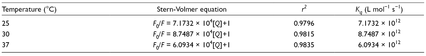
To study the interaction behavior between baicalein and α-glucosidase, the effect of baicalein on the fluorescence spectrum (FS) of α-glucosidase at different temperatures (25°C, 30°C, and 37°C) was investigated (Figure 4). Compared with its initial fluorescence intensity (F 0), the fluorescence intensity (F) of α-glucosidase gradually decreased with increasing concentrations ([Q]) of baicalein. The quenching constant (K q) between baicalein and α-glucosidase could be calculated based on the Stern-Volmer equation, where τ 0 (the average life of proteins) was about 10–8 s 14 :
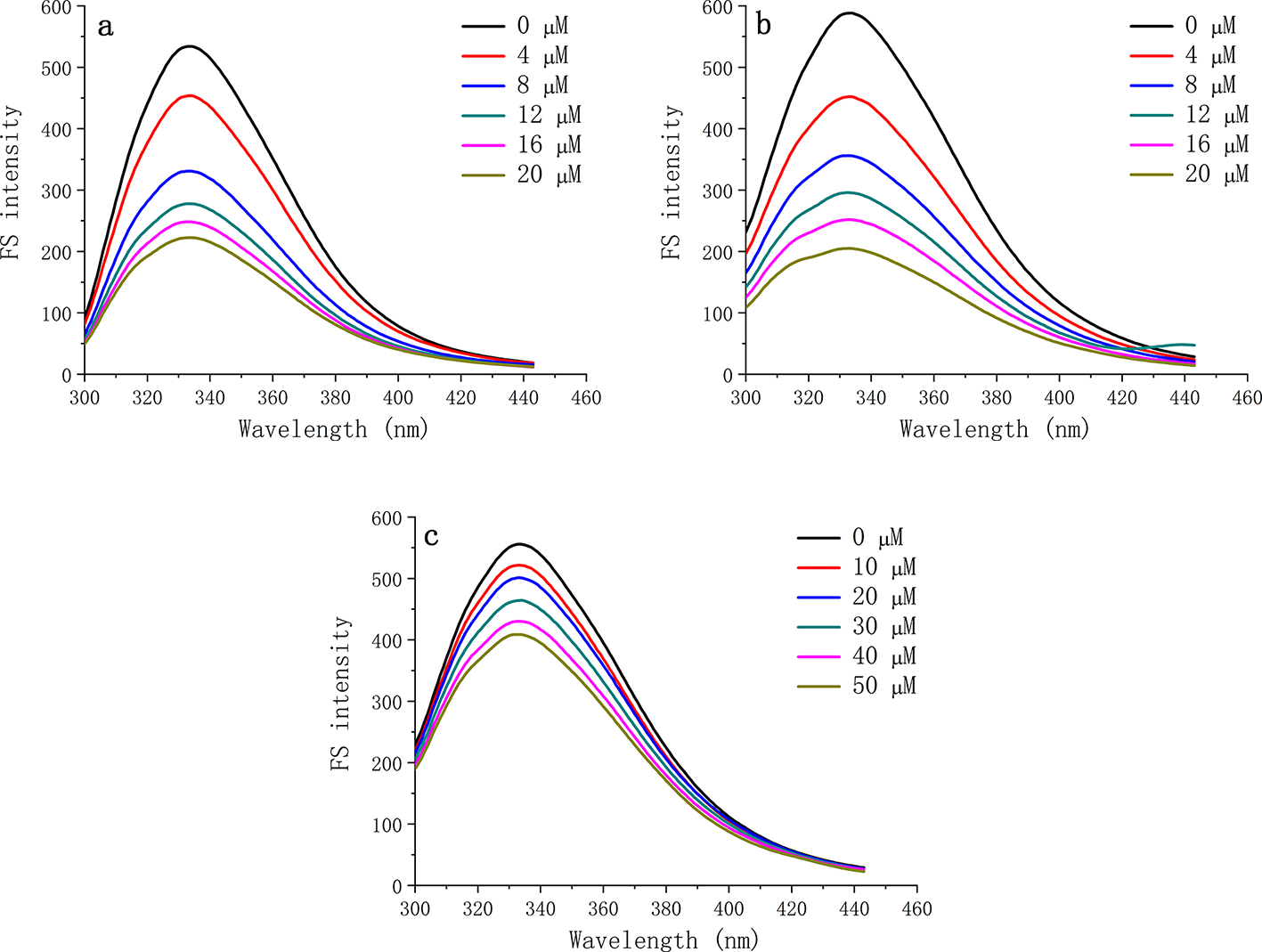
Effects of baicalein on the fluorescence spectrum of α-glucosidase at 25°C (a), 30°C (b), and 37°C (c).

At the designed temperatures, all the K q values were higher than 2 × 10¹⁰ L mol−1 s−1 (Table 1), which indicated that baicalein could cause static fluorescence quenching by forming a complex with α-glucosidase. Then, the corresponding binding constant (K A) and binding site number (n) were obtained (Table 2) by the double-logarithm equation 14 :
Stern-Volmer Quenching Constants Between Baicalein and α-Glucosidase.
Binding Parameters Between Baicalein and α-Glucosidase.
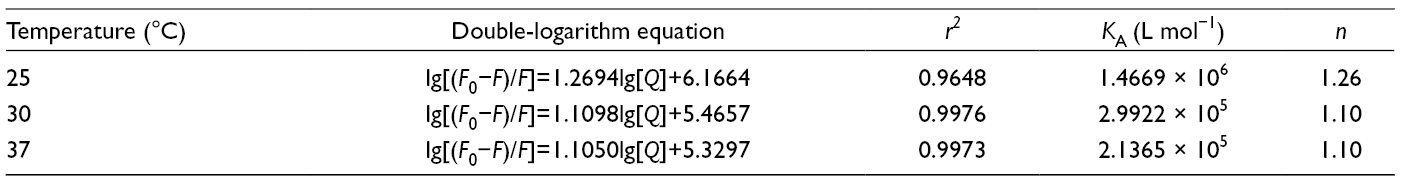

It was found that all the binding site numbers were near to 1. However, the K A values significantly decreased with the increase in incubation temperature, which also proved the formation of the complex since the high temperature was harmful for the non-covalent interaction.
The thermodynamic parameters describing the interaction were further calculated based on the Vant Hoff equation and Gibbs free energy equation 14 :


In Table 3, all the free energy changes (ΔG) under the test temperatures were negative, suggesting that the formation of the complex was a spontaneous process. The negative enthalpy change (ΔH) and entropy change (ΔS) indicated that the formation of the complex was driven by hydrogen bonding and van der Waals force.
Thermodynamic Parameters Between Baicalein and α-Glucosidase.

Based on the above experimental results, molecular docking was carried out to establish the binding mode. Figure 5(a) provided the binding mode with the lowest binding energy (−7.17 kcal mol−1). Baicalein formed 6 hydrogen bonds with Trp391 (2), Arg428, Gly566, and Glu771 (2) of α-glucosidase. The lengths of these hydrogen bonds were in the range from 1.8 to 3.3 Å. Figure 5(b) shows the hydrophobic interaction around baicalein. The hydrophobic interaction between baicalein and some amino acid residues (Phe385, Phe389, Arg387, Glu429, Phe444, Trp789, and Trp710) could be found, which coincided well with the fluorescence results. Our results show that baicalein is a potential α-glucosidase inhibitor for controlling the postprandial blood glucose level of diabetics, which may be used in the fields of functional foods and medicines.

Binding mode (a) and noncovalent interaction (b) between baicalein and α-glucosidase provided by molecular docking analysis.
Experimental
Chemicals
Baicalein, acarbose, and p-nitrophenol were purchased from Aladdin (Shanghai, China), and α-glucosidase (from Saccharomyces cerevisiae) and pNP-G from Sigma-Aldrich (St Louis, MO, USA). Ultrapure water from a Thermo Gen-Pure UF/UV pure water system (Waltham, MA, USA) was used in the whole experiment.
α-Glucosidase Inhibition Assay
In total, 0.25 mL of 0.3 U mL−1 α-glucosidase solution (0.1 M PBS, pH 6.8), 0.25 mL of baicalein at different concentrations (0.05-0.3 mg mL−1), and 0.25 mL PBS (0.1 M, pH = 6.8) were mixed and incubated at 37°C for 10 minutes. Then, 0.25 mL of 1 mM pNP-G was added. The obtained mixture was oscillated at 37°C for 20 minutes before 1.0 mL ethanol was added to terminate the reaction. The decrease of pNP-G and the formation of PNP were detected at 405 nm at 25°C by using an Agilent 1260 HPLC system (Santa Clara, CA, USA) with a DIKMA C18 Diamonsil Plus column (250 × 4.6 mm, 5 µm particle size, Beijing, China). The mobile phase consisted of methanol and water (60:40) with a flow rate of 1.0 mL min−1. The injection volume was 10 µL. Both the PNP areas of sample (S sample) and control (S control) were recorded. The α-glucosidase inhibitory activity of the sample was calculated by using the following equation:

Fluorescence Spectra
These were obtained according to a previous report. 15 One milliliter of baicalein at different concentrations (0-50 μM) and 4 mL 0.4 U mL−1 α-glucosidase solution were mixed and incubated at different temperatures (25°C, 30°C, and 37°C) for 10 minutes. Then, the FS in the range of 290 to 450 nm was recorded at the excitation wavelength of 280 nm. Both the slit widths of excitation and emission were 5 nm.
Molecular Docking
The chemical structure of baicalein was optimized by the PM6-D3H4 method of MOPAC 2016, and the crystal structure of α-glucosidase (PDB ID: 4J5T) was used. The molecular docking was performed by using Auto-dock 4.2 software (The Scripps Research Institute, La Jolla, USA). 16 The grid was run with Auto-grid module. The grid spacing was set at 0.375 Å, and all the numbers of grid points in X, Y, and Z directions were 60 Å. The Lamarck genetic algorithm was used to search the possible docking modes.
Statistical Analysis
The results are expressed as mean ± standard deviation of triplicate measurements. Student’s t-test was applied for the statistical comparison; a value of P < 0.05 was considered to be significant.
Footnotes
Declaration of Conflicting Interests
The author(s) declared no potential conflicts of interest with respect to the research, authorship, and/or publication of this article.
Funding
The author(s) disclosed receipt of the following financial support for the research, authorship, and/or publication of this article: This work was supported by the National Natural Science Foundation of China (No. 31771941).