
Abstract
Gout is a metabolic arthritis that originates from increased accumulation of monosodium urate (MSU) crystals in joints. This work aimed to evaluate the antioxidant and anti-inflammatory activities of the hydromethanolic extract of Gnidia kraussiana (HEGK) using model of Gouty arthritis on mice. The total polyphenol, flavonoid, tannin content and the antioxidant activity of HEGK were also evaluated. MSU-injected mice were treated daily for 3 days with HEGK (25, 50 and 100 mg/kg). Indomethacin and colchicin were used as reference drugs. Paw oedema and body temperature were measured at different time intervals post-injection. Malondialdehyde, acid phosphatase, β-Galactosidase, catalase, superoxide dismutase and glutathione levels were evaluated. HEGK is rich in polyphenol (129.93 mg/100 g), flavonoid (67.78 mg/100 g) and tannin conferring it a high antioxidant activity. Acute oral toxicity of HEGK resulted in LD50 greater than 2000 mg/kg. Oral administration of HEGK induced a significant decrease in the oedema of legs injected with urate crystals and reduced the release of acid phosphatase and β-Galactosidase. A model of oxidative damage was successfully established, revealing a significant increase in malondialdehyde and inhibition of antioxidants, including superoxide dismutase, catalase and glutathione activity. Thus, HEGK can actively inhibit the effect of inflammatory mediators in gouty arthritis.
Introduction
Gouty arthritis is an inflammatory rheumatism caused by a deposit of monosodium urate at joints and on a vast number of tissues. It is a frequently occurring metabolic disease linked to a metabolic disorder of purines inducing hyper-uricaemia, 1 which can be secondary due to an excess of purines production or a deficit of uric acid elimination. 2 The accumulation of urate crystals in the joint cavity activates synovial cells, resulting in the recruitment of a large number of neutrophils, which in turn activate phagocytosis of these crystals. The interaction between monosodium urate crystals and phagocytic cells induces the synthesis and release of lysosomal enzymes, reactive oxygen species (ROS), E2 prostaglandins, chemokines such as IL-8 and cytokines comprising tumour necrosis factor TNF-α and IL-1β. 3 IL-1β production in the knee joint is the first step in recruiting neutrophils that produce other hyperalgesia mediators. 4 In general, gouty arthritis is well controlled if it is managed properly. Despite this, gout can trigger acute flare-ups which lead to clinical consultation. In fact, acute gout flare-ups are recognised as one of the most painful experiences, along with childbirth or visceral colic. 5 Gout flare-ups in humans are self-limited with spontaneous resolution within 7 to 10 days, 6 while in mice, resolution occurs within 24 to 36 h, given that MSU crystals are rapidly destroyed by uricases. 7 However, if left untreated, the continuous deposition of MSU crystals do not only cause further flare-ups, but also, and most importantly, promotes irreversible joint damage accompanied by chronic symptoms and disability. 5 In fact, patients with advanced gout often experience chronic joint pain, swelling, movement limitation and recurrent flare-ups. 8 Its incidence is constantly increasing and associated to great comorbidities.
However, previous findings revealed that uric acid plays a vital role in the gouty arthritis pathogenesis and has been associated with an increase in the prevalence of many chronic diseases such as hypertension, diabetes, kidney diseases and cardiovascular diseases. 9 Additionally, oxidative stress could be responsible for chronic inflammations and several lesions of tissues 10 as it possess both antioxidant and oxidant properties whose limit is not well defined.10,11 Moreover, the prevalence of gouty arthritis ranges from 4 to 6.8% worldwide 12 based on age, notably 7% at 65 years old and 3% at 85 years old in men and women, respectively.
In Sub-Saharan Africa and before 1980, this disease was less frequent because of its scarcity and the scientific research on this pathology focused on anecdotic series.13,14 In Cameroon, Singwé-Ngandeu et al.
9
recognised an increment of this pathology frequency in the year 2007. Nowadays, the treatment of gout mainly relies on the association of many hypo-uricemiant (allopurinol), anti-inflammatory agents (indomethacin, colchicin…) with interleukin (IL-1) inhibitors. This therapeutic management remains limited due to the diversity and the gravity of their side effects such as fever, cutaneous eruption, hypersensitivity reactions, hepatitis, nephropathy and gastro-intestinal bleeding.
15
Due to limits of existing therapies, an alternative treatment approach using medicinal plants provide good and long-term source of new bioactive compounds with fewer side effects. Recent findings showed that plants are rich in bioactive compounds such as polyphenols conferring them analgesic, antiseptic and anti-inflammatory properties. About 80 % of the African population use medicinal plants as the main source of drug to treat ailments.16,17 Gnidia kraussiana is traditionally recognised by local population of ‘Tokombéré’ in the Far-North region and used to treat various diseases such as oedema, fever, bronchitis, hepatitis, snakebite, earworms, leprosies and acute gout.18,19 It is also used in the treatment of gouty arthritis. Phytochemical screening of this plant revealed a high flavonoid, coumarins and terpenes content.
20
Another study revealed that phenolic compounds of this plant extract stimulate immunoglobulines synthesis in case of immune deficient, pulmonary infections of children, chronic bronchitis in adult, HIV/AIDS, leukemia and cancer.18,21 There is no published data to the best of our knowledge concerning its anti-gout potential coupled with its anti-inflammatory potential. Therefore, the present work aimed to investigate the effect of the HEGK on gouty arthritis induced in mice by monosodium urate.
Experimental Part
Plant Material and Extract Preparation
G. kraussiana roots were collected in ‘Tokombéré’ subdivision (Far-North region of Cameroon: longitude E 14°08’35”; latitude N 10°52’18”; altitude 746 m). The fresh material was transported in polystyrene bags to the Laboratory of Biochemistry and Biological Chemistry of the Faculty of Science of the University of Maroua where Pr Tchobsala, botanist in the same University, achieved identification. A voucher specimen was deposited at the Cameroon National Herbarium and compared to specimen N° 322643/HNC. Roots were washed 3 times with tap water and dried at room temperature (35 ± 3°C). The dry material was ground. The powder obtained was sieved (.5 mm) and kept until extraction. The extraction was achieved by macerating 300 g of powder in 3000 mL of methanol (80%) for 72 hours while shaking at room temperature. 22 The mixture was filtered using Watman filter paper (N°1) and the filtrate was collected. The solvent of the filtrate obtained was concentrated with a rotary evaporator set at 70°C. The residue was kept at −4°C.
Experimental Animals
The study was conducted on male albino Swiss mice weighing between 25 and 30 g, purchased from LANAVET (‘Laboratoire National Vétérinaire’, Garoua branch). All animals aged 4 months were kept in the animal breeding facility of the Department of Biological Sciences (University of Maroua). Animals were housed in polyacrylic cages (5 mice/cage) with free access to water and food ad libitum. They were acclimatized for 14 days at room temperature, with natural light/dark cycle. Mice were treated following the guidelines of the Cameroonian Bioethics Committee (reg N° FWAIRB00001954) and in accordance with NIH-Care and Use of Laboratory Animals (8th edition).
Phytochemical Analysis
The total polyphenols content of the hydromethanolic extract was evaluated using Folin-Ciocalteu as described by Singleton et al. 23 In a test tube containing .2 mL of the sample previously diluted in 80% methanol, 1 mL of Folin-Ciocalteu (10%) was introduced. The mixture was stirred for 5 min. Then, .8 mL of sodium bicarbonate (7.5%) was added in the tube before incubating at room temperature for 30 min. The absorbance of the mixture was measured at 745 nm using UV-VIS spectrophotometer (PRIM Light and Advanced, Germany). Quantification of polyphenols was achieved using the standard curve of gallic acid (0–250 μg/mL) and the result was expressed as milligrams of gallic acid equivalent per 100 g of dry weight (mg GAE/100 g).
The aluminium chloride (AlCl3) colorimetric method was used to estimate flavonoid content according to Mimica-Dukic et al. 24 Briefly, 1 mL of the extract and 1 mL of AlCl3 reagent were mixed into a tube and stirred for 5 min before adding two drops of acetic acid. The absorbance of the mixture was measured at 430 nm. The standard curve of optical density against quercetin concentration (0–100 μg/mL) was used to express results in quercetin milligram equivalent per 100 g of dry weight (mg QE/100 g).
The tannins content was evaluated as described by Bainbridge et al. 25 where .2 mL of the hydromethanolic extract and 2 mL of vanillin (1 g vanillin/100 mL of HCl conc.) were introduced, respectively, into a tube and incubated for 30 min in a dark place. The absorbance of the mixture was read at 500 nm. The calibration curve was established with catechin (0–50 μg/mL) and results were expressed in catechin milligram equivalent per 100 g of dry weight (mg CatE/100 g).
Evaluation of the In-Vitro Antioxidant Activity
The ferric reducing antioxidant power (FRAP) was determined as described by Benzie and Strain. 26 Briefly, 1 mL of sample or standard (Trolox) solution was mixed with 3 mL of FRAP reagent (Fe (III)-2,4,6-Tri (2-pyridyl)-s-triazine). The mixture was shaken for 60 s and incubated for 40 min at room temperature. The absorbance was measured at 593 nm using UV-VIS spectrophotometer. The calibration curve was established with ascorbic acid (0–25 μg/mL) and results were expressed in ascorbic acid milligram equivalent per 100 g dry weight (mg AAE/100 g).
The radical scavenging activity of 2,2-diphenyl-1-picrylhydrazyl (DPPH) was carried out according to the method described by Sun et al. 27 with some modifications. The sample (1 mL) or standard (Trolox) was mixed with 3 mL of 1 mM DPPH. The mixture was stirred for 2 min and incubated for 40 min in the dark at room temperature. Absorbance was measured at 517 nm using UV-VIS spectrophotometer. The calibration curve was established with Trolox (0–200 μM) and results were expressed in Trolox millimole equivalent per 100 g of dry weight (mmol TE/100 g).
The radical scavenging activity of 2,2’-azino-bis (3-ethylbenzothiazoline-6-sulfonic acid) (ABTS) was performed as described by Re et al. 28 Briefly, 1 mL of each sample or standard (Trolox) were mixed with 3 mL of 7 mM ABTS+. The mixture was shaken for 2 min and incubated for 40 min in the dark at room temperature. Then, absorbance was read at 734 nm. The calibration curve was established with Trolox (0–200 μM) and results were expressed in Trolox millimole equivalent per 100 g of dry weight (mmol TE/100 g).
Acute Toxicity Test
The toxicity test was conducted according to guideline 423 of the Organisation for Economic Co-operation and Development (OECD). 29 Three groups (n = 3) of female Wistar rats weighing 150–200 g were used. Distilled water (10 mL/kg) was given to one control group; the other one group received a high dose (2000 mg/kg) of the HEGK. Behaviour and lethality of rats were observed during the first 4 hours after the administration of the extract, then on a daily basis for 14 days.
Evaluation of the Anti-inflammatory Activity Induced by Monosodium Urate
Preparation of Monosodium Urate Crystals
The monosodium urate crystals (MSU) were prepared according to Schorn et al. 30 method with slight modifications. Briefly, 3 g of uric acid salt was dissolved in 600 mL of .03 M NaOH. The mixture was incubated in a water bath for 2 hours and allowed to cool down before adjusting the pH to 7.5. To the mixture, 6 mL of 5 M NaCl was added and stirred for 24 hours at room temperature. The mixture was then centrifuged and the urate crystals were collected, washed and sterilised in a Pasteur furnace at 180°C for 2 h.
Inflammation Induction
A total of 35 mice were randomly divided into seven groups (n = 5 each) as follows: 2 control groups (normal and negative control) treated with distilled water (2 mL/kg); 2 positives controls groups treated with indomethacin (3 mg/kg) and colchicine (1 mg/kg); and 3 other groups were treated with HEGK (25, 50 and 100 mg/kg). All treatments were given orally, once per day. An hour after the first treatment administration, an aliquot (.15 mL) of MSU (26.6 mg/mL) was injected in the intra-articular region of the left paw of animals of all groups except the normal control, which received just .15 mL of physiological water. 31 The diameter of the paw was measured at different time intervals (0 h, 4 h, 24 h, 48 h and 72 h) after oedema induction. The evolution of rectal temperature (°C) was also registered using a thermometer (CHICCO) and paw thickness was evaluated using an electronic calliper. At the end of the experiment, mice were sacrificed using an intraperitoneal injection of thiopental (50 mg/kg, i. p). Blood was collected in heparin tube, centrifuged at 3000 r/min for 15 min and the serum was collected in microtubes for lysosomal enzymes analysis. Liver and spleen were isolated and their sections homogenised in Tris HCl buffer (50 mM; pH 7.4). The supernatant obtained from homogenate centrifugation was used for the evaluation of the lysosomal enzymes activities and antioxidant parameters (malondialdehyde, superoxide dismutase, catalase and glutathione). Posterior paws were sectioned for histological studies.
Liver and Spleen Homogenate Preparation
The liver and spleen were isolated and homogenised in Tris-HCl buffer (50 mM; pH 7.4). The slurry was centrifuged at 3000 r/min for 10 min at 4°C and the supernatant was collected and kept at −20°C for the assessment of lysosomal enzymes, lipid peroxidation and antioxidant status.
Total Protein Content Evaluation
Liver and spleen homogenates protein content were evaluated according to Bradford method. 32 Coomassie blue reagent solution (2 mL) was added to 100 μL of each sample before mixing. The absorbance of the mixture was read at 595 nm using UV-VIS spectrophotometer (PRIM Light and Advanced, Germany). Proteins content was calculated from the standard curve of bovine serum albumin (BSA) (0–100 μg/mL) and results were expressed in BSA microgram equivalent per mL of homogenate (μg BSAE/mL).
Acid Phosphatase Activity
Total phosphatase acidic (TPA) evaluation in serum, spleen and liver was realised using the enzymatic Linear Chemicals kit following the manufacturer’s instruction and the result was expressed in U/L using the following formula:

ΔA/mn = Absorbance variation per min; 853 = conversion factor of the enzyme.
β-Galactosidase Activity Evaluation
Serum β-galactosidase activity, spleen and liver was evaluated according to the method described by Kawai and Anno. 33 Briefly, 450 μL of O-nitrophenol galactosidase (ONPG) and 500 μL of the homogenate or the serum were incubated in a water bath at 37°C in 10 min. Then, 2 mL of sodium carbonate was added just to stop the reaction. The absorbance of the reaction medium was read at 290 nm. The standard curve was established by using O-nitrophenol (ONP) solution, and the amount of ONP was determined and expressed in μg ONP/mL of homogenate. β-galactosidase activity was expressed in μmole ONP/min/mg of protein.
In-Vivo Antioxidant Capacity Evaluation
Superoxide dismutase (SOD) activity was evaluated using the method descried by Misra and Fridovish. 34 In a spectrophotometric curve, 134 μL of homogenate (20%) was mixed with 1.666 mL of carbonate buffer (.05 M; pH 10.2). Then, .2 mL of adrenalin (.3 mM) was added and the mixture was homogenised. The increase of the absorbance was evaluated between 20 and 80 s at 480 nm. Specific SOD activity was expressed in SOD Unit/mg of proteins. Specific SOD activity = Number of SOD units/mL/mg of proteins x dilution factor.
Catalase activity was evaluated according to the method described by Sinha. 35 Glutathione (GSH) content of organs homogenate was evaluated using Ellman method. 36 Briefly, .02 mL of homogenate supernatant and 3 mL of Ellman reagent (DTNB: 5,5’-dithiobis-nitrobenzoic acid) were introduced in a test tube. The mixture was stirred and kept at room temperature for 60 min. .02 mL of Tris-HCl buffer (50 mM; pH 7.4) and 3 mL of Ellman reagent were pipetted in a control tube. The absorbance was read at 412 nm and the blood GSH level was expressed in nmol/g of organ.
The malondialdehyde content (MDA) was determined according to Wilbur et al. 37 method. Each organ sample (2 Ml), 1 mL of 20% trichloroacetic acid (TCA) and 2 mL of .67% thiobarbituric acid (TBA) were mixed in a test tube and control tube was prepared using 2 mL of Tris HCl buffer (50 mM; pH 7.4). These tubes were incubated for 10 min at 90°C in a water bath. After that, tubes were allowed to cool and centrifuged at 3000 r/min for 10 min. The supernatant was collected for analysis and the absorbance was measured at 532 nm and the result was expressed in nmol/L.
Paw Histological Analysis
At the end of the experiment, the posterior paw was sectioned and fixed in formaldehyde (10%), then decalcified for 2 weeks with EDTA solution (14%) and incorporated in paraffin for analysis. Paraffin representation were coloured in haematoxylin and eosin. Each representation was sectioned, photographed and analysed to identify cellular infiltration, pannus formation and cartilage damage.
Statistical Analysis
Results were expressed as mean ± standard error of mean of five animals per group. Data were analysed using one-way analysis of variance (ANOVA) performed by GraphPad Prism 8.01. Data was assessed using Dunnett’s post tests and significance was considered at P < .05.
Results
Phytochemical Characteristics and Toxicity of the Hydromethanolic Extract
Extraction Yield, Total Polyphenols (Gallic Acid Equivalent), Flavonoids (Quercetin Equivalent), Tannins (Catechin Equivalent) Contents, Ferric Reducing Antioxidant Potential (FRAP: Ascorbic Acid Equivalent) and ABTS, DPPH Scavenging Activities (Trolox Equivalent) of Hydromethanolic Extracts of Roots From Gnidia Kraussiana. Values are Giving According to the Dry Weight.

Values are means ± standard deviation of 4 repetitions.
Acute Toxicity of the Hydromethanolic Extract of Gnidia Kraussiana
Oral administration of a single dose of 2000 mg/kg of HEGK did not result in any lethality or signs of characteristic toxicity such as decreased sensitivity to pain, noise, and locomotion during the 14 days of observation. Four hours after the administration of HEGK, all rats treated with 2000 mg/kg showed signs of moderate toxicity, including drowsiness, isolation, hair straightening and hypo-activity. On the second day, a doughy stool was observed with an insignificant change in weight. On the third day and 1 week after extract administration, rats recovered normal behaviour comparable to the control group. The LD50 of the extract is, therefore, higher than 2000 mg/kg.
Variation of the Paw Oedema
Paw oedema measurements were recorded before (0 hours) and 4, 24, 48 and 72 hours after every treatment (Figure 1). In negative control mice, a significant (P < .001) increase in paw oedema was observed 4 and 24 hours after the injection of MSU. On the last 2 days (at 48 and 72 hours), a successive decrease in oedema was noted but remained significantly (P < .001) higher than the normal control. Moreover, the successful decrease of oedema (48 h and 72 h after paw oedema induction) was observed and remained significantly (P < .001) higher than that observed among the normal control group. However, a drastic and significant (P < .001) decrement of oedema was recorded in groups that received the HEGK (25, 50 and 100 mg/kg) and reference drugs (indomethacin and colchicine). This effect was revealed 24 h post-treatment and significant at 48 h and 72 h. Effect of hydromethanolic extract of Gnidia kraussiana, indomethacin and colchicine on paw oedema of MSU crystal-induced arthritis. Values were recorded 4, 24, 48, and 72 hours after treatment (Mean ± SEM, n = 5, ***P < .001, compared to the normal control. #P < .05, ##P < .01, ###P < .001 compared to the negative control). HMGK: hydromethanolic extract of G. kraussiana; Col: colchicine; Indo: indomethacin.
Temperature Variation Rectal
Rectal temperature of mice at different time points were recorded to assess the different groups treated with HEGK and standard drugs vs the negative control group (Figure 2). An antipyretic effect was observed in the different groups treated with HEGK, indomethacin and colchicine between 24 and 48 h after treatment. The rectal temperature of mice was significantly (P < .001) decreased 24 h and 48 h after injection of MSU crystal. Effect of hydromethanolic extract of Gnidia kraussiana, indomethacin and colchicine on body temperature in mice MSU crystal-induced arthritis. The values are expressed in mean ± SEM, (n = 5), *P < .05, **P < .01, ***P < .001 significant difference compared to normal (N). #P < .05, ##P < .01; ###P < .001 significant difference compared to negative control (NC). HEGK: hydromethanolic extract of G. kraussiana; Col: colchicine; Indo: indomethacin.
Lysosomal Enzymes Activities
Effect of Hydromethanolic Extract of Roots from Gnidia Kraussiana, Colchicine and Indomethacin (Indo) on the Activity of Lysosomal Enzymes (Acid Phosphatase and β-Galactosidase) of Plasma, Liver and Spleen of Monosodium Urate Crystal-Induced Arthritis.

The values are expressed in mean ± SEM, (n = 5), *P < .05, **P < .01, ***P < .001 Significant difference compared to normal groups (N). £ P < .05, ££ P < .01; £££ P < .001 significant difference compared to negative control groups (NC) β-galactosidase (μmole ONP/min/mg of protein), Acid phosphatase (U/L). HEGK = hydromethanolic extract of G. kraussiana.
Oxidative Stress Markers
Figure 3 depicts that MSU crystals injection significantly increased oxidative stress (P < .001) by decreasing the CAT, SOD and GSH levels in the negative control group in contrast to the normal control group. However, MDA expression decreased by standard drugs treated groups (indomethacin: 3 mg/kg and colchicine: 1 mg/kg) and HEGK (25, 50 and 100 mg/kg). The decrease in CAT and SOD levels was considerably (P < .001) noticed in HEGK, indomethacin and colchicine treated groups. Additionally, the treatment of mice with HEGK (25, 50 and 100 mg/kg) and standard drugs could restore and maintain GSH level at values close to those of the normal control group. Effect of hydromethanolic extract of Gnidia kraussiana, colchicine and indomethacin on oxidative parameters of liver and spleen. MDA: Malondialdehyde; SOD: Superoxide dismutase; CAT: Catalase; GSH: Glutathion. The values are expressed in mean ± SEM, (n = 5), *P < .05, **P < .01, ***P < .001 and ****P < .0001 significant difference compared to normal (N). ŧ P < .05, ŧŧ P < .01; ŧŧŧ P < .001 and ŧŧŧŧ P < .0001 significant difference compared to negative control (NC). HEGK: hydromethanolic extract of G. kraussiana; Col: colchicine; Indo: indomethacin.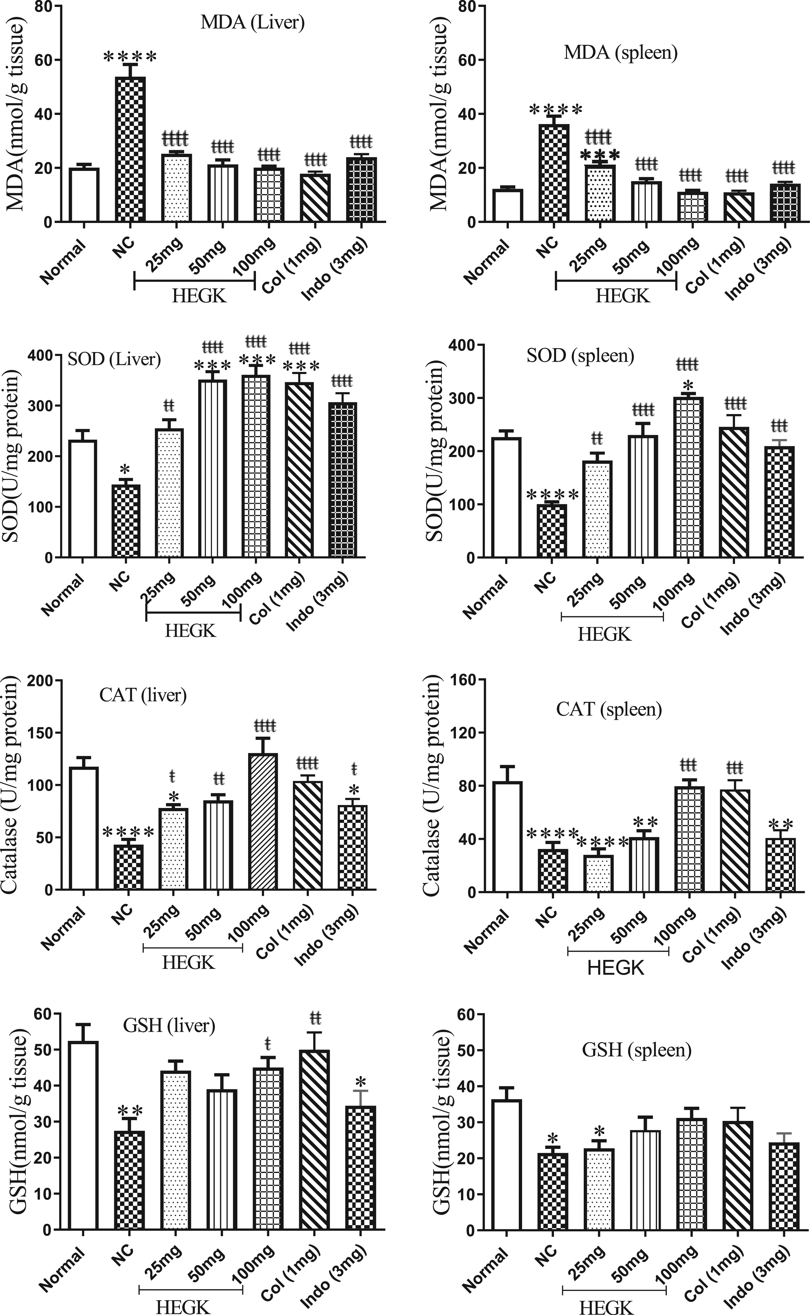
Study of the Left Hind Paw
Histopathological analysis of mice paws (Figure 4) showed that a normal morphology was observed in the group of normal mice. While in the negative control group, inflammation and destruction of the synovial membrane, infiltration of immune cells and tissue damage were observed. Photomicrographs (H&E staining100×; section thickness5 µm) of paw transverse of normal, arthritic, and treated mice with colchicine, indomethacin, and hydromethanolic extract of G. kraussiana. Normal = Animals receiving distilled water; Negative control = Arthrosis induced animals receiving distilled water; Indo: 3 mg = Arthrosis induced animal receiving indomethacin 3 mg/kg; Col: 1 mg = Arthrosis induced animals receiving colchicine 1 mg/kg, HEGK = Arthrosis induced animals receiving hydromethanolic extract of G. krausiana 25, 50, and 100 mg/kg doses. MS = Membrane synovial, In = Inflammatory cells; TL = Tissue lesion.
In different groups treated with HEGK (50 and 100 mg/kg) doses, there was no inflammation of the synovial membrane or tissue damage. With the 25 mg/kg dose, inflammation of the synovial membrane and tissue damage was observed, but no cell infiltration or destruction of the synovial membrane was noticed. Treatment of animals with diclofenac limited synovial membrane destruction and cell infiltration. In animals treated with colchicine, infiltration of immune cells was observed, but treatment limited inflammation and destruction of articular cartilage and tissue damage (Figure 4).
Discussion
Gout is a painful acute inflammatory disease induced by the deposition of monosodium urate (MSU) crystals in joints and peri-articular tissues. The interaction between these crystals with synovial fluid and synovial cells leads to the release of many pro-inflammatory mediators. 38 These mediators act with endothelial cells promoting a vast migration of leukocytes, especially neutrophils which represent 90% of leukocytes. The interaction between crystals and neutrophils leads to an inflammatory reaction called acute gout crisis. The present study aimed to evaluate the antioxidant and anti-inflammatory potentials of the hydromethanolic extract of G. kaussiana roots an in-vivo model of gouty arthritis.
The phytochemical analysis of the extract showed an important amount of polyphenols, flavonoids and tannins. The obtained values of tannin contents were similar to those previously observed by Dif et al. 39 and Mohammedi 40 in the methanolic extract of Daphne gnidium (2.40 mgCE/g) leaves, another member of Themeliacea family. However, the obtained values of polyphenols content in this present finding were lower than those obtained in recent works. 41 This difference may be attributed to the geographical, climatic and environmental factor, genotype, harvesting seasons, conservation time, plant maturity, cultural practices, temperature and solvents used for extraction.42,43 Polyphenols and tannins are endowed with a great antioxidant potential. 44
Results on antioxidant activities revealed that the HEGK has free radical scavenging ability revealed by DPPH and ABTS. Additionally, the extract exhibited a strong capacity to reduce ferric ions. Previous findings showed that antioxidant activities of plant material were related to its polyphenol content45,46 which exert their antioxidant potential by acting as a reducing agent, hydrogen or electron donor and metal chelator. 47
To mimic gout physiopathology, MSU was injected in the left hind paw and some parameters such as oedema size and body temperature at 4 h, 24 h, 48 h and 72 h. At the 4th h after the injection of MSU, a significant increase (P < .001) of oedema size was noted in the negative control group that reached its maximum value at 24 h. A slight decrease of oedema was noticed at the 48th h but smaller than its initial state at 72th h. This may stipulate that MSU induced an inflammatory reaction. Lima et al. 48 obtained similar results. In fact, MSU injection led to the release of many chemical mediators which amplified the inflammation.
Among groups which received the HEGK (25, 50 and 100 mg/kg), a significant (P < .001) decrease of oedema was observed 24 h after treatment. At the 4th h, a significant (P < .05) decrease of oedema was obviously present in mice that received 25 mg/kg and 100 mg/kg doses of extract. This increment remained significantly (P < .001) high compared to the control group. In fact, the metabolism of the extract might be achieve very late, and compounds with anti-inflammatory properties such as polyphenols and flavonoids reached their efficient doses on the inflammation site at 24 h. A similar result was reported by Yanik et al. 49 This effect could also be indirectly related to the administration route that might have not permitted the extract’s bioactive compounds to be rapidly released into the bloodstream. 50 Between 48 h and 72 h, a significant decrease of the oedema among tested groups with the HEGK 50 mg/kg and 100 mg/kg was observed compared to groups which received reference drugs (colchicine and indomethacin). These drugs acted differently on the same target organ. Colchicine acted by inhibiting NLRP3 inflammasome, Cas-1, by producing IL-1β and by recruiting leucocytes on the inflammation zone. 51 They also decreased the amount of histamine released by mastocytes leading to total disappearance of oedema. 52 The reduction of oedema induced by MSU after treatment with indomethacin may be linked to many mechanisms including the inhibition of COX2. 53 Indomethacin reduces prostaglandins effects 54 and prevents the infiltration of lymphocytes, monocytes and macrophages in the synovial cavity. 55 HEGK may have acted via the same mechanisms as these drugs in exerting its anti-inflammatory effect. During inflammation, macrophages and monocytes secrete pro-inflammatory cytokines such as IL-1 which excite the thermoregulatory centre and increase the patient’s body temperature. 56 Thus, the significant increase in body temperature in arthritic mice could probably be related to the secretion and action of IL-1.
An inflammatory reaction is followed by a rise in body temperature and fever among negative control group members. However, the HEGK significantly reduced the rise in the body temperature of mice suggesting that HEGK is antipyretic. Either by inhibiting the production of cytokines or by limiting its secretion, the interaction between urate microcrystals and cellular membrane can change the cellular metabolism and cause the secretion of inflammatory mediators 57 facilitating phagocytosis, inflammation and tissue damage 58 with a reduction of lysosomal enzymes level. In the present work, intra-articular injection of urate crystals on the left hind paw in the negative control group led to a significant release of lysosomal enzymes. This result was similar to findings of other researchers. 59 Treatment with reference drugs such as colchicine and indomethacin revealed a significant (P < .001) decrease in lysosomal enzymes activity. These two drugs have various anti-inflammatory mechanisms which are well defined by many authors. 60 Moreover, animals treated with HEGK revealed a low level of acid phosphatase. This decrement was similar to that achieved by both reference drugs used in this study. Thus, it is possible that the G. kraussiana plays the same role as the two reference drugs. Therefore, G. kraussiana has probably inhibited the release of this lysosomal enzyme due to the presence of diverse phenolic compounds capable to stabilise the biological membrane.
It is well known that inflammation can be associated with the increase of oxidative stress and the production of radical oxygenated species which are involved in inflammatory pathology complications. 61 The present work revealed that MDA level in the liver and spleen was high among arthritic mice. This was also noticed by Ostałowska et al. 62 and Umar et al. 63 The increment of MDA level could be directly related to the enhancement of lipid peroxidation which plays an important role in the arthritis pathogenesis. 64 A dose dependent reduction of MDA by HEGK and reference drugs was observed in the liver and spleen. This may be explained by their lipid peroxidation inhibition effect as suggested by Umar. 65
Antioxidant enzymes such as catalase, superoxide dismutase and glutathione peroxidase combat reactive oxygen species (ROS). It is well known that MSU-induced arthritis results in oxidative stress state. Free radicals and reactive oxygen species induce the production of a great number of phagocytosis leading to cells damage. The resulting toxicity on liver and spleen cells is due to redox cycle and toxic ROS generated.
To prevent these damages, many endogenous defense mechanisms involving SOD, GSH and CAT are implicated. 66 In this study, SOD, GSH and CAT activities were significantly (P < .05) decreased among mice which developed arthritis compared to normal control group mice. This can be due to their involvement in the restauration of oxidative stress balance. Treatment with HEGK (25, 50 and 100 mg/kg) has significantly improved antioxidant status among mice injected with MSU crystals. HEGK could chelate free radicals, reduced lipid peroxidation and increase antioxidant enzymes activities. These findings are similar to previous works.67,68 The present results suggest that the HEGK can further prevent MSU crystals induced gouty arthritis by its antioxidant effects.
Inflammation can bring about the shrinkage of the articular space leading to the degradation of cartilage. A treatment which can reduce inflammation by blocking cytokines could help to overcome articular space shrinking and bone degradation problem. 69 In the present work, HEGK administered per os limited inflammation in mice and prevented articular space shrinking, infiltration of inflammatory cells and cartilage degradation.
Conclusion
In summary, the study demonstrated that the hydromethanolic extract of G. kraussiana is rich in polyphenolic compounds. These bioactive compounds showed an important antioxidant activity and inhibited oedema development, the release of lysosomal enzymes and oxidative stress markers (SOD, CAT, GSH, MDA). Thus, G. kaussiana extract has an anti-inflammatory effect against gouty arthritis in mice MSU-induced inflammation model.
Footnotes
Acknowledgments
We thank all members of ‘Medicine et Partage’ for their contributions.
Declaration of Conflicting Interests
The author(s) declared no potential conflicts of interest with respect to the research, authorship, and/or publication of this article.
Funding
The author(s) disclosed receipt of the following financial support for the research, authorship, and/or publication of this article: This study was financially funded by ‘Medicine et Partage’ group in Toulouse, France.