
Abstract
Spherical selenium-oxide and copper-oxide nanoparticles (SeO-NP with mean diameter 51 ± 14 nm and CuO-NP with mean diameter 21 ± 4 nm) were found to be cytotoxic for human fibroblast-like cells in vitro, as judged by decreased ATP-dependent luminescence. Compared with SeO-NP, CuO-NP produced a somewhat stronger effect of this kind. Along with cell hypertrophy developing in response to certain doses of SeO-NP and CuO-NP, our experiment also revealed doses causing a decrease in cell and cell-nucleus sizes. We observed both monotonic and different variants of nonmonotonic dose-response relationship. For the latter, we have succeeded in constructing adequate mathematical expressions based on the generalized hormesis paradigm that we had considered previously in respect of CdS-NP and PbS-NP cytotoxicity for cardiomyocites. It was demonstrated as well that combined toxicity of SeO-NP and CuO-NP is of different types depending on the outcome.
Introduction
Previously, we demonstrated 1 that dose-response relationships in lead sulfide and cadmium sulfide nanoparticles (PbS-NP and CdS-NP) cytotoxicity studied on a culture of cardiomyocites could be monotonic for some effects and nonmonotonic for others. The relationship of the first type means that as the impact intensifies, the effect grows stronger or weaker in the entire range of doses. In the relationship of the second type, the direction of the effect changes at least twice with growth in impact, and it may be termed as hormesis in its broader sense considered in detail in.2,3 In these publications of ours, one can find a sufficiently comprehensive review the relevant scientific literature dealing with the hormesis paradigm.
Besides, it was demonstrated that the type of combined action of the above nanoparticles depends on the toxicity outcome it is characterized for.
The purpose of this paper is to demonstrate that the above features of dose-response relationship are not a special case which holds only for the above nanoparticle species and cell type. To this end, we have analyzed results of experiments on a culture of fibroblast-like cells exposed to selenium-oxide (SeO-NP) or copper-oxide (CuO-NP) nanoparticles.
Materials and Methods
Preparation and Characterization of Nanoparticles
Suspensions of nanoparticles (NP) were prepared by laser ablation of 99.9% pure CuO and SeO targets in deionized water and visualized under a scanning electron microscope (SEM), Merlin (Carl Zeiss, Germany). The size distribution functions were obtained by a statistical analysis of the SEM images of several hundreds of respective NPs. The chemical nature of these NPs was characterized using the energy-dispersive X-ray spectroscopy.
Cell Line Characterization, in vitro Exposure Technique and Parameters, Cytotoxicity Estimates
Cell Cultures
The experiments were performed on a FELCH-104 stable cell line from BioloT Ltd. (Saint-Petersburg, Russia), which presents a culture of fibroblast-like cells derived from an 8-week human embryo. The cell culture was kept at 37°С under a 5% CO2 atmosphere in a DMEM medium containing L-glutamine, 1 g/L glucose, 10% embryonic bovine serum, and .5% gentamicin. We waited till a monolayer has been formed having investigated the preparation under inverted microscope each 24 hours after seeding the cells. For assessing NP cytotoxicity, cells were seeded in a 96-well plate (ТРР Techno Plastic Products AG, Trasadingen, Switzerland), 70 000 cells per well in 100 mcL medium, and maintained under standard conditions for 48 hours until a monolayer was obtained. Then suspensions of CuO and SeO nanoparticles (diluted to a needed concentration with DMEM medium) were added to the wells and incubated for 24 hours under standard conditions before performing an ATP assay. The final concentration of each NP type in the medium was 25-50-100 mcg/mcL. Nanoparticles (ether of one species or their combination) were added to the culture medium in these concentrations.
Cytotoxicity Effects Assessment
To estimate quantitatively cytotoxic effects produced by the nanoparticle species in the concentrations used, we determined the ATP content of the culture by the luminescent signal and measured cell and cell-nucleus size.
The Bioluminescence Assay
An ATP bioluminescence assay was performed using CellTiter-Glo reagents (Promega Corporation, USA). A working solution was obtained by reconstituting lyophilized CellTiter-Glo Substrate in CellTiter-Glo Buffer and warmed up to room temperature on a water bath. A 100 mcL portion of this solution was added to each well, and then the plate was rotated in one plane for 2 minutes to cause cell lysis. Upon incubation for 10 minutes at room temperature, we measured cell luminescence using an LM-01T luminometer with Kilia software (Immunotech, Beckman Coulter Company, Praha, Czech Republic). The measurement results were presented in relative luminescence units (RLU).
Cell Size Estimation
For morphometric study, we used microphotographs of cells suspended in the DMEM. To this end, 10 mcL of suspension were put on the microscope slide. In this way, 10 samples for each experimental condition were prepared and 8–10 cells in each sample were measured. For size measurements, the cells were removed from the plate and transferred onto a glass slide to perform morphometry under an optical microscope, 3D Cell Explorer (Nanolive, Switzerland). The professional image processing program ImageJ 1.48v (by Wayne Rasband, National Institutes of Health, USA) was used to measure the cell and cell-nucleus area in mcm. 2
The assaying techniques used are described in more detail in our previous publication. 1
Mathematical Description of the Experimental Results
In order to identify the type of dose-response relationship, experimental data for a particular outcome index should be approximated with an appropriate functional expression. However, the choice of approximating functions is not uniquely determined even where the dependence of the response on agent dose is monotonic. The problem becomes even more complex where the dependence is nonmonotonic.1,3
Monotonic dose-response relationships are often described with the Hill function (1), proportional to the cumulative function of the log-logistic distribution4-6

Another mathematical model for monotonic dependence presents a hyperbolic function (2) associated with the Michaelis–Menten equation, which is used, for example, to describe the rate of enzymatic reactions
7

Here, variable y also represents the outcome and the variable x is the acting agent doses.
However, often these functions are not enough to ensure good approximation of experimental data even in the case of monotonic dose-response dependence.
It is still more difficult to find a suitable mathematical model for a nonmonotonic dose-response relationship. Although there are lots of functional expressions for describing relationships of this type,1,3,8 none of them is universal, being confined to a certain limited area of research only.
At the same time, nonmonotonic relationship is often associated with manifestations of hormesis in cases where the effect is directed oppositely in 2 adjacent intervals of doses. This renders theoretical generalization of different variants of a nonmonotonic dose-response dependence a lot more complex problem since this type of dependence can feature more than 2 phases of this kind.1,2,9,10 The objective of approximating such multiphase relationships of a hormesis-associated type becomes a difficult challenge which requires using more complex power1,2 or special functions. 3
Like in the previous studies (for instance1,3,11 and many others), the type of combined cytotoxicity was estimated in a model based on the Response Surface Methodology (for example12,13). In this methodology, equation (3) describing the response surface
Y = Y (x1, x2) can be constructed by fitting its coefficients to experimental data

Today, the RSM is one of the most important general methods used in the analysis of combined effects produced by mixtures of bioactive substances, including toxic ones. This method enables the potentialities of effective experimental design to be used for approximating a response function. Constructing such approximation requires choosing an analytical model whose parameters would be determined by fitting to experimental data using the ordinary least squares method.
The quality of approximation by the proposed models was estimated by the usual and adjusted coefficient of determination. It turned out to be quite high for all models (both coefficients are at least .6). The evaluation of the statistical significance of the model parameters also shows their high significance (P < .001). In addition, it is obvious from the presented figures that the proposed models describe the observed experimental values well.
Results and Discussion
Characterization of the Nanoparticles
NPs of both types were found to be nearly spherical in shape (Figure 1). The mean diameters were 51 ± 14 nm for SeO-NP and 21 ± 4 nm for CuO-NP (Figure 1). Energy-dispersive X-ray spectroscopy performed in conjunction with SEM showed the chemical composition of the NPs to be exactly SeO-NP and CuO-NP. SEM visualization of (A) SeO-NPs and (B) CuO-NPs (left panels) and respective size distribution functions (right panels).
The absence of any noticeable changes in the zeta potential as well as in the shape and position of the plasmon resonance peak 2 weeks after suspension preparation confirmed the satisfactory stability of the suspensions.
Decrease in ATP-dependent Luminescence
The experimental data on the relationship between decrease in ATP-dependent luminescence and SeO-NP dose in a culture of fibroblast-like cells display a monotonic character, though with the presence of a marked plateau in the middle of the range.
An adequate model in this case is a linear combination of sines. For this endpoint, the model predicts the kinetics of its decrease as shown by the curve in Figure 2. Approximation by model 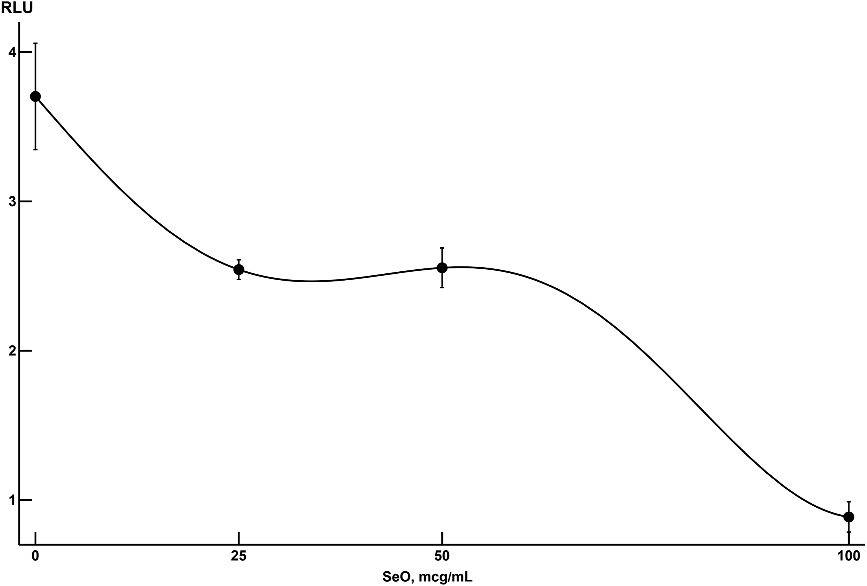
A similar dependence for CuO demonstrates an even more explicitly monotonic character, although here as well the quality of the approximation by models (1) and (2) proves to be insufficient. At the same time, quite a satisfactory approximation of this dependence is ensured by the model that is shown in the caption to Figure 3. Approximation by model 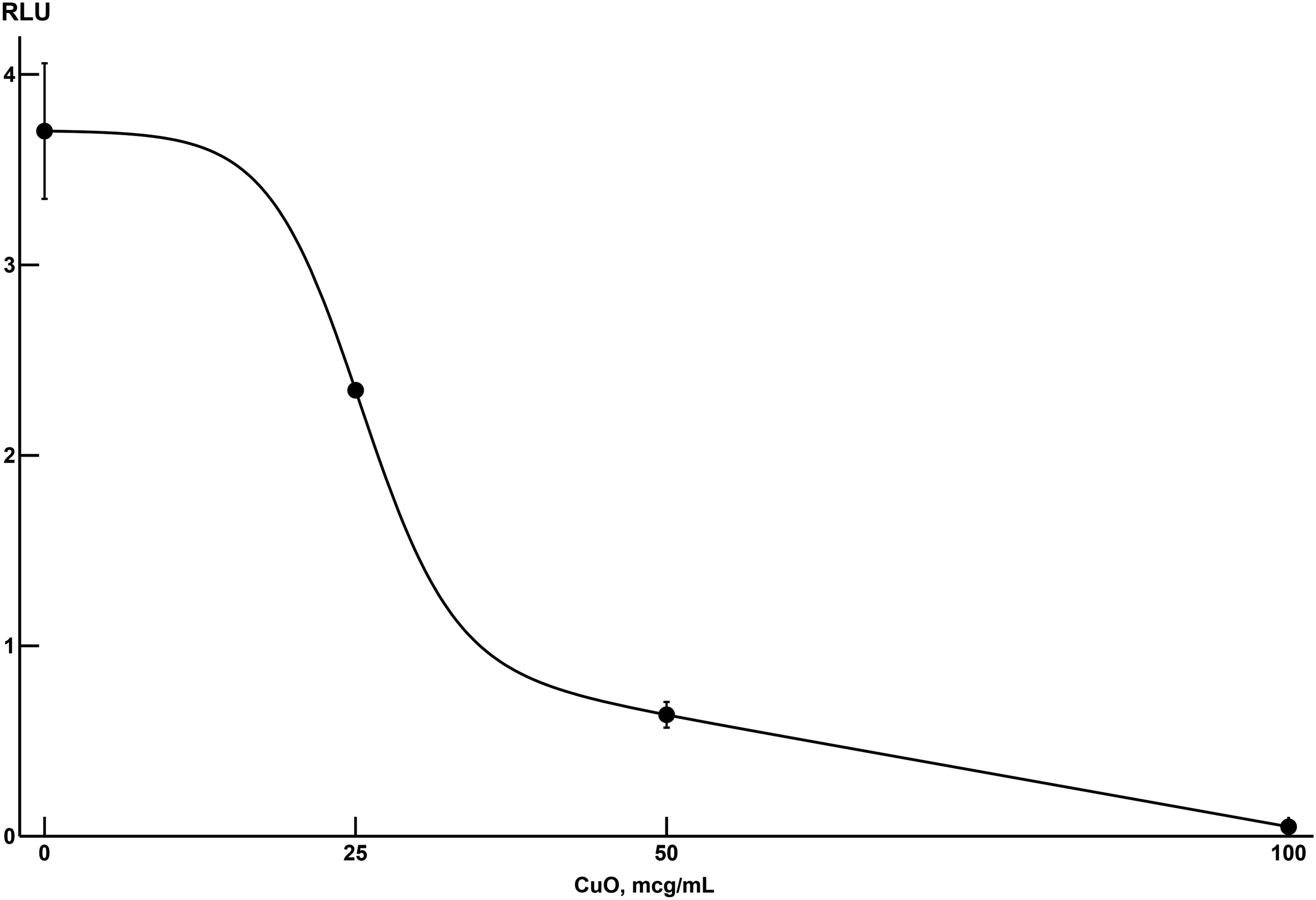
Note also that, whereas at minimal NP concentrations the intensity of the cytotoxic impacts produced by SeO-NP and CuO-NP are virtually similar, at concentrations over 25 mcg/dL CuO-NP appears to be more cytotoxic. Assumingly, in this range of doses the adverse impact of SeO-NP on the cells is partly set off by the cytoprotective effect of selenium as an essential trace element, shared by its nanoparticles as well (e.g., see14). In the meantime, we have not come across any mentions in the literature of a similar protective effect of copper-containing ones.
Morphometric Data
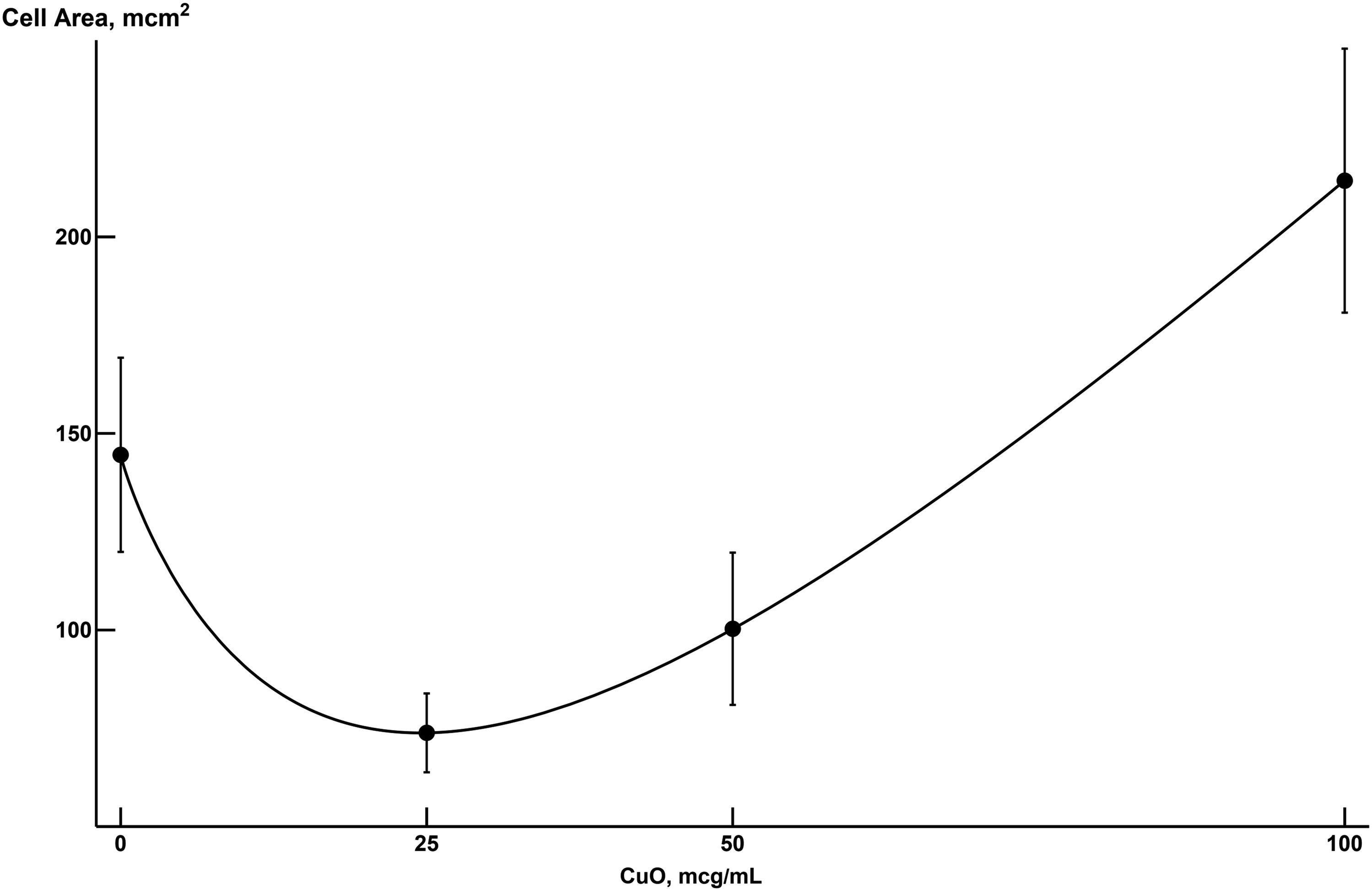
The dose-response relationship for cell area under exposure to nanoparticles of both species proved to be nonmonotonic and required special models for describing them (Figure 4 and 5). The approximation of the cell area by the model Approximation of cell area by the model 
It is clear that in the case of SeO-NP we deal with hormesis, though considered in the broader sense as was discussed in detail in Ref. 2, 3, while traditional approximations of two-phase hormesis1,8 are insufficient in this case. Meantime, the dependence given in the figure caption proved to be quite satisfactory and, at the same time, not too complex.
At the same time, exposure to CuO nanoparticles revealed a picture of hormesis corresponding to its traditional understanding (Figure 5), which can be described by the well-known dependence. 8
In a similar assessment, the dependence of cell-nucleus area on dose for both SeO and CuO nanoparticles proved to be of the same type, а three-phase one, which is likely to reflect the dissimilarity of the mechanisms responsible for the increase in cell and cell-nucleus size.
The quality of approximation by the proposed models of dose-response relationships for both luminescence and morphometric indices, as estimated by the coefficient of determination (see Mathematical Description of the Experimental Results), proved to be quite high for all models (both usual and adjusted coefficients were no lower than .6). The evaluation of the statistical significance of the model parameters also shows their high significance (P < .001). In addition, it is obvious from the presented Figures 6 and 7 that the proposed dose-response models describe the observed experimental values well. Approximation of cell-nucleus area by the model Approximation of cell-nucleus area by the model 

It should be noted that in a study involving exposure to CdS and PbS nanoparticles, 1 the dose-dependent suppression of cell viability as measured by ATP-luminescence was also monotonic, while the dependence of the morphometric characteristic proved to be nonmonotonic by the hormesis-associated type despite the fact that two studies of ours involved NPs of different compositions and cells of essentially different type (cardiomyocites and fibroblasts). We do not yet have sufficient experimental data to regard this coincidence as a regular feature and make suggestions concerning its mechanisms. However, it would be reasonable to make a mental note of it and revisit this issue when new data become available.
At the same time, it should be stressed that, even if the cell hypotrophy may seem an expected and better understood effect of cytotoxic impacts, it is not for the first time that we found that it was typical only for certain dose ranges while under the impact of other doses, on the contrary, the cell hypertrophy was evident. In particular, we observed the same in our in vitro experiments on cardiomyocites under toxic impacts of CdS-NP and PbS-NP. 1 The possible molecular or other mechanisms of this paradox are no more clear than the mechanisms of hormesis phenomenon in general and need special investigation for each particular response. Let us mention in this connection that, as it was many times demonstrated long ago (e.g.,15, 16) the products of macrophages breakdown caused by different agents have both in vivo and in vitro a stimulating effect on these cells differentiation and functions, so that a resulting response can be directed oppositely depending on a balance between a damage and its compensation.
In the same vein, the data that we have gained so far enable us to suggest that the type of dose-response relationship, whether monotonic or nonmonotonic, is not predetermined by the cell type or chemical nature of the impacting nanoparticles; rather, it depends on the impact’s outcome assessed as a response. The same can be said about the studied nanoparticles’ combined action type.
Modeling of Combined Action
As may be seen from isoboles presented by the Figure 8 ATP-dependent luminescence, the combined action type proved the same as in the case of CdS-NP plus PbS-NP combination.
1
Meantime, for the same combination, this type assessed for a morphometric index was different as compared with the present study. Isobolograms characterizing the combined toxic action of CuO-NP and SeO-NP on an fibroblast-like cell culture as estimated by its effects on (A) ATP-dependent luminescence (additivity), (B) cell area and (C) cell nuclear area (superadditivity). Numbers at the axes are respective NP concentrations in mcg/mL; numbers at the isoboles are the values of the effect to which they correspond.
In general, if considered together with our previously described experiments, which gave principally similar results, this study suggests once more that: (a) dose-response relationships for one and the same toxic agent but for different outcomes can be of both monotonic and nonmonotonic type, the latter corresponding to the hormesis paradigm in its generalized form; (b) a diversity of types of joint action characteristic of one and the same pair of toxic agents is one of the important assertions of the general theory of combined toxicity.
Conclusions
(1) Selenium-oxide and copper-oxide nanoparticles when acting on fibroblast-like cells in vitro display cytotoxicity manifesting itself as a decrease in ATP-dependent luminescence, CuO-NP producing a somewhat stronger effect than SeO-NP. (2) Along with cell hypertrophy under the action of certain doses of SeO-NP and CuO-NP, our experiment also revealed doses causing a decrease in cell and cell-nucleus size. (3) We obtained both monotonic and different variants of nonmonotonic dose-response relationships, and for the latter we managed to construct adequate mathematical expressions based on the generalized hormesis paradigm considered by us previously in respect of the cytotoxicity of CdS-NP and PbS-NP for cardiomyocites. (4) In general, the analysis of data obtained in the above-considered studies enable us to suggest that variability of dose-response relationship types displayed by different nanoparticle cytotoxicity effects is a common rule not accidental which makes it worthy of collecting further experimental evidence for establishing it as a common nanotoxicological rule. (5) It was demonstrated as well that combined toxicity of SeO-NP and CuO-NP is also of different types depending on the outcome for which it is assessed.
Footnotes
Declaration of Conflicting Interests
The author(s) declared no potential conflicts of interest with respect to the research, authorship, and/or publication of this article.
Funding
The author(s) received no financial support for the research, authorship, and/or publication of this article. The equipment of the Ural Center for Shared Use “Modern nanotechnology” Ural Federal University (Reg.& numero; 2968) was used.