
Abstract
Hormesis, or biological effects of low level exposures (BELLE), is characterized by nonmonotonic dose response which is biphasic, displaying opposite effects at low and high dose. Its occurrence has been documented across a broad range of biological models and diverse type of exposure. Since hormesis appears to be a relatively common phenomenon in many areas, the objective of this review is to explore its occurrence related to dermatology and its public health and risk assessment implication. Hormesis appears to be a common phenomenon in in-vitro skin biology. However, in vivo data are lacking and the clinical relevance of hormesis has yet to be determined. Better understanding of this phenomenon will likely lead to different strategies for risk assessment process employed in the fields of dermatologic toxicology and pharmacology. We believe that hormesis is a common phenomenon and should be given detailed consideration to its concept and its risk assessment implications, and how these may be incorporated into the experimental and regulatory processes in dermatology. The skin, with its unique characteristics, its accessibility, and the availability of non-invasive bioengineering and DNA microarray technology, will be a good candidate to extend the biology of hormesis.
Keywords
INTRODUCTION
Biphasic dose response, namely a low-dose stimulatory and a high-dose inhibitory response, also called hormesis, or biological effects of low level exposures (BELLE) in the field of toxicology, has been noted in a wide range of biological model systems from immunology to cancer biology (Calabrese and Baldwin 2001, 2003; Calabrese 2005a, 2005b). Calabrese has been the mainstay in bringing the attention of the scientific community to this interesting and a not uncommon phenomenon (Calabrese and Baldwin 2001, 2003; Calabrese 2005a, 2005b). As noted by Calabrese, the quantitative features of the hormetic-like biphasic dose response were remarkably similar with respect to the amplitude of the stimulatory response, the width of the stimulation, and the relationship of the maximum stimulatory response to the zero equivalent point (ZEP, i.e. threshold). Typically, the low-dose hormetic biphasic dose response stimulation is modest, with maximum stimulation between 30 to 60% greater than controls, and has a rather similar appearance in different cell types with various chemicals (Calabrese 2005b). Most stimulatory ranges were less than 100-fold (averages 10- to 20- fold) measuring back from the ZEP. The low-dose stimulatory response often occurs following an initial disruption in homeostatis and appears to represent a modest overcompensation response. It is believed that the modest stimulatory responsiveness is due to the result of a compensatory process that “slightly” overshoots its goal of the original physiological set-point, ensuring that the system returns to homeostasis without unnecessary and excessive overcompensation (Calabrese 2001). Therefore, it is important to follow the dose-response relationships overtime in order to better define its quantitative features. While initial interest focused on the hormetic effects of pollutants and toxic substances on biological systems (Calabrese and Baldwin 1997), the interest expanded to include pharmacological agents, phyto-compounds, as well as endogenous agonists (Calabrese 2005b). The hormetic- like biphasic dose response relationships appears to be highly generalizable; that is, such responses do not appear to be restricted by biological model, endpoint, or chemical/physical stressors (Calabrese 2005b).
Many investigations attempted to assess mechanisms that could account for the hormetic-like biphasic dose-response relationship. In general, there is no single mechanism that accounts for the plethora of hormetic relationships. Nonetheless, a common molecular tactic by which biphasic dose-response relationships are displayed involves the presence of two receptor subtypes affecting cell regulation, one with high and the other with low affinity for the agonist but with notably more capacity (i.e. more receptors) (Calabrese 2005b). Such an arrangement may lead to the biphasic dose response, with the high-affinity receptor activated at low concentrations, which stimulates DNA synthesis and cellular proliferation; and the low affinity/high-capacity receptor becoming dominant at higher concentrations decreasing the cell proliferative response. This is a general pharmacological mechanism in that it is employed for a large number of receptor-based responses from cancer cells to neutrophil chemotaxis and many others.
This article reviews hormetic effects of various agents on in vitro skin biology. Recognition of this emerging biological phenomenon in dermatology should lead to markedly improved integrative assessments of animal/human skin responses to toxic substances, pharmacological agents, as well as endogenous agonists.
EVIDENCES OF HORMESIS IN SKIN CULTURES
Skin is a complex biological model but highly approachable. Models exist for dermatologic research which include animal vs human skin models, in vitro vs in vivo models, regional variation, stem cell biology and hair follicle biology. Many pharmaceutical preparations in dermatology affect cell regulation. Nonetheless, the FDA sometimes exempts dose justification for dermatologic preparations. As a result, the presence of any hormetic effect might have been missed.
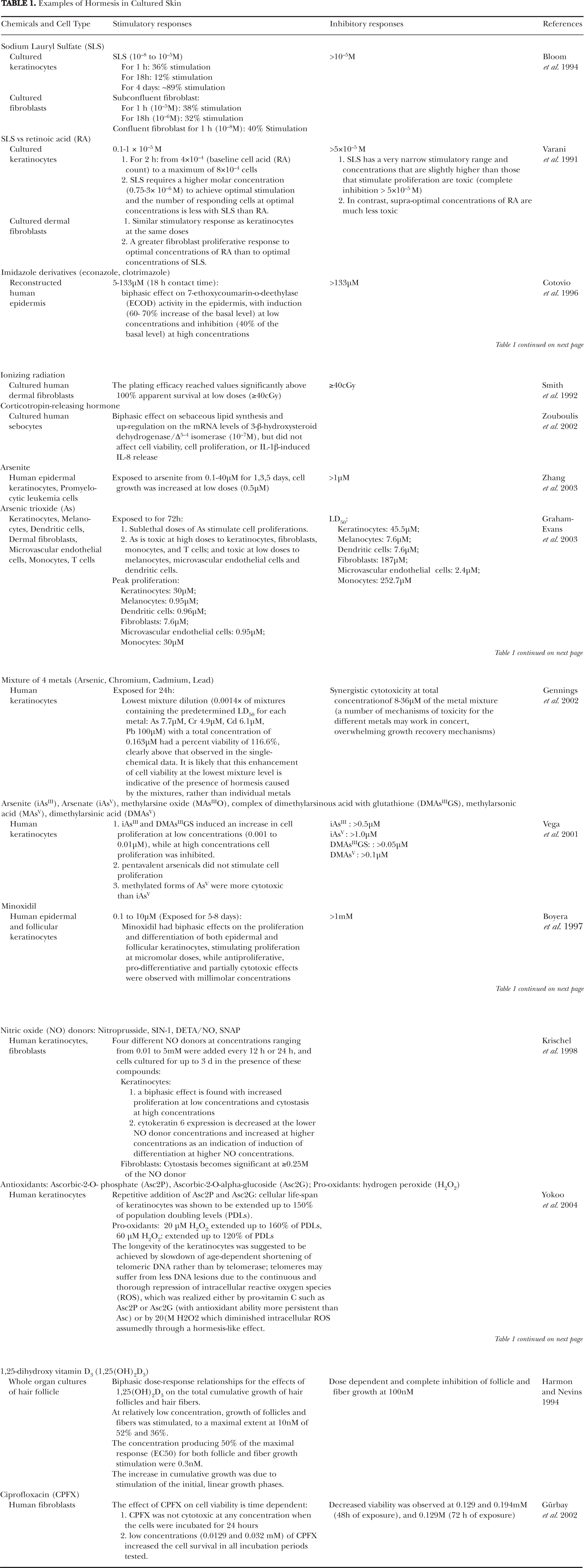
The literature in dermatology indicates that several cell types in the skin provided evidence of hormetic-like biphasic dose/concentration-response relationships. A brief listing of the cell types showing hormetic relationships and the quantitative features of dose responses is presented in Table 1.
Examples of Hormesis in Cultured Skin
MELANOMA AND TUMOR CELL LINES DISPLAY HORMETIC DOSE RESPONSES
Perhaps a more important issue regarding hormesis is its relationship to cancer biology. The existence of hormetic dose responses in many tumor cell lines has been noted and reviewed by Calabrese (2005b). 12 melanoma cell lines (M4Beu, B16, M24, MNT, SK-MEL, H1144, SK-MEL28, Cal 1, Cal 4, Cal 23, Cal 24, Cal 32) have been shown to display hormetic dose responses to various chemicals (guanine or guanosine derivatives, mistletoe extract, salsolinol, tetrahydropapaveroline, dopamine, resveratrol, thrombin and suramin) (Calabrese 2005b). Numerous endogenous agonists, drugs, environmental contaminants, and phytochemicals, some relevant to dermato-toxicology and dermato-oncology, have also been noted to exert hormetic dose responses in various tumor cell lines (Calabrese 2005b). Examples and the proposed mechanistic explanations are listed in Table 2.
Examples of dermatology-relevant chemicals displaying hormetic dose-response relationship in tumor cell lines

HORMESIS AND ITS IMPLICATION IN SKIN AGING
Detailed molecular mechanisms of the hormetic effects on cell aging are being increasingly understood. Studies have shown that repeated mild heat stress (RMHS) has anti-aging effects on serially passaged human fibroblasts throughout their replicative lifespan in vitro. Treatment with RMHS at 41° C, for 1 hour twice a week, increased the levels of various chaperones and antioxidant enzymes, increased the phosphorylation-mediated activities of various stress kinases, reduced the accumulation of oxidatively and glycoxidatively damaged proteins, stimulated proteasomal activities for the degradation of abnormal proteins, improved cellular resistance to ethanol, hydrogen peroxide, and UV-B rays. RMHS-treated aging human fibroblasts are also better protected against glucose- and glyoxal-induced growth inhibition and apoptosis. Various hormetic effects of RMHS were also noted on normal human epidermal keratinocytes, including increased replicative life span, increased proteasomal activity, and enhanced levels of Na/K-ATPase pump. (Rattan 2004a; Rattan and Ali, 2007). While some important issues with respect to establishing the optimal hormetic conditions are yet to be resolved by future research, hormesis could be a promising approach for modulating aging (Rattan 2004b; Rattan 2004c).
DISCUSSION
Despite the acknowledgement of the existence of hormesis, its importance has been disregarded by the scientific community, with one of the reasons being the close association of the concept of hormesis with the highly controversial medical practice of homeopathy (Burgdorf and Happle 1996; Clement 1997; Cook and Calabrese 2006).
Calabrese and Blain (2005) developed a hormesis database, containing 5600 hormetic-like dose response relationships over approximately 900 agents from a broadly diversified spectrum of chemical classes and physical agents, stressing the general robustness of published studies to establish support for the hormetic dose-response hypothesis. Table 1 showed that clear examples of hormesis do exist in dermatology, and Table 2 suggested that the presence of hormesis in cancer biology may be an important phenomenon not to be overlooked.
Despite the extensive observation of hormetic dose-response relationships for numerous agents across the biological spectrum, most studies assessed cellular responses. Few studies followed up in animal and human models—normal or disease, or assessed the simultaneous responses of different systems to the same agent. We believe in vivo studies are necessary to provide an integrative assessment of the whole animal/human responses to various agents, to document any discrepancies between the in vitro and in vivo responses, and to clarify the clinical implication of hormesis.
Studies on the mechanism of action and the exact definition of the low-dose to be applied are essential to achieve a better understanding of hormesis. Another important issue to discuss in the field of hormesis, as proposed by van der Woude et al (2005), is the need for risk assessment paradigms to be modified to take hormesis into account. Note that research methods may need to be modified to take into account that using lower doses and getting smaller responses may mean that a substantial increase in the number of animals or test subjects may be needed to get statistically significant results. Rietjens and Alink (2006) also suggested that more focus should be redirect from looking only at adverse effects at high levels of exposure to characterizing the complex biological effects, both adverse and beneficial, at low levels of exposure. Low-dose toxicology and pharmacology will contribute to better methods for low-dose risk assessment of chemical compounds and their effect on carcinogenesis, taking into consideration that the ultimate biological effect of a chemical may vary with its dose, the endpoint or target organ considered, cellular interactions, and/or the combined exposure with other chemicals.
We believe skin is an excellent candidate to gain entrance into this biology due to its accessibility; its complex nature, with highly differentiated cell types and various subsystems (keratinocytes, melanocytes, Langerhans’ cells, fibroblasts, epidermis, dermis, hair follicle, eccrine, apocrine and sebaceous units); and the availability of specialized non-invasive technology for in vivo studies (Maibach 1996; Elsner et al. 2001). In addition, skin has been among the first organs analyzed using DNA microarrays in various topics from skin cancers, melanomas, basal cell carcinomas, squamous cell carcinomas, psoriasis and other inflammatory disorders, to stem cell biology, the biology of epidermal keratinocytes, and so forth (Table 3) (Blumenberg 2006). DNA microarray studies will be an excellent tool to elucidate the mechanisms of hormesis in skin biology. In short, better understanding of hormesis will likely lead to different strategies for risk assessment process employed in the fields of dermatologic toxicology and pharmacology.
Targets for DNA microarray studies in dermatology and skin biology (Blumenberg 2006)

CONCLUSION
Hormesis is a common phenomenon in dermatology and other fields. Detailed consideration should be given to its concept, its risk assessment implications, and its clinical significance. However, without additional mechanistic insight, the consequences of hormesis for risk assessment and the possibilities for in vitro to in vivo extrapolation will remain limited.
Skin can be an excellent candidate to study hormesis and its underlying mechanisms because of its accessibility; its repertoire of inflammatory and immunomodulating cytokines, hormones, vitamins, unique responses to ultraviolet light, toxins, and physical injury; and the availability of non-invasive bioengineering and DNA microarray technology. Artificial skin substitutes are also available to study the effects of harmful or dangerous agents. In essence, the skin has everything: from stem cells, signaling and cellular differentiation, to inflammation, diseases, and cancer. All these facets could become excellent models to further study hormesis and its clinical implications following exposure to a variety of toxic compounds and pharmaceutical agents.