
Abstract
The goal of this investigation was to evaluate the effects of gestational administrations of arsenic trioxide (ATO; As2O3) on fetal neuroendocrine development (the thyroid-cerebrum axis). Pregnant Wistar rats were orally administered ATO (5 or 10 mg/kg) from gestation day (GD) 1 to 20. Both doses of ATO diminished free thyroxine and free triiodothyronine levels and augmented thyrotropin level in both dams and fetuses at GD 20. Also, the maternofetal hypothyroidism in both groups caused a dose-dependent reduction in the fetal serum growth hormone, insulin growth factor-I (IGF-I), and IGF-II levels at embryonic day (ED) 20. These disorders perturbed the maternofetal body weight, fetal brain weight, and survival of pregnant and their fetuses. In addition, destructive degeneration, vacuolation, hyperplasia, and edema were observed in the fetal thyroid and cerebrum of both ATO groups at ED 20. These disruptions appear to depend on intensification in the values of lipid peroxidation, nitric oxide, and H2O2, suppression of messenger RNA (mRNA) expression of nuclear factor erythroid 2-related factor 2 and peroxisome proliferator-activated receptor gamma, and activation of mRNA expression of caspase-3, nuclear factor kappa-light-chain-enhancer of activated B cells, cyclooxygenase-2, Bcl-2–associated X protein, and inducible nitric oxide synthase in the fetal cerebrum. These data suggest that gestational ATO may disturb thyroid-cerebrum axis generating fetal neurodevelopmental toxicity.
Introduction
Arsenic, a natural metalloid, is a known ubiquitous environmental contaminant 1 –4 found in human foods (meat and fish), drinking water, soil, and air. 5 –8 In particular, the effective harmful source of arsenic is in drinking water. 9 In 8 states of America, the levels of arsenic in drinking water are 50 to 100 ppb above the approved maximum contaminant level, 10 ppb. 10 In addition, arsenic trioxide (ATO; As2O3) is a potent toxic agent used in agriculture as herbicide and pesticide, and in the manufacturing of many industrial materials such as glass, wood, dyes, and pigments. 11 –13 Arsenic is a harmful toxin to human 6,14,15 and animal health 4,11 causing a wide range of diseases, including skin cancer and cognitive dysfunction, 16 diabetes mellitus and cardiovascular diseases, 17 and abdominal problems. 18 These abnormalities are amplified during the fetal developmental period. 11,19
Thyroid hormones (THs) are susceptible to disruption by arsenic, 20 particularly during the fetal development. Exposure of guinea pigs to 50 ppm arsenic in the basal diet decreased the levels of THs. 13 However, rats exposed to 100 mg arsenic/L for 4 weeks exhibited an increase in the level of serum T3. 21 Epidemiological and animal studies have proposed that arsenic penetrates the placenta 22 causing miscarriage, death, and several developmental toxicities. 23,24 In human, low values of arsenic (0.8-2 ppb) can increase the risk of spontaneous abortion 25 and fetal mortality. 26 In utero arsenic exposure caused morbidity in rural Bangladesh. 27 In pregnant rats, Holson et al 28 reported that a reduction in the food intake for the dams and body weight of both dams and their fetuses was progressed from doses 1 to 10 mg ATO. In addition, gestational arsenic can accumulate in the fetal brain of rats 29 causing several brain disorders. 30 Administration of rats to 50 mg/L inorganic arsenic (arsenite) as a chronic dose induced apoptosis in the brain. 31 Exposure of mice to 0.5 and 5 ppm ATO via the drinking water for 6 weeks induced apoptosis in the neuronal cells of the cerebral cortex. 32 Additional investigations have shown that exposure to arsenic causes neuronal apoptosis 30,33 and neural dysfunction. 34 In clinical trials and animal models, chronic exposure to arsenic causes neurotoxicity 35,36 and cognitive impairments. 37 These disturbances could be attributed to the ability of arsenic to deplete sulfhydryl groups, increase reactive oxygen species (ROS), 4,38 and disrupt the activity of peroxisome proliferator-activated receptor gamma (PPAR-γ), 39 Nrf-2 expression, 40 and phosphoinositide 3-kinase (PI3K)/AKT phosphorylation pathways. 41 These variations can disrupt the neural gene expression and brain activities. 42,43 However, the mechanisms by which gestational ATO induces developmental neuroendocrine disruption are still unclear.
Thyroid hormones, 44 growth factors, 45 and gene expression factors 46 display dynamic roles during brain development. As the sensitivity of the developing neuroendocrine axis to any stress is found to be obviously critical, 47,48 the purpose of the current investigation was to follow the novel action of gestational administrations of ATO (5 or 10 mg/kg) from gestation day (GD) 1 to 20 on the fetal thyroid-cerebrum axis.
Materials and Methods
Chemicals
Arsenic trioxide (As2O3; 99.99% purity) was procured from Sigma-Aldrich (St. Louis, MO, USA). The T4, T3, thyrotropin (TSH), growth hormone (GH), insulin growth factor-I (IGF-I), and IGF-II kits were purchased from Millipore (St. Charles, MO, USA) ELISA Kit. All chemicals were of the optimum analytic grades.
Ethics Statement
All animal experimentations were approved with the overall rules of Egyptian Animal Care and Animal Ethics Committee in Beni-Suef University, Faculty of Science, Zoology Department (BSU/FS/ 2016/9). All efforts were made to ensure the proper care and use of animals; http://www.bsu.edu.eg/Content.aspx?section_id=874&cat_id=43&.
Animals and Experimental Design
The experiment was conducted on 24 mature virgin female Wistar rats (Rattus norvegicus) weighing 155 to 165 g. Also, 12 adult males were used for the coupling only and were otherwise not part of the study. Rats were obtained from the animal house of VACSERA (Holding Company for Biological Products & Vaccine) in Helwan, Egypt. Female and male rats were given tap water and food daily ad libitum, and kept in stainless steel cages at 25°C ± 2°C temperature, 50% ± 5% relative humidity, and light/dark cycle of 12 hours each (lights on at 7:00 hours) 49 –53 for 2 weeks to get rid of any intercurrent infection and familiarization. Then, pregnancy was carried out in a separate cage by coupling 1 male with 2 proestrous females overnight for 1 or 2 days 54,55 and confirmed by the presence of a copulatory plug or sperms in the vaginal smears (GD 0). Pregnant rats were then weighed and housed separately in cages. Notably, rats are used in this experiment because their erythrocytes are extremely prone to retain arsenic. 56
Arsenic trioxide was dissolved in deionized water. Then, 1 M NaOH was added to form a clear solution. HCl of 1 M was added to adjust pH between 7.0 and 8.0. Then, a suitable volume of deionized water was added to each preparation to achieve the desired concentration. Aqueous solutions of ATO were freshly prepared and administered to pregnant rats by gavage (0, 5, or 10 mg ATO/kg) from GD 1 to GD 20. These dosages were designated according to Holson et al 28 The control pregnant was orally administrated deionized water only as a vehicle. The maternal mortality and abortion were observed throughout the experimental period, and the maternal weight gain, fetal weight, fetal death, and fetal brain weight were recorded at GD 20.
Anesthetized dams and fetuses by mild diethyl ether were killed at GD 20. The quantity of the mild diethyl ether was very low with very short exposure-time to avoid any irritation to the eyes, nose, and respiratory airways according to the local ethics committee and several papers. 48,49,51 –53,55,57 –62 Maternal blood samples were obtained from the jugular vein whereas blood samples of fetuses were obtained directly from the umbilical cord. 57,59,60,63 Samples of coagulated blood were centrifuged at 3000 rpm (1006.2 g) for 20 minutes, and the clear supernatants were retained at −20°C for the following biochemical examinations. On the other hand, thyroid and cerebrum of dissected fetuses at embryonic day (ED) 20 were rapidly excised and fixed in 10% neutral buffered formalin for the histopathological examination. The remaining cerebrum tissue was perfused in ice-cold phosphate-buffered saline (PBS). Homogenates of frozen cerebrum samples in chilled PBS (10% wt/vol) were centrifuged at 3000 rpm (1006.2 g) for 20 minutes. The homogenates were retained at −70°C for subsequent quantitative reverse transcription polymerase chain reaction (qRT-PCR) assay. Furthermore, homogenates of other-remaining cerebrum samples in 0.9% NaCl (10% wt/vol) were prepared for further biochemical assay.
Enzyme-Linked Immunosorbent Assay Examination
The concentrations of maternal and fetal serum T3, T4, and TSH, and fetal GH, IGF-I, and IGF-II were assessed by ELISA (Spectra Max 190-Molecular Devices, USA) in Egyptian Cairo University, Faculty of Medicine, Department of Biochemistry, using a spectrophotometer at 450 nm. The standard plots were assembled using standards and used to calculate the concentrations of unknown samples.
Histological Examination
After fixation of the thyroid and cerebral tissues in 10% neutral buffered formalin for 1 day at room temperature, the samples were transferred to the Histopathological Department, Veterinary Faculty, Beni-Suef University, Egypt for additional processing, paraffin blocking, sectioning at 6 µm, mounting on glass slides, deparaffinized, and staining with hematoxylin and eosin (H&E) stain. 64 The stained slides were evaluated under the light microscope.
RNA Isolation and qRT-PCR Examination
The homogenate of frozen cerebrum samples was transferred to the Department of Biochemistry, Faculty of Medicine, Cairo University, Egypt for additional processing. A Fermentas RNA isolation kit was used to isolate the total RNA. The concentrations were measured at 260 nm and the selection of RNA samples was A260/A280 ratios ≥1.7. A reverse transcription Fermentas kit was used in reverse transcription of RNA to complementary DNA (cDNA) with 1 µg RNA. A SYBR Green master mix was used to augmentation the produced cDNA with a total volume of 20 µL. Table 1 shows the primer set of examined PPARγ, nuclear factor erythroid 2-related factor 2 (Nrf2), cysteine-aspartic acid protease-3 (caspase-3); cyclooxygenase-2 (Cox2), nuclear factor kappa-light-chain-enhancer of activated B cells (NF-κB), Bcl-2–associated X protein (BAX), and inducible nitric oxide synthase (iNOS). A 96-well plate was used for the sample seeding reactions. The PCR thermal cycles involved initial denaturation at 95°C for 10 minutes and 35 cycles of denaturation at 95°C for 30 seconds, annealing at Tm-5 for 60 seconds and extension at 72°C for 30 seconds. Nonspecific amplifications were detected by nontemplate and water controls. Each standard and experimental sample was examined in duplicate. The acquired amplification results were assessed by the 2−ΔΔCt method. 65 Notably, β-actin and glyceraldehyde-3-phosphate dehydrogenase, 2 reference genes, were utilized for normalization of all values.
Primer Sequences for the Quantitative RT-PCR Analyses.
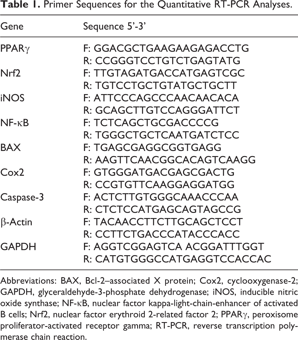
Abbreviations: BAX, Bcl-2–associated X protein; Cox2, cyclooxygenase-2; GAPDH, glyceraldehyde-3-phosphate dehydrogenase; iNOS, inducible nitric oxide synthase; NF-κB, nuclear factor kappa-light-chain-enhancer of activated B cells; Nrf2, nuclear factor erythroid 2-related factor 2; PPARγ, peroxisome proliferator-activated receptor gamma; RT-PCR, reverse transcription polymerase chain reaction.
Biochemical Assay
The homogenates of frozen cerebrum samples were centrifuged at 3000 rpm (1006.2 g) for 20 minutes. The levels of ROS were assayed in the Zoology Department, Faculty of Science, Beni-Suef University, Beni-Suef, Egypt. Lipid peroxidation (LPO) level was estimated by the formation of the malondialdehyde (MDA) as per the methodology of Preuss et al. 66 In addition, nitric oxide (NO) and H2O2 levels were determined as per the procedures of Dutta et al, 67 and Sergiev et al, 68 respectively.
Statistical Analysis
The data were stated as a mean ± standard error. Difference between the experimental groups was analyzed by PC-STAT program software (version 1A(C) PC-STAT, University of Georgia). 69 The groups were compared using analysis of one-way of variance and the least significant degree (LSD). The general variations between the experimental groups were recognized by F-probability. The number of considered samples/marker/group was 6. The variations between the experimental groups were P <.01 and P < .001.
Results
Gestational ATO-Perturbed the Maternofetal Thyroid Functions and Development
In 5 and 10 mg maternal ATO-treated groups, the concentrations of serum T3 and T4 were considerably (LSD; P < .01) decreased while the concentration of serum TSH was profoundly (LSD; P < .01) increased at GD 20 in both dams and fetuses (Figure 1A and B). These disorders indicate a maternofetal hypothyroid state. Fetal serum GH, IGF-I, and IGF-II levels were decreased by 3.56, 4.64, and 2.1-fold, respectively in the maternal ATO-low treated group and by 6.09, 9.26, and 5.47-fold, respectively in the maternal ATO-high treated group, respectively in relative to the control group (Figure 1C). In both maternal treated groups, there was a substantial drop in the maternal body weight gain (−32.22% in the As2O3-low treated group and −54.35% in the As2O3-high treated group), fetal body weight (−38.98% in the As2O3-low treated group and −54.47% in the As2O3-high treated group), and fetal brain weight (−11.65% in the As2O3-low treated group and −21.69% in the As2O3-high treated group; Table 2). Otherwise, both administrations of ATO caused a death in only 1 pregnant rat. However, abortion was only observed in 1 pregnant rat in the ATO-high treated group only. The fetal death was folded in the maternal ATO-high treated group compared to the maternal ATO-low treated group at ED 20 (Table 2).

Administrations of As2O3 caused (A) maternal hypothyroidism at GD 20, (B) fetal hypothyroidism at ED 20, and (C) Fetal growth retardation at ED 20. Data are expressed as mean ± SE. Number of animals in each group is 6. Where * is a highly significant (P < .01) change between the control and treated group. ED indicates embryonic day; GD, gestation day; SE, standard error.
Gestational Administrations of As2O3 Changed the Maternal and Fetal Markers at GD 20.a

Abbreviations: ANOVA, analysis of one way of variance; GD, gestation day; LSD, least significant degree; SE, standard error.
aData are expressed as mean ± SE. Number of animals in each group is 6.
bIndicates a highly significant (P < .01) change between the control and treated group and P < .001 is very highly significant change for whole experiment.
Gestational ATO-Induced a Fetal Thyroid and Cerebrum Dysgenesis
In the control group, the follicles of the fetal thyroid gland appeared normally in their distribution at ED 20 (Figure 2A). Fetal thyroid sections of maternal ATO-low treated group showed a flattened cell lining epithelium, hyperplasia, edema, atrophy, and destructive degeneration (Figure 2B). In addition to the previous lesions, colloid vacuoles were observed in the fetal thyroid sections of maternal ATO-high treated group (Figure 2C). On the other hand, the fetal cerebrum sections showed a normal histological architecture at ED 20 (Figure 3A). Vacuoles (Figure 3B 1), congested blood vessels and degenerative changes (Figure 3B 2), and pericellular edema (Figure 3B 1,2) were noticed in the fetal cerebrum of maternal ATO-low treated group. Moreover, Figure 3C 1 showed a vacuolar degenerative and hyperplastic proliferation, and Figure 3C 2 appeared severe congested blood vessels and several degenerative changes in the fetal cerebrum of maternal ATO-high treated group.

Sagittal sections in the thyroid gland of fetal rats at ED 20 in control (A), 5 mg As2O3 (B), and 10 mg As2O3 (C). Hematoxylin and eosin stain. Where, A indicates atrophy; C, colloid; CCLE, cuboidal cell lining epithelium; CV, colloid vacuoles; DD, destructive degeneration; ED embryonic day; FCLE, flattened cell lining epithelium; HYP, hyperplasia; O, edema; PFC, parafollicular cell.

Sagittal sections in the cerebrum of fetal rats at ED 20 in control (A), 5 mg As2O3 (B1,2), and 10 mg As2O3 (C1,2). Hematoxylin and eosin stain. Where, CBV indicates congested blood vessel; DC, degenerative changes; ED, embryonic day; HP, hyperplastic proliferation; PCO, pericellular edema; PYC, pyramidal cell; V, vacuoles, and the arrows refer to the apical dendrites.
Gestational ATO-Disturbed the Gene Expression and Increased ROS in the Fetal Cerebrum
In both maternal ATO-treated groups, qPCR analysis exhibited a considerable (LSD; P < .01) downregulation of messenger RNA (mRNA) expression of Nrf2 and PPARγ in the fetal cerebrum at ED 20 (Table 3). On the contrary, both maternal administrations of ATO showed significant (LSD; P < .01) upregulation of mRNA expression of caspase-3, NF-κB, Cox2, BAX, and iNOS in the fetal cerebrum, where their means in the ATO-high treated group were 5.16, 2.91, 2.65, 2.80, and 3.25, respectively compared to their means in the ATO-low treated group (4.50, 2.45, 1.96, 1.54, and 2.47, respectively) or in the control group (3.68, 1.89, 1.14, 0.98, and 1.97, respectively; Table 3). On the other hand, LPO (MDA) level of both maternal ATO-treated groups was considerably (LSD; P < .01) elevated in the fetal cerebrum at ED 20 in comparison with the control group (Figure 4). Nitric oxide and H2O2 levels displayed a parallel pattern where their levels were significantly increased in the fetal cerebrum at ED 20, as described in Figure 4.
Gestational Administrations of As2O3 Disrupted the Relative mRNA Expression of PPARγ, Nrf2, iNOS, NF-κB, BAX, Cox2, and Caspase-3 in the Fetal Cerebrum at ED 20.a

Abbreviations: ANOVA, analysis of one-way of variance; BAX, Bcl-2–associated X protein; Cox2, cyclooxygenase-2; ED, embryonic day; GAPDH, glyceraldehyde-3-phosphate dehydrogenase; LSD, least significant degree; iNOS, inducible nitric oxide synthase; mRNA, messenger RNA; NF-κB, nuclear factor kappa-light-chain-enhancer of activated B cells; Nrf2, nuclear factor erythroid 2-related factor 2; PPARγ, peroxisome proliferator-activated receptor gamma; SE, standard error.
aExpression levels were normalized using β-Actin and GAPDH as the reference genes. For each gene, levels are expressed relative to the average level in the control group. Data are expressed as mean ± SE. Number of animals in each group is 6.
bIndicates a highly significant (P < .01) change between the control and treated group and P < .001 is very highly significant change for whole experiment.

Gestational administrations of As2O3 increased LPO, NO, and H2O2 levels in the fetal cerebrum at ED 20. Data are expressed as mean ± SE. Number of animals in each group is 6. Where * is a highly significant (P < .01) change between the control and treated group. ED indicates embryonic day; LPO, lipid peroxidation; NO, nitric oxide; SE, standard error.
Discussion
The gestational administrations of ATO (5 or 10 mg/kg) from GD 1 to 20 induced a hypothyroidism/thyrotoxicity in both dams and fetuses at GD 20. This state was evident by elevated level of serum TSH, along with the declined levels of serum T4 and T3. Also, some histological alterations such as a flattened cell lining epithelium, hyperplasia, edema, atrophy, colloid vacuoles, and destructive degeneration were observed in the fetal thyroid gland at ED 20. Previously, ATO accumulates in the thyroid follicles 70 and binds the protein sulfhydryl group of thyroid peroxidase (THs synthesis) 20,71 decreasing THs levels, 13,72 altering THs-metabolism, 73 perturbing thyroid receptors (TRs), 72,74 and thus initiating goiter. 75 Another possible mechanism is that exposure to arsenic can decrease the levels of selenium (necessary for conversion of T4 to T3) in blood and tissue causing a negative action on thyroid functions. 76,77 These results suggest that gestational ATO may cause maternofetal thyroid dyshormonogenesis, fetal thyroid dysgenesis, and fetal pituitary-thyroid axis dysregulation.
The current study demonstrated that administrations of ATO during pregnancy induced a marked reduction in the weight of both dams and fetuses at GD 20. This disturbance may reduce the uteroplacental transfer, interrupt the pregnancy, and delay the fetal development. Maternal and fetal death was observed in both ATO-treated groups while abortion was only recorded in the ATO-high treated group. Alternatively, the maternofetal hypothyroidism in both ATO-experimental groups resulted in a substantial reduction in the values of fetal serum GH, IGF-I, and IGF-II at ED 20. Interestingly, gestational ATO-high dose group (10 mg/kg) effects were more severe than those in the gestational ATO-low dose group (5 mg/kg). These findings may reflect the toxicity and teratogenic outcomes of gestational ATO. In parallel, rats exposed to 1, 2.5, 5, or 10 mg/kg ATO displayed a diminution in the maternal food intake and in the body weight of both dams and their fetuses.
28
Epidemiological and animal studies have proposed that arsenic penetrates the placenta
22
causing a spontaneous abortion, low maternal food consumption, stillbirth, low maternofetal body weight, preterm delivery, and developmental retardation in human,
24,27
in rats,
19,78,79
and in mice.
11,79
These complications could be attributed to the following causes: (1) accumulation of arsenic in the placenta might reduce the placental blood flow and disturb the placental vasculogenesis causing growth retardation
11,19,23
; and (2) growth retardation due to exposure to arsenic might be mediated, at least partly, by inhibiting mRNA expression of IGF-I and its activity.
80
Also, the maternofetal hypothyroidism described here may be associated with a reduction of GH and IGF-I/IGF-II, and intensification in the ROS levels that may retard the fetal growth. On the basis of these data, it can be inferred that
The histopathological findings showed an ATO dose-dependent vacuolar degeneration, hyperplastic proliferation, congested blood vessels, pericellular edema, and destructive changes at ED 20 in the fetal cerebrum sections of maternal ATO-treated groups. The observed cortical deformation and neurodegeneration may be attributed to
Conclusion and Future Direction
Gestational exposure of ATO initiated a hypothyroidism/thyrotoxicity in both dams and fetuses indicating a stress-responsive aspect for a fetal thyroid-cerebrum axis and in general prenatal development. Gestational ATO accumulated and concentrated in the fetal thyroid and cerebrum causing several histopathological lesions in these tissues. These disruptions appear to rely on augmentation of ROS (LPO, NO, and H2O2), suppression of mRNA expression of Nrf2 and PPARγ, and upregulation of mRNA expression of caspase-3, NF-κB, Cox2, BAX, and iNOS in the fetal cerebrum. A novel interpretation of this study is that the gestational ATO may disrupt thyroid-cerebrum axis generating fetal neurodevelopmental toxicity (Figure 5). The variations were dose-dependent for all estimated markers except for the dead of pregnant dams, where the alterations were similar in both treated groups. However, these disorders may be dependent on the experimental design, developmental period, concentration, absorption, and metabolism of ATO. Additional studies are warranted to discovering the detailed molecular mechanisms of the gestational ATO-induced apoptosis to avoid any disturbance in the fetal and neonatal development.

Diagram about the gestational As2O3 and fetal neuroendocrine disruptions.
Footnotes
Acknowledgments
The authors acknowledge all the staff in their departments for general assistance. The authors also pleasantly thank Dr Manushree Bharadwaj (National Institute of Environmental Health Sciences, Durham, North Carolina) and Prof Dr Douglas B. Learn (Director of Photobiology and Cellular Therapeutic Safety at Charles River Laboratories, Pennsylvania) for their assistance.
Declaration of Conflicting Interests
The author(s) declared no potential conflicts of interest with respect to the research, authorship, and/or publication of this article.
Funding
The author(s) received no financial support for the research, authorship, and/or publication of this article.