
Abstract
X-rays have been the gold standard for diagnosis, evaluation, and management of spinal scoliosis for decades as other assessment methods are indirect, too expensive, or not practical in practice. The average scoliosis patient will receive 10 to 25 spinal X-rays over several years equating to a maximum estimated dose of 10 to 25 mGy. Some patients, those getting diagnosed at a younger age and receiving early and ongoing treatments, may receive up to 40 to 50 X-rays, approaching at most 50 mGy. There are concerns that repeated radiographs given to patients are carcinogenic. Some studies have used the linear no-threshold model to derive cancer-risk estimates; however, it is invalid for low-dose irradiation (ie, X-rays); these estimates are untrue. Other studies have calculated cancer-risk ratios from long-term health data of historic scoliosis cohorts. Since data indicate reduced cancer rates in a cohort receiving a total radiation dose between 50 and 300 mGy, it is unlikely that scoliosis patients would get cancer from repeated X-rays. Moreover, since the threshold for leukemia is about 1100 mGy, scoliosis patients will not likely develop cancers from spinal X-rays. Scoliosis patients likely have long-term health consequences, including cancers, from the actual disease entity itself and not from protracted X-ray radiation exposures that are essential and indeed safe.
Introduction
Scoliosis is a curvature of the spine in the coronal plane with a simultaneous rotation of the affected vertebrae. 1 A curve is considered definitive scoliosis when it is measured greater than 9° by the gold standard Cobb angle of measurement on the anterior–posterior or posterior–anterior radiograph (Figure 1). 2

Cobb angle as measured in the coronal plane. The top of the superior uppermost vertebra and the bottom of the lowermost vertebra are the end points of the curve. The intersection of these lines are off the image and therefore are measured by the angle formed by 2 right-angled lines drawn off each.
Radiography has been the gold standard in quantifying the magnitude of spinal deformity including scoliosis for over at least 70 years and continues to be so.
3
In fact, according to the Society On Scoliosis Orthopaedic and Rehabilitation Treatment (SOSORT), In the era of evidence-based medicine, radiographic evaluation of scoliosis continues to be the most expedient, cost-effective, and reliable assessment method. Historically, it has been the standard for determining Cobb angle, curve pattern, apices, end points, rotation, vertebral body shape, and structural anomalies. It is vital in making clinical decisions.4(p4)
More specifically, the scoliosis diagnosis has different nomenclature depending on the age of the patient (ie, infantile, juvenile, adolescent, adult), etiology (ie, congenital, idiopathic, neuromuscular, degenerative), its pattern classification including the side of convexity (ie, right or left), and spinal area (ie, thoracic, lumbar, thoracolumbar, or combined thoracic and lumbar; Figure 2). Regardless of the scoliosis parameters specific to a given patient, the traditional and current gold standard for its assessment and management continues to be spine radiography. 3,4

Patterns of scoliosis. Left: normal; middle three: single curves; right: double curve.
Due to the unpredictability of scoliosis progression despite common treatments, patients with scoliosis often undergo many repeated radiographic procedures throughout the course of their treatment or nontreatment (watchful waiting). Because of this, there are concerns for the long-term well-being of scoliosis patients, not from their medical condition, but from the iatrogenic exposures to radiation from repeated radiography. 5 -12
The purpose of this article is to review many uses of spinal radiography in the screening, treatment, and management of scoliosis and to provide a brief update on the scientific evidence supporting the safety of radiography and rationale for its continued use as the gold standard in daily clinical practice.
Scoliosis Screening
Although alternative tests for “quick” scoliosis screening are available and used, they are just that—simple generalized tests. The classic scoliosis test is the Adam’s forward bend test (Figure 3). 13 This test involves the patient to stand facing away from the assessor and to bend forward keeping the knees straight and hanging the arms down; a positive test is when the back appears asymmetric.
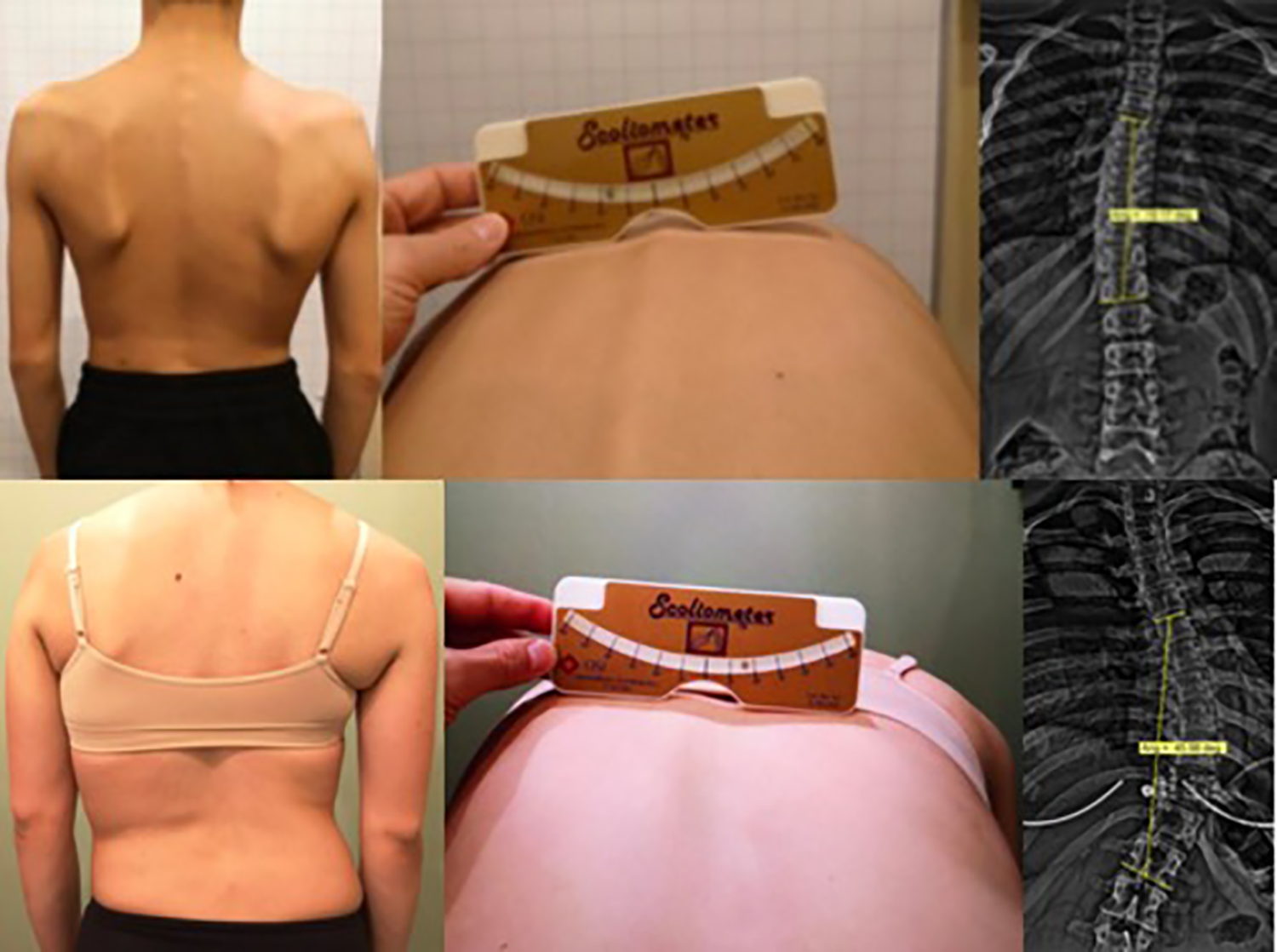
Demonstration of the specificity of X-ray versus imprecision of the angle of trunk rotation (ATR) by scoliometer measurement. Left: Posterior view of patient; Middle: Adam’s forward bend test with scoliometer measurement; Right: Anteroposterior spinal X-ray. Top: Patient has a mild T6-T12 scoliosis of 19° Cobb angle and an 8° ATR; Bottom: Patient has a moderate-severe T6-L1 curve of 39° Cobb angle and an 8° ATR.
The Adam’s test may be done subjectively, or objectively measured using a Scoliometer, a special ruler measuring the angle between the horizontal and the amount of rotation of the surface of the back termed “angle of trunk rotation” (ATR; Figure 3). 14 If the ATR measures ≥5°, it is likely that a subject has a scoliosis curvature >10°. In screening for adolescent idiopathic scoliosis (AIS), Bunnell found that 80% of high school students had at least a 3° ATR and therefore suggested an ATR of 7° at any level of the spine suffices as an appropriate criterion warranting referral to a specialist for a more thorough (radiological) assessment. 15
Other alternative tests for scoliosis are more complex and require trained users. Skin markers, for example, although may be estimated with reasonable accuracy for sagittal plane assessment of the thoracic spine, are shown to systematically underestimate spinal angles in the coronal plane due to positional and structural deformities of scoliotic vertebrae. 16
Scoliosis screening tests are indirect and poorly correlate with directly measured Cobb angles of spine curvature. 16,17 Although alternative imaging methods to measure the curvature of the spine accurately by the Cobb method are possible, for example, by MRI, 18 it must be done in the standing position and standing MRI units are rare, expensive, and simply not practical for daily clinical practice. Newer EOS X-ray imaging systems are now available that reduce radiation levels, 19 but these systems are expensive, not widely used, and not cost-effective, 20 raising doubts as to whether they will become the future standard. For these reasons, standard plain film X-ray examination of the spine remains the essential gold standard. 3,4
X-Ray Use in the Diagnosis, Evaluation, and Management of Scoliosis
Spinal radiography in scoliosis is essential for an accurate initial diagnosis; in fact, the definition of scoliosis revolves around the radiographic mensuration of the spinal curvature in the coronal plane of at least 10° (Cobb angle) with rotation of the involved vertebrae. 1 Further, classification of scoliosis severity is also based on the coronal Cobb angle measurement with classifications of mild, moderate, and severe corresponding to Cobb angle ranges of 10° to 25°, 25° to 40°, and greater than 40°. 21
Radiographs also help determine the skeletal maturity of the patient. The Risser sign is assessed by observing the degree of ossification of the iliac apophysis indicative of developmental stage. 22 Risser sign assessment has a 6-point grading system (0-5) correlating to no ossification started (Risser 0) to full ossification (Risser 5); grades 1 to 4 indicate different percentages of ossification along the iliac crest, and this grading varies depending on whether one uses the US or French Risser grading system. 23 Although an alternate developmental indicator of X-raying the left hand exists (Tanner-Whitehouse method), 24 it is more difficult and time-consuming. 25 The Risser sign has proven to be a simple, reliable, and clinically useful orthopedic classification system that remains widely used 26 and is readily seen on the standard lumbar-pelvic coronal X-ray view.
The estimated developmental maturity of the skeleton is important information and may indicate risk of potential worsening of spinal deformity while a child is still growing (Risser 0-3), or give reassurance of little risk of worsening that is associated near or at end-stage of skeletal development (Risser 4-5).
X-rays are also used for differential diagnosis such as in the diagnosis of congenital scoliosis. Congenital scoliosis involves the asymmetrical development of the architecture of a vertebra, for example, a “hemivertebra” is an anomaly where one-half of the vertebral body does not fully form and may be impossible to treat nonsurgically. Another differential diagnosis is the newer understanding of differentiating true scoliosis from “pseudoscoliosis,” 27 -29 which is a postural thoracolumbar subluxation that mimics aspects of scoliosis but responds better to different treatment approaches. 30,31
X-rays are essential in the follow-up management of scoliosis to monitor treatment progress. Often a change in treatment will be indicated if the deformity progresses (ie, “watch and wait” to bracing; bracing to surgery). Alternatively, a follow-up radiograph displaying a similar spinal curve measurement is a positive outcome in treating scoliosis particularly if the patient has gone through a growth spurt (ie, “stabilization”). Treatment cessation may also be indicated based on treatment success (ie, reduced curvature or straightened spine 32 ) or developmental indicators signaling end of growth (Risser 4-5).
The ultimate goal of treating scoliosis, and AIS in particular, is to prevent surgery. Recent evidence has substantiated that contemporary patient-specific, customized treatment approaches including intensive exercise programs (ie, Schroth methods 33 -35 ), spinal traction techniques (CBP approach 36,37 ), and 3-dimensional types of corrective spinal braces (ie, ScoliBrace, 38 Spinecor brace, 39 -41 Gensingen brace 42 ) point to successful outcomes of curve magnitude reduction strategies that were not yet available or popular in the past. In fact, it has been suggested that spinal curves even greater than 40° to 45°, the traditional surgical threshold, may be successfully treated nonsurgically, 41 -44 and some question whether surgery even has a role in the treatment of AIS. 45
With the great challenge of treating scoliosis, many of the factors pertaining to the age of the patient, skeletal maturity, magnitude of curvature, pattern of curvature, presence or absence of anomalies, and response to treatment all substantiate the essential reliance on radiographic examinations. This begs 3 important questions: (1) How many X-rays will a scoliosis patient receive during their treatment and management; (2) How much radiation exposure will this translate to; and (3)Will these cumulative radiation exposures result in future iatrogenic cancer risks.
How Many X-Rays Do Scoliosis Patients Receive?
Whether treated or only monitored (watch and wait), a child or adolescent with scoliosis is repeatedly X-rayed at least every 6 months until skeletal maturity (Risser sign grade 4/5). According to the consensus paper by SOSORT, “The universal desire is to minimize the amount of x-ray exposures; however, the x-ray is an important diagnostic and monitoring tool essential for assessing the need for bracing and for monitoring its effectiveness.” 4(p7) For skeletally immature or maturing patients, SOSORT recommends X-rays every 6 to 12 months, and this recommendation varies according to the age and skeletal maturity of the patient (Table 1). 4
Scoliosis Diagnosis, Maturity, and SOSORT X-Ray Frequency Recommendations by Age. 4

Abbreviations: AIS, adolescent idiopathic scoliosis; SOSORT, Scoliosis Orthopaedic and Rehabilitation Treatment.
In reality, however, scoliosis patients who receive treatment (bracing and/or exercise rehabilitation programs) are X-rayed much more frequently than the SOSORT guidelines in order to assess treatment response. Further, patients who end up getting surgery are X-rayed even more frequently. 6 Overall, the greater the curve, the more imaging necessary to monitor the progression. 6
Generally, a full spine series is recommended at the initial screening and diagnosis with only coronal images taken at follow-up assessments to minimize radiation exposures. Patients receiving brace treatment require in-brace images to assess treatment efficacy, as well as may receive initial lateral bending views to assess flexibility, and/or recumbent views to assess “correction potential.” Other reasons for extra imaging are for stress films while the patient performs a corrective exercise or lays over a traction block. In total, it is unknown how many radiographs a scoliosis patient may receive as the number may vary dramatically based on treatment versus monitoring or a patient receiving various treatments who has had continued spinal curve progression throughout childhood and adolescence.
Although the reported frequency of radiography use in scoliosis management is limited, 4 there are some cohort studies that can be used to estimate the total number of X-rays that scoliosis patients receive in actual clinical settings. Hoffman et al reported an average of 41.5 X-rays in a sample of 1030 females with scoliosis who attended medical facilities between 1935 and 1965. 6 Doody et al reported an average of 25 spinal X-rays in a cohort of 5573 female patients who were diagnosed prior to the age of 20 years between 1912 and 1965. 8 Nash et al reported an average of 22.5 X-rays in 13 females treated with a brace over a 3-year period. 5 Simony et al reported an average of 16 X-rays in a cohort of 215 consecutive AIS patients treated between 1983 and 1990. 12 Levy et al reported an average of 10 and 12 X-rays received by males and females, respectively, in a group of 2039 patients diagnosed with scoliosis between 1965 and 1979. 7
In clinical practice, it seems that different patients will get different numbers of X-rays based on the clinical progression of their spinal curve and from assessment of various treatments potentially given. Treatment assessment from a brace would add in several more over the course of treatment; thus, a 12-year-old who skeletally matures at age 16 will receive about 10 to 20 X-rays (2-4/yr, x 5yrs), whereas a child aged 9 years skeletally maturing at 16 will receive about 16 to 32 X-rays (2-4/yr, x 8yrs). A safe estimate, therefore, is about 10 radiographs as a minimum, and up to 25 radiographs seem typical. Some patients may receive up to 40 or 50 radiographs at a maximum. Clinical practice dictates many more X-rays are taken than is ideally recommended for the purpose of minimizing radiation exposures; thus, clinicians are always weighing the necessity of X-ray imaging based on the universal acceptance of the assumption that X-rays are harmful. We will provide evidence that they are not.
What Is the Cumulative Effective Radiation Dose for a Scoliosis Patient?
Although some patients with progressive scoliosis who are treated from an earlier age may get exposed to a higher number of X-rays (ie, 40-50), the majority of cases will fall in the range of 10 to 25 depending on treatment. Assuming a coronal plane spinal radiograph is 0.92 mGy 46 (ranges from <0.5–1 mGy), then radiation exposures can be estimated for children and adolescents receiving 10 to 50 images upon initial diagnosis (Table 2).

As can be seen the average cumulative dose for a patient receiving 10 X-rays may approach 10 mGy, whereas a patient receiving 25 X-rays may approach 25 mGy. These estimates are in the range calculated by Law et al who estimated a 15 mSv cumulative dose per scoliosis patient during the course of their treatment and management between the ages of 5 and 30 years by digital radiography. 11 So, it seems that most patients with scoliosis who get repeated radiographic exposures receive a cumulative dose of up to about 25 mGy and at most up to 50 mGy. Smaller patient masses (ie, a small 7-year-old) will receive about 50% less radiation than older teens, and images taken in the PA position may also reduce radiation exposures by about 50% 47 ; thus, the amounts demonstrated in Table 2 are conservative as actual patient exposures are likely less than shown.
Are Cumulative Radiation Doses From Scoliosis X-rays Harmful?
To answer this question, we examine the 2 ways that researchers have taken to estimate carcinogenic risk to scoliosis cohorts. The first is by using linear no-threshold (LNT) estimates and weighting factors to estimate theoretical future cancer incidence based on estimated total radiation exposure doses, and the second is from actual follow-up data from old patient files who were questioned about cancer incidence or had documented cancer deaths as compared to expected cancer incidence or mortality rates. We briefly discuss representative results from these types of studies and the faults with each approach.
Flaws With LNT-based Radiogenic Cancer-Risk Estimates
Theoretical cancer-risk estimates are calculated using the LNT model that is currently still recommended by all national and international bodies (ie, NAS BEIR, NCRP, etc). This model extrapolates high-dose data down to low-dose radiation exposures (<200 mSv) for which no data of carcinogenicity exists. 48 -50 Therefore, according to the LNT concept, any amount of radiation is harmful and also cumulative with dose; thus, dose is used as a surrogate for risk. 51
Studies that have incorporated this type of radiogenic risk estimation in scoliosis patients receiving repeated X-rays include Nash et al 5 and Levy et al. 7 Nash et al estimated an increase of 3.4 to 15 per million in organ cancer and an increase of 140 to 290 per million in breast cancer from assessing 13 females receiving 22 X-rays over a 3-year period. 5 Levy et al estimated an excess cancer risk of 42 to 238 cases per 100 000 for women, and 14 to 79 cases per 100 000 for men from assessing 2039 patients referred to a large pediatric hospital during the years 1925 to 1965 who received an average of 12 and 10 X-rays, respectively. 7
Since no clear data exist to support the contention that low-dose radiation exposures <200 mGy are harmful, 48 -50 we believe that the LNT model used to estimate potential future cancer risks from scoliosis patients receiving X-rays is invalid. Further, as pointed out by Jaworowski 52 and Cuttler, 53 -57 data from the 1958 UNSCEAR report 58 indicate that a J-shaped (hormetic) dose–response relationship exists (not linear) for radiation exposures and leukemia incidence in atomic bomb survivors (Figure 4). Cuttler 57 has pointed out that a threshold of about 1100 mGy exists; since leukemia would be the first cancer expected to occur after detrimental radiation exposure, other cancers would have higher thresholds. 29,55,56 Thus, X-rays in general (single exposures), and X-rays as used in serial fashion on scoliosis patients (protracted exposures) do not come close to the 1100 mGy threshold and therefore cannot be considered as carcinogenic. Finally, we believe that cancer-risk estimates for scoliosis patient populations based on LNT theory are not accurate and that they are predictions based on an inappropriately used model for low-dose exposures.

1958 UNSCEAR data indicate a threshold of about 1.1 Gy (1100 mGy; assuming RBE = 1) for radiogenic leukemia in 97 000 persons exposed to A-bomb radiation from Hiroshima. 57
Flaws With Cancer Relative Risk Ratios in Scoliosis Cohorts
Seemingly more robust data exist utilizing relative risk ratios for both incidence (standardized incidence ratio [SIR]) and mortality (standardized mortality ratio [SMR]) as has been reported in the long-term follow-up of scoliosis cohorts. 6,8,10,12 These are simple ratios of incidence or mortality in scoliosis cohorts as compared to the general population or the expected number (Table 3). Hoffman et al reported on 1030 females with scoliosis given an average of 41.5 X-rays over 8.7 years. 6 Upon 26 years of follow-up, 11 patients had reported breast cancer versus an expected number being 6; this resulted in an SIR of 1.82 (90% CI = 1.0-3.0) and 2.4 (90% CI = 0.9-5.0) for those followed for over 30 years. 6 Doody et al assessed mortality data linked to 5573 female patient files diagnosed with scoliosis and X-rayed between the years 1912 and 1965 and determined the females followed up for 40 years had an SMR for breast cancer of 1.69 (95% CI = 1.3-2.1). 8 In the same data set, reporting an average follow-up of 47 years, Ronckers et al reported an SMR of 1.08 (95% CI = 0.97-1.2) for any cancer and an SMR of 1.68 (95% CI = 1.38-2.02) for breast cancer. 10 Of note, however, death rates from many other cancers were consistent with no increase, including the organs that would be expected to be have greater SMRs resulting from radiation exposures including leukemia (SMR = 0.87, 95% CI = 0.42-1.6) and thyroid cancer (SMR = 0.8, 95% CI = 0.0-4.58). Simony et al reported an SIR of 4.8 (CI = 2.3-5.8) for developing any cancer compared to the general Danish population in assessing 215 scoliosis patients receiving 16 X-rays at a 24.5-year follow-up after being treated by brace or surgery during the years 1983 to 1990. 12

Abbreviations: SIR, standardized incidence ratio; SMR, standardized mortality ratio.
In the above studies, scoliosis patient radiation doses were estimated from historic exposures, thus these are not estimated future cancers but are actual increases in cancers in scoliosis patient cohorts. It must be mentioned that because these are long-term follow-up studies, the dose rates the patients were exposed to are much more than would be given compared to current technology. The studies using the data from the US scoliosis cohort study includes data from patient files from 1912 to 1965. 8 -10 The radiation exposures from X-rays in the early 20th century could not be accurately measured and great estimations were made; the cohort was assumed to have received a total accumulated dose of 100 to 150 mGy. 10 Assuming fairly accurate estimations of the historic radiation doses, the question of concern is: Is the increased rates of cancer in scoliosis cohorts attributable to the radiation exposure from the X-rays received at a younger age?
As previously mentioned, in the assessment of leukemia incidence of 97 000 Hiroshima atomic bomb survivors, Cuttler has illustrated an apparent dose threshold of about 1100 mGy. 57 Since blood-forming bone marrow is most sensitive to ionizing radiation, other types of cancers would likely have a higher dose threshold. 29,55,56 Since all of the mentioned studies showed total radiation doses to scoliosis patients being well below 1100 mGy (even with greater exposures from older X-ray equipment), it would not be expected that scoliosis patients would succumb to carcinogenic effects greater than background.
At such dose rates, the scoliosis cohorts may actually be expected to get less cancers due to activation of adaptive protection systems. For example, in a reassessment of the Canadian breast fluoroscopy study, 59 Cuttler and Pollycove demonstrated that females receiving a total dose of about 50 to 300 mGy had a breast cancer incidence of up to one-third less than the background incidence. 60 Thus, because the studies assessing historic radiation exposures in scoliosis patients demonstrate total doses within this range of exposure, radiation-induced breast cancers would not be expected to occur. Even assuming a dose threshold value of 500 mSv, Cuttler and Welsh state: “No predictions or hints of excess cancer risk (or any other health risk) should be made for an acute exposure below this value until there is scientific evidence to support the LNT hypothesis.”55(p202)
Why Would Scoliosis Patients Have Greater Cancer Rates If Not From Radiation Exposures?
Any increase in breast or other cancers in scoliosis cohorts are likely due to the disease entity itself as scoliosis of the spine has direct biomechanical consequences on internal organs (ie, heart and lungs) as well as the spinal cord and nerves, both factors being critical issues of concern in evaluating scoliosis surgery necessity as these factors influence long-term health and longevity. There is also a possible association of increased physiological aging with spinal deformity. Studies using mouse gene knockout models showing premature aging also demonstrate hyperkyphosis spinal deformity. It is found, for example, that mice with mutation in the XPD gene (a gene responsible for DNA helicase, which works as a repair and transcription mechanism) show normal development but with premature aging and hyperkyphosis. 61 Similarly, mice having mutations in either p53 genes 62 (tumor suppressor genes) or Kotho genes 63 (phenotypical aging suppressor genes) show defects in the joining of DNA double-strand breaks and present with simultaneous rapid aging and hyperkyphosis—both leading to increased mortality rates. Hence, spinal deformity may be associated with underlying genetic mutations that enhance cellular senescence predisposing the organism to cancer. 64
Any study finding increased mortality rates in scoliosis cohorts are confounded by the variable of radiation exposures from repeated X-rays. The spinal condition of thoracic hyperkyphosis (THK, ie, hunchback), however, is a condition of the spine where there have been several well-conducted studies substantiating increased mortality rates in these patients from all-causes as well as specifically cancer. 65 -70 These studies do not have the same confounding factor of repeat X-rays as these typically include older patients with progressive deformity with ageing. Thoracic hyperkyphosis and scoliosis are 2 types of subluxation patterns 71 referred to as adult spinal deformity (ASD), where recently Pellisé et al brought attention to the scientific community that ASD is a global burden that causes great suffering as surprisingly these patients were found to have worse health-related quality-of-life scores than patients having several major chronic diseases including arthritis, chronic lung disease, diabetes, and congestive heart failure. 72 Also, as per Kado et al, spinal deformity (THK) is associated with a 2.4 times greater mortality rate from factors such as atherosclerosis. 67 Pellisé et al urge that ASD warrants the same health policy attention as other common chronic diseases. 72 Thus, it would be expected that scoliosis patients would have a similar fate as THK patients (increased mortality; increased cancers) from prolonged postural subluxation (ASD)—not resulting from increased radiation exposures that are well below the cancer induction threshold.
Radiation Exposures From Scoliosis X-Rays Are Not Harmful
The average scoliosis patient may get 10 to 25 X-rays equating at most, up to 25 mGy. Even a child getting diagnosed at an early age and receiving treatments over a period of several years who receives a higher number of X-rays than the typical scoliosis patient, say 50 X-rays equating to as much as 50 mGy, the total exposure remains greater than an order of magnitude below the dose threshold for radiogenic cancer (leukemia).
Another essential consideration is that scoliosis patients receive repeated imaging over several years or in fractionated (protracted) exposures. “Cumulative dose estimates” are therefore misleading as it is known that low-dose exposures including the doses received by scoliosis patients (1-3 mGy/imaging session) will have physiological adaptive response mechanisms engaged to overcompensate for any damage caused. In fact, for low LET radiation (X-rays), the first radiation track through a cell (approximately 1 mGy) produces the maximum increase in DNA repair capacity and other protective effects. 73,74 Thus, by the time the next imaging occurs (typically 3-6 months), the body is at the same or more likely better level of health status.
Mitchel argues that the assumption of dose additivity is not supported by the literature and states: at the low doses and dose rates, typical of public and occupational exposures, the radiation protection principle of dose additivity, and the concept that risk can only increase as dose increases are not justified. In general, the use of dose as a surrogate for risk needs re-evaluation.
51(p287)
Thus, a cumulative dose of 25 mGy for an average scoliosis patient does not at all equate to a 25-mGy single acute exposure (even if it did, this is 55 times lower than the dose threshold for radiogenic cancer). Since the body’s adaptive response will repair damage done at each X-ray event, X-ray exposures of about 1 to 3 mGy will always remain at a level that is 367 to 1100 times below the radiogenic dose threshold. Thus, the safety margin to scoliosis patients getting repeated X-ray imaging is substantial, and there will be no long-term carcinogenic harm.
Conclusion
Increased cancer mortality ratios from long-term studies of scoliosis patient cohorts are likely documenting the consequence of the disease process itself rather than phantom radiogenic effects from low-dose radiation exposures. Ethical concerns for improving pediatric health care should be directed at the diagnosis and treatment of the scoliotic disease entity itself rather than using valuable resources that, although well-intended, create a fear avoidance mentality from anti or limited imaging campaigns. Due to the growing body of evidence for new and effective nonsurgical scoliosis treatments, a contemporary evidence-based risk-to-benefit ratio points to routine pediatric radiographic scoliosis screening as this would avoid false negative test results by inaccurate non-radiographic screening methods and allow for routine early diagnosis to achieve best patient outcomes without any risk.
Footnotes
Authors’ Note
D.E.H. teaches spine rehabilitation methods and sells products to physicians for patient care that require radiography for biomechanical analysis.
Declaration of Conflicting Interests
The author(s) declared no potential conflicts of interest with respect to the research, authorship, and/or publication of this article.
Funding
The author(s) disclosed receipt of the following financial support for the research, authorship, and/or publication of this article: P.A.O. is a paid research consultant for CBP NonProfit, Inc. This work was supported by funds from CBP NonProfit, Inc.