
Abstract
In this paper, we combine electrospinning with laminar flow theory to verify the influence of the spinning needle length on the internal structure of PVDF (polyvinylidene fluoride) nanofibers. The long spinning needle is referred as a reference like long ducts in the body of spiders. We also discussed the effect of different needle length on the micro-morphology, mechanical properties, electrical properties, porosity, and hydrophobic properties of PVDF nanofiber membranes. The results showed that when the needle length increased, more PVDF macromolecular chains will have sufficient time to be straightened and aligned, resulting in an increasingly ordered internal structure of the prepared PVDF nanofibers. The different length of the long needle has a certain influence on the morphology, mechanical properties, electrical properties, porosity, and hydrophobic properties of PVDF nanofiber membrane obtained by electrospinning. The more ordered the internal structure of nanofibers, the worse the membrane’s mechanical properties, but the lower its electrical resistivity.
Introduction
Spider silk is a fascinating biomaterial due to its light weight, elasticity, malleability, UV resistance, biocompatibility, and biodegradability.1–3 In particular, it has excellent mechanical properties that are unparalleled among industrial materials.4,5 Therefore, spider silk is regarded as one of the future’s most promising industrial materials in the fiber industry.6–8 Scientists have realized the potential for mass production of spider silk for commercial applications based on the favorable characteristics of spider silk. Due to the fact that spiders are territorial carnivores, it is impractical to domesticate them for the production of spider silk.9–12 For a long time, the limited availability of spider silk hindered its scientific progress.
Due to the superior mechanical properties and biomedical benefit of spider silk, scientists are attempting to apply it to a variety of technical and biological disciplines. In order to meet the demands of these high-performance applications, finding a reasonable and cost-effective way to mass-produce spider silk has become a crucial concern.3,13–15 Spiders have the characteristics of territoriality and cannibalism. In addition, they are difficult to accurately separate the filaments produced by the seven glands of spiders and to meet specific production needs.16–19
Faced with numerous difficulties and problems in the production of spider silk, researchers deftly shifted their attention to the investigation of imitation spider silk.20–22 At present, the methods of imitating spider silk mainly include the following: the biotechnology production method, the chemical synthesis method of multi-block copolymer, the microstructure bionic method, the bionic method based on a solution of natural spider protein, and bubble electrostatic spinning.23–25
Before spinning of spiders, the protein solution needs to pass through a long tube inside the spider’s body. Tian et al. imitated the spider’s long tube spinning4,26,27 and combine it with electrospinning using the spinning mechanism of spider silk.5,28–30 Using low viscosity spinning solution of PVA, PAN and other high polymer as the object of this study, their research demonstrated that long tube spinning can alter the internal structure of nanofibers and influence of their properties (membranes).31,32 Based on the research of Tian et al., this study examined the effect of various long needle length on the micro-morphology, mechanical properties, electrical properties, porosity, and hydrophobic properties of PVDF nanofiber membranes produced by electrospinning.
Materials and methods
Raw materials and reagents
PVDF (Polyvinylidene Fluoride, molecular weight of 300,000, Analytical reagent) was purchased from Aladdin Biochemical Technology Co., Ltd., DMF (N-N Dimethyl formyl, molecular weight of 75, Analytical reagent) was provided by ShangHairun Jie Chemical Reagent Co., Ltd.
Long needle electrospinning principle
When the pipe diameter, fluid density, fluid viscosity, and other conditions remain unchanged, the flow rate is related to the flow state of the fluid. If the flow rate is less than a specific value, the flow pattern is laminar. Inspired by the laminar flow theory and long tube spider spinning, we used high-viscosity spinning solution as the spinning object, which is comparable to the high viscosity of spider protein spinning solution, and long needle electrospinning technology to spin nanofibers. Figure 1 depicted the apparatus and schematic diagram for long needle electrospinning, the spinning needle in Figure 1 can be replaced. For high viscosity spinning solution, we use PVDF as the solute and DMF as the solvent to prepare the 15% mass fraction of PVDF/DMF spinning solution. Under standard atmospheric conditions, the HSY-265N rapid viscosity tester (Shanghai Haogao Instrument Co., Ltd.) was used to test the viscosity of the spinning solution. After five times of testing, the viscosity of the spinning solution were determined to be 2761 (±32), 2854 (±43), 2843 (±36), 2788 (±45), and 2794 (±31) mPa·s, respectively, with an average viscosity of 2808 (±38) mPa·s. The average density of the spinning solution was determined to be 2.562 (±0.35) kg/m3, while the injection speed was set to 0.8 ml/h and the inner diameter of the long needle was 0.7 mm. According to formula (1), the Reynolds number of 15% PVDF/DMF spinning solution in the long needle was calculated as follows 23 :


Long needle spinning device and schematic diagram.
In the above equation, ρ is the fluid density (kg/m3); V is the average flow velocity of the fluid (m/s); D is the diameter of the pipeline (m); µ is the viscosity of the fluid (Pa·s); V is the kinematic viscosity of the fluid (m2/s), and v = µ/ρ; Q is the volumetric flow rate of the fluid in the pipeline (m3/s); A is the cross-sectional area of the pipeline (m2).
When the flow in the pipe gradually increases from zero, the flow pattern will change from laminar flow to turbulent flow, which corresponds to a upper critical Reynolds number (Rec). After repeated testing of Rec, the lower Rec in the circular pipe is 2320. That is to say, when of the actual fluid is less than 2320, the flow state of the fluid in the pipeline is laminar, and when Re is greater than 2320, the flow state of the fluid in the pipeline is turbulent.
Since 3.68 < 2320, the flow pattern of PVDF/DMF spinning solution in the long needle pipe was determined to be laminar. Figure 2 depicted the velocity distribution of laminar flow and the path of nanoparticles under the influence of laminar flow, which can be used to explain the path of PVDF macromolecules in the spinning needle. Under laminar flow, due to the fastest fluid velocity in the center, particles tend to arrange in the middle under pressure driven by flow velocity. The PVDF macromolecules move randomly when the PVDF/DMF spinning solution just enter the long needle. As shown in Figure 2, with the further penetration of the spinning solution into the needle tube, the PVDF macromolecules near the center of the tube moved faster, resulting in a gradual straightening of the macromolecules. Alternatively, the movement speed of PVDF macromolecules at the edge of the inner wall of the pipeline is slow; however, under the influence of pressure in the pipeline, the other section with slow movement speed of PVDF macromolecules will also be close to the center of the pipeline. All PVDF macromolecules in the pipeline will be aligned along the length direction and flow in a straight line as the pipeline length increased gradually. When the length of the spinning needle is increased, the PVDF macromolecules will be thoroughly stretched and more regularly aligned in a straight line. When the PVDF/DMF spinning solution emerge from the front end of the needle, it is further stretched and refined under the influence of a high-voltage electrostatic field, forming nanofibers to cover the collection device, and the macromolecules within the PVDF nanofibers are arranged in a good orientation.

Laminar velocity distribution and motion path of nanoparticles under laminar flow: (a) laminar velocity distribution and (b) motion path of nanoparticles in laminar flow.
Preparation of nanofibers by long needle electrospinning with high concentration spinning solution
The 15% mass fraction mixture was prepared in a glass container with a sealable lid. After sealing, the container was placed on a heat-collecting, constant-temperature magnetic stirrer and heated at 75°C for 8 h, until the PVDF was completely dissolved in the DMF. After cooling, PVDF/DMF spinning solution was injected into a 10 ml syringe, and the required length needle was attached to the syringe. Then, the syringe containing the solution was attached to the injection pump. In the preparation process, we employed needles of varying lengths: 6.5, 11.5, 16.5, 21.5, 26.5, 36, 60, 100, and 150 mm; the needle’s inner diameter is 0.7 mm; and the following electrostatic spinning parameters are set: The injection rate is 0.8 ml/min. The distance between the front end of the needle and the collecting device is 18 cm. The front end of the needle is connected to the positive electrode of the high-voltage electrostatic generator, while the collecting device is connected to the negative electrode and grounded simultaneously. The spinning voltage is set to 20 kV. During spinning, a layer of aluminum foil paper is applied to the collecting device to facilitate the collection of nanofibers and their peeling after film formation.
Performance characterization
SEM (JSM-IT100, Japan Electronics Co., Ltd.) was used to characterize the morphology of the nanofiber membrane, and the diameter of a single nanofiber was measured by ImageJ software (National Institute of Mental Health, USA), with 150 nanofibers chosen for measurement and the average value taken. The micrometer (WD outer diameter digital display micrometer caliper) was used to measure the thickness of the nanofiber membrane. The tensile and bursting properties of nanofiber membranes were evaluated by a universal testing machine (INSTRON-3365, USA). Cut the nanofiber membrane into rectangular sample of 1 cm × 4 cm before testing its mechanical properties, then the thickness of the nanofiber membrane was measured with a micrometer. The two ends of the rectangular nanofiber membrane was then wrapped in tin foil, and the two ends of the wrapped nanofiber membrane were clamped on the machine. The distance after clamping was 2 cm, and the stretching speed was set to 20 m/min. After testing each sample five times, the average value was calculated. The thickness of nanofiber membranes were measured with a micrometer in 10 different locations, and the average value was calculated.
The POROMETER 3G (Anton Paar Trading Co., Ltd.) was used to test the porosity of the nanofiber membrane. The water contact angle on the surface of the nanofiber membrane is measured by the water drop angle tester (PZ-300SD, Beijing Pinzhi Chuangsi Precision Instrument Co., Ltd.).
The megohmmeter (FT-400AHXM, Ningbo Rui Keweiye Instrument Co., Ltd.) A with three electrode system was used to measure the electrical resistance of the nanofiber membrane, preheat the megohmmeter for half an hour before testing to maintain the stability of the instrument. Cut the sample with high-quality nanofiber membranes with good appearance and spinning quality into a circular shape with a diameter of 10 cm and place on the circular testing platform, and select different parts of each sample for testing three times and calculate the average value.
Results and discussion
Surface morphology of nanofiber membrane
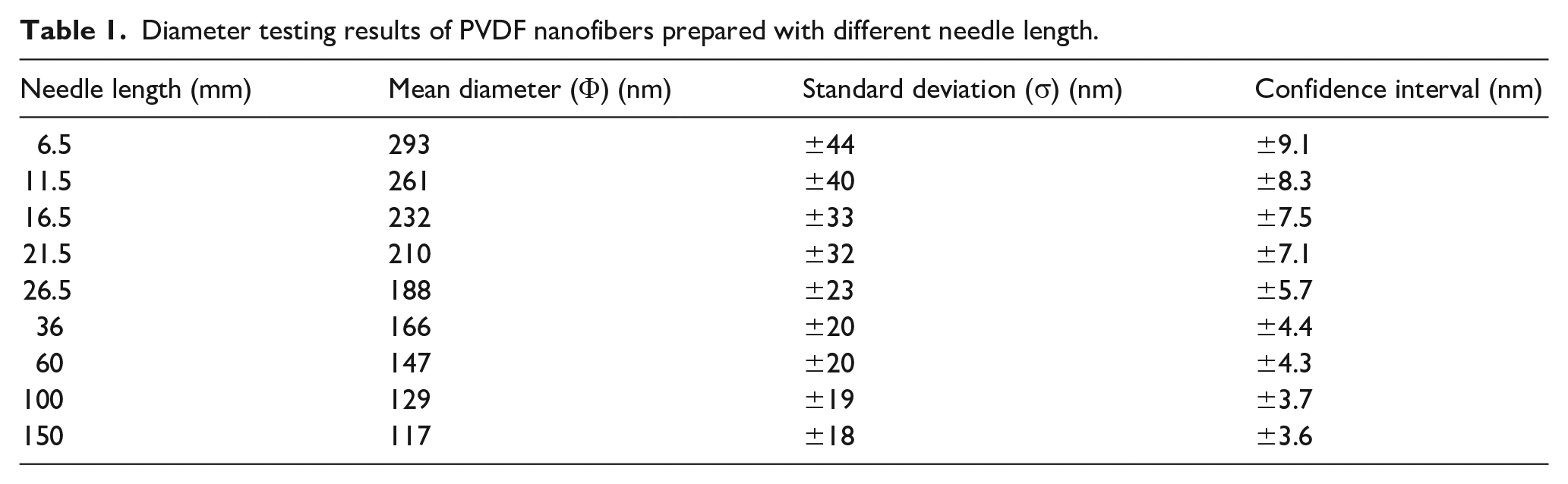
The SEM of PVDF nanofibers prepared with different needle length was shown in Figure 3, and the diameter testing results of PVDF nanofibers prepared with different needle length was shown in Table 1. It can be seen from Figure 3 that the most direct influence of different spinning needle length on PVDF nanofiber membranes was the diameter of nanofibers. With the increase of spinning needle length, the diameter of PVDF nanofibers tend to decrease, but the smoothness of the surface of nanofibers always maintain a good level. In addition, it can be found from Table 1 that the standard deviation of nanofibers had been decreasing while the diameter of nanofibers was decreasing, which showed that the uniformity of the diameter of nanofibers had been improved to a great extent.

SEM images of PVDF nanofiber membranes spun with different needle length: (a) 6.5 mm, (b) 11.5 mm, (c) 16.5 mm, (d) 21.5 mm, (e) 26.5 mm, (f) 36 mm, (g) 60 mm, (h) 100 mm, and (i) 150 mm.
Diameter testing results of PVDF nanofibers prepared with different needle length.
Liff et al. 22 and Zuo and Liu 23 pointed out that the ordered arrangement of the internal structure of nanofibers had a great relationship with the reduction of the diameter. Combining with the law of conservation of mass and using MATLAB software (Math Works), the fiber diameter tested was fitted. The fitting curve between the diameter (D) of nanofibers and the length (L) of spinning needle was shown in Figure 4, and the fitting equation was as follows:


The relationship between the spinning needle length and the nanofiber diameter (the five-pointed star scatter is the testing value, and the pink curve is the fitting curve).
In the above equation, e is a constant. The unit of D is nm, and the unit of L is mm.
As shown in Figure 4, the diameter of nanofibers and the curve had a high degree of correspondence. When the length of the spinning needle increased gradually, the diameter of PVDF nanofibers decreased rapidly at first, then gradually slowed down, and eventually tend to stabilize, that is, the diameter of nanofibers tend to remain constant at a given diameter. The reason for this was that initially, as the arrangement of macromolecules became increasingly straight and organized, the arrangement between macromolecules became increasingly close. All macromolecules within nanofibers completed their orderly movement when the needle length exceeded a certain threshold.
Tensile and bursting properties of nanofiber membranes
We tested the tensile and bursting properties of PVDF nanofiber membranes to further characterize that the length of spinning needles (the ordered arrangement of the internal structure of nanofibers) had a significant effect on the mechanical properties of nanofiber membranes. Figure 5 depicted the testing results for the tensile and bursting properties of PVDF nanofiber membranes. Figure 5 (left) demonstrated that when the needle length was increased from 6.5 to 26.5 mm, the mechanical properties of the nanofiber membranes became unstable and generally exhibited a downward trend. When the needle length was increased from 26.5 to 150 mm, the membrane’s mechanical properties were obviously diminished. The reason analysis demonstrated that the alignment of macromolecules in the nanofiber membrane contributed to its decreased tensile fracture strength. The greater the alignment, the lower the stress caused by sliding resistance (mutual entanglement) between chains of macromolecules. Therefore, as the length of the spinning needle increased, the stress decreased, thereby diminishing the nanofiber membrane’s fracture strength proportionally. In addition, the nanofiber’s macromolecules were easily dislodged, which would inevitably increase the membrane’s fracture strain (toughness). From Figure 5 (right), we can see that the bursting stress and bursting displacement are gradually decreasing. However, by comparing Figure 5 (left) and Figure 5 (right), we can see that the maximum bursting stress of PVDF nanofiber membrane was clearly greater than the maximum tensile stress, which was related to the breaking principle of nanofiber membrane bursting. The bursting primarily affected the stress of the macromolecular chain and the entanglement force generated by nanofiber (macromolecular chain) accumulation, so disorder and entanglement in nanofibers resulted in a greater bursting stress than tensile stress. In contrast, when the macromolecules in nanofibers were arranged in an orderly fashion, the entanglement force and bursting stress would be reduced.

Mechanical property of PVDF nanofiber membranes prepared with different spinning needle length: (a) tensile stress-strain curves and (b) bursting stress-displacement curve.
Utilizing the optimized capstan friction equation, the tensile force and bursting force of nanofiber membrane were characterized;

In the above formula,
We used MetLab software to fit the test data of tensile and bending properties of PVDF nanofiber membrane, and the fitting equation of tensile stress and bursting stress was:


In the above equation,
Figure 6 depicted the relationship between spinning needle length and nanofiber membrane (tensile stress and bending stress). We can see from Figure 6 that the comparison of the fitted curve to the experimental testing data demonstrate that the fitted curve is essentially consistent with the experimental testing data. There was two distinct relationship between tensile stress, bursting stress, and needle length. When the spinning needle length reached a certain length, the tensile stress of the PVDF nanofiber membrane stabilized at a level. However, the bursting stress of the PVDF nanofiber membrane continued to decrease with the increase of spinning needle length, but at a slower rate than the tensile stress. Numerous factors can be seen to influence the bursting stress of nanofiber membranes, and even after the macromolecular chains were aligned in a straight orientation, other resistance changes would lead to additional bursting stress changes.

The relationship between the length of the spinning needle and the nanofiber membrane (tensile stress and bending stress) (scattered points are the experimental data, and the curve is the fitting curve of the experimental data).
Electrical properties of nanofiber membranes
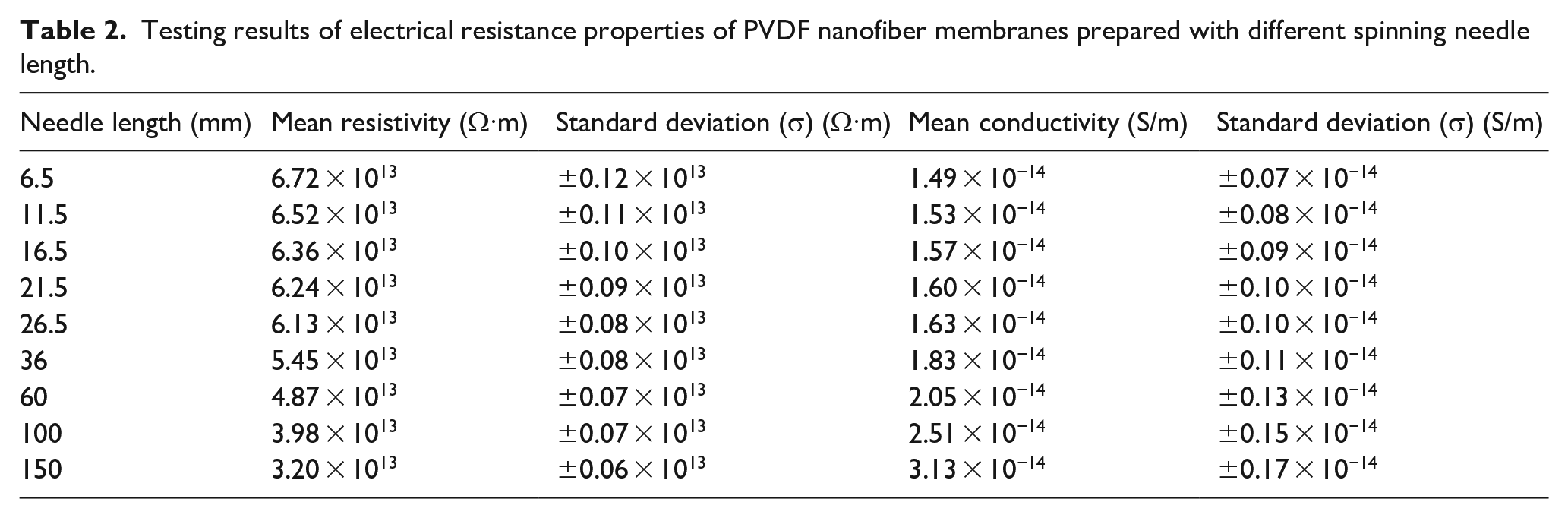
Qian and He 24 noted that long needle spinning can affect the nanofibers’ crystallinity, and that the change in crystallinity can also affect the nanofibers’ conductivity (membranes). To verify that PVDF/DMF spinning solution can alter crystallinity after spinning with different needle length, we indirectly characterized it by analyzing the electrical properties of nanofiber membranes. Table 2 displayed the electrical resistance testing results of PVDF nanofiber membranes prepared with different needle length. As shown in Table 2, as the length of the spinning needle increased, so did the electrical conductivity of the PVDF nanofiber membrane, confirming the findings of He et al., namely, the higher the crystallinity (the degree of macromolecular arrangement order) within the nanofibers, the higher the electrical conductivity of the nanofiber membrane.
Testing results of electrical resistance properties of PVDF nanofiber membranes prepared with different spinning needle length.
Pore analysis of nanofiber membranes
The pore structure of PVDF nanofiber membranes was tested and analyzed to further characterize the influence of the spinning needle length on the porosity of the nano-fiber membrane. Table 3 displayed the pore size testing results for PVDF nanofiber membranes. Figure 7 depicted the pore distribution of PVDF nanofiber membranes prepared using various spinning needle length. Table 3 showed that as the length of the spinning needle increased, the average pore diameter of PVDF nanofiber membrane decreased. Some studies have demonstrated that the diameter of the nanofiber on the nanofiber membrane has a direct influence on the pore diameter on the nanofiber membranes (in a positive correlation, i.e. the smaller the diameter, the larger the pore diameter), which is consistent with our experimental results. The diameter of the nanofiber decreased as the length of the spinning needle increased, and the decrease in average pore diameter lead to a larger generated pore size in the same area; as a result, the porosity had been significantly increased. The diameter of the prepared nanofiber was thicker and the pore size distribution of the nanofiber membrane was wider when the length of the spinning needle was shorter, as shown in Figure 4. The diameter of the prepared nanofiber was smaller and the pore size distribution of the nanofiber membrane was narrow when the length of the spinning needle was up to 150 mm. This also implied that the average diameter difference of the spun nanofibers was currently small.
Pore size testing results of PVDF nanofiber membrane prepared with different spinning needle length.


The pore distribution of PVDF nanofiber membrane prepared with different spinning needle length.
Analysis of surface hydrophobicity of nanofiber membrane
The hydrophobicity of the nanofiber membrane surface is closely related to their material and surface structure. Since the PVDF chosen for this study is a hydrophobic material, the surface structure of the nanofiber membrane will play a crucial role. The water contact angle of nanofiber membranes spun with various needle length were measured in order to characterize the observable change in hydrophobicity. Different length of spinning needle had a direct effect on the nanofiber diameter, so we combined the nanofiber diameter distribution and water contact angle in PVDF nanofiber membranes into a single diagram. Figure 8 depicted the nanofiber diameter distribution and water contact angle in PVDF nanofiber membranes with different needle length (the blue curve in the figure is a Gaussian fitting curve of nanofiber diameter distribution). Figure 8 clearly demonstrated that when the length of the spinning needle was short (6.5 mm), the diameter variation of nanofibers was quite different (the difference between the shortest and coarsest diameters was close to 180 nm), that is, the diameter distribution was broad. Increasing the length of the spinning needle narrowed the distribution of PVDF nanofibers and reduced the diameter difference between nanofibers. Changes in the diameter difference of nanofiber have a direct effect on the surface hydrophobicity of nanofiber membranes, with the water contact angle increasing from 94.6° to 140°.

Diameter distribution and water contact angle of PVDF nanofibers prepared with different spinning needle length.
Conclusion
Although the viscosity of high-viscosity spinning solutions is relatively high, the calculated Reynolds number of high-viscosity spinning solutions such as PVDF is still less than 2320, so PVDF high-viscosity spinning solutions continue to flow in a straight line through the spinning long needle pipe. In this study, PVDF high-viscosity spinning solution was used as the research object, and the effect of various long needle lengths on the micro-morphology, mechanical properties, electrical properties, porosity, and hydrophobic properties of PVDF nanofiber membranes obtained by electrospinning were investigated. The results demonstrated that the length of long needles also exhibited obvious changes in the aforementioned properties, and that the results of Tian et al. regarding the length changes of long needles spun in low-viscosity solutions were generally consistent.
Footnotes
Acknowledgements
Lei Zhao and Ting Zhu are co-first authors of this article.
Declaration of conflicting interests
The author(s) declared no potential conflicts of interest with respect to the research, authorship, and/or publication of this article.
Funding
The author(s) disclosed receipt of the following financial support for the research, authorship, and/or publication of this article: This work is supported by Provincial Scientific Research Platform Open Project Funding of Yancheng Polytechnic College (YGKF202011). This research is funded by Jiangsu Higher Vocational College Teachers’ Professional Leaders’ High and Training (Team Visit) Project (2022TDFX008). The work is also funded by Qing Lan Project of Jiangsu Colleges and Universities for Excellent Teaching Team in 2023, the doctoral research initiation fund project of Yancheng Polytechnic College (2023), Jiangsu Province Higher Vocational Education High-level Major Group Construction Project-Modern Textile Technology Major Group (Grant number: Jiangsu Vocational Education 2020. No. 31). Brand Major Construction Project of International Talent Training in Colleges and Universities-Modern Textile Technology Major (Grant number: Jiangsu Foreign Cooperation Exchange Education 2022. No. 8) also supports the research of this subject. Key technology innovation platform for flame retardant fiber and functional textiles in Jiangsu Province (2022JMRH-003) also supports this research.