
Abstract
Background and Objective. The likelihood of regaining independent walking after stroke is of concern to patients and their families and influences hospital discharge planning. The objective of this study was to explore factors that could be combined in an algorithm for predicting whether and when a patient will walk independently after stroke. Methods. Adults with new lower limb weakness were recruited within 3 days of having a stroke. Clinical assessment, transcranial magnetic stimulation, and magnetic resonance imaging were completed 1 to 2 weeks poststroke. Classification and regression tree (CART) analysis was used to identify factors that predicted whether a patient achieved independent walking by 6 or 12 weeks, or remained dependent at 12 weeks. Results. We recruited 41 patients (24 women; median age 72 years, range 43-96 years). The CART analysis results were used to create the Time to Walking Independently after STroke (TWIST) algorithm, which made accurate predictions for 95% of patients. Patients with a trunk control test score >40 at 1 week walked independently within 6 weeks. Patients with a trunk control test score <40 only achieved independent walking by 12 weeks if they also had hip extension strength of Medical Research Council grade 3 or more. Neurophysiological and neuroimaging measures did not predict independent walking after stroke. Conclusions. In this exploratory study, the TWIST algorithm accurately predicted whether and when an individual patient walked independently after stroke using simple bedside measures 1 week poststroke. Further work is required to develop and validate this algorithm in a larger study.
Introduction
The ability to walk independently is the most common rehabilitation goal after stroke. 1 While up to 85% of all stroke survivors are able to walk independently by 6 months, 2 only 60% of those who require assistance to walk early after stroke regain independent walking.2-4 Whether a patient is expected to achieve independent walking influences decisions about the type and duration of rehabilitation and likely discharge destination. 4 Several factors in the first 2 weeks poststroke predict walking outcomes, including age, lower limb weakness, sensory loss, hemianopia, sitting balance, and trunk control.4-11 Previous studies have typically predicted walking outcome at a single time point,5,11-13 using group data to produce regression equations that are not easily implemented in clinical practice.8,10,11 While clinicians may be able to predict whether a patient will regain independent walking ability, predicting when this will occur is even more difficult 14 as motor recovery is a nonlinear process.2,3,11,15,16 Predicting when a patient will become independently mobile could be clinically useful and allow early anticipation of the level and duration of support required after discharge from hospital.
Neurophysiological and neuroimaging biomarkers can be useful for predicting motor outcomes in the upper limb, particularly for patients with severe initial impairment.17-19 Transcranial magnetic stimulation (TMS) and magnetic resonance imaging (MRI) can be used to examine the integrity of descending motor pathways.17,18,20,21 Few studies have investigated TMS or MRI as predictors for recovery of walking,12,13,22-26 and none have used these biomarkers to predict when a patient will walk independently.
The aim of this exploratory study was to identify predictors of whether and when an individual patient will regain independent walking after stroke, and to combine these in an algorithm that could be used in clinical practice.
Methods
Participants
Patients aged 18 years or more and presenting within 3 days of stroke (ischemic or intracerebral hemorrhage) were eligible if they had new lower limb weakness (<100 on lower limb Motricity Index score) and required supervision or assistance for walking. Patients with previous stroke and those who were treated with intravenous thrombolysis and endovascular thrombectomy could be enrolled. Exclusion criteria included the requirement for supervision or assistance to walk prior to admission, cerebellar or bilateral stroke, not expected to survive for the duration of the study, communication or cognitive deficits precluding informed consent, and contraindications to TMS or MRI. Written informed consent was obtained from all patients. This study was approved by the national ethics committee.
Clinical Assessments
Demographic and stroke characteristics were recorded at day 3 poststroke (Table 1). Neurologic impairment was assessed with the National Institutes of Health Stroke Scale (NIHSS). 27 NIHSS subscores for items 4 (hemianopia), 8 (sensory), and 11 (inattention) were used to categorize stroke type as either motor (M), motor-sensory (MS), or motor-sensory-hemianopia (MSH) for analysis. 11 Walking ability was graded with the Functional Ambulation Category (FAC) scale. 28 FAC is a 6-point scale with a score of 0 indicating the patient is nonambulatory or requires the assistance of at least two people to walk. An FAC score of 1 to 3 indicates either assistance or supervision from one person is required to walk. An FAC score of 4 indicates that the patient is able to walk indoors on level surfaces without hands-on assistance or supervision, and a score of 5 indicates that the patient is able to walk up and down stairs, slopes and outdoors without assistance or supervision. Independent walking was defined as an FAC score of 4 or 5. Participants were allowed to use walking aids such as a stick, quad stick, or an ankle support. FAC scores for each participant were obtained on day 3, and again at 1, 4, 6, and 12 weeks poststroke. FAC scores were dichotomized at each time point to classify participants as being able to walk independently (FAC ≥4) or as dependent (FAC <4).4,5 The primary outcome was the time poststroke at which the participant achieved independent walking (FAC ≥4).
Demographic and Clinical Characteristics.
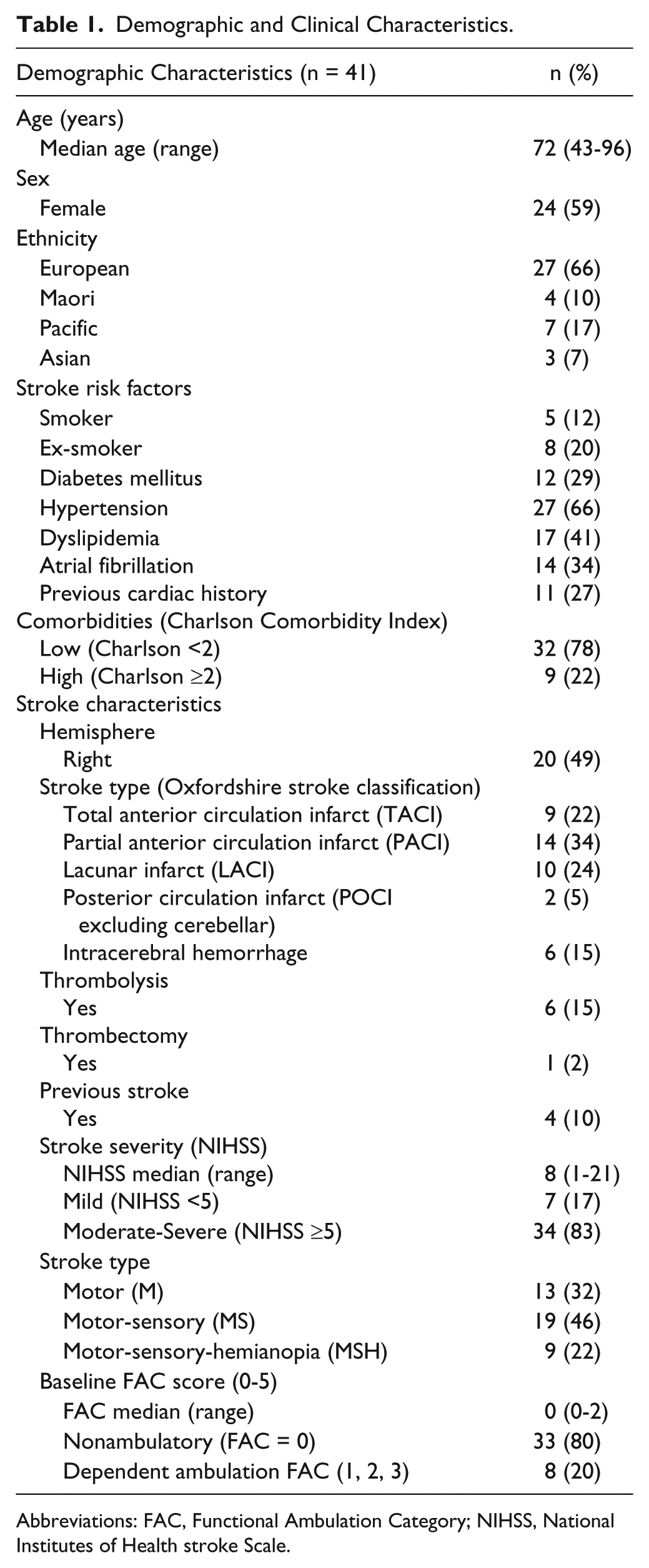
Abbreviations: FAC, Functional Ambulation Category; NIHSS, National Institutes of Health stroke Scale.
All participants completed full clinical assessments one week after stroke (Table 2). These included the following: walking ability (FAC); trunk control (Trunk Control Test [TCT]); lower limb power (Motricity Index); and lower limb muscle strength graded with the Medical Research Council (MRC) grades for hip flexion, extension and abduction, knee flexion and extension, and ankle dorsiflexion and plantarflexion. The TCT is a validated 4-item scale measuring both static and dynamic trunk control.29,30 Rolling to each side in bed, moving from lying to sitting, and sitting unsupported for 30 seconds, are each scored out of 25, totaling 100 points. Clinical assessments were conducted by an experienced research physiotherapist trained in the assessments, and not involved in patient care.
One-Week Clinical, Neurophysiological, and Magnetic Resonance Imaging Measures.
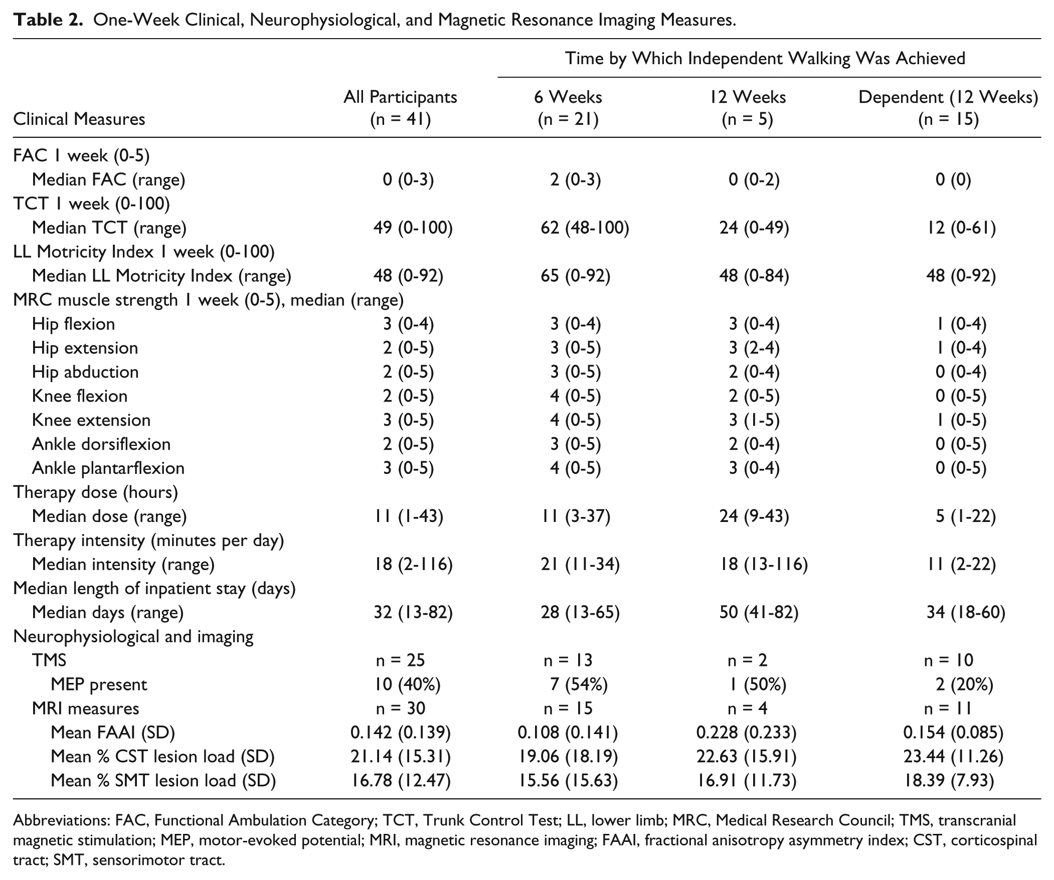
Abbreviations: FAC, Functional Ambulation Category; TCT, Trunk Control Test; LL, lower limb; MRC, Medical Research Council; TMS, transcranial magnetic stimulation; MEP, motor-evoked potential; MRI, magnetic resonance imaging; FAAI, fractional anisotropy asymmetry index; CST, corticospinal tract; SMT, sensorimotor tract.
Neurophysiological and Imaging Measures
TMS was used to evaluate the functional integrity of descending motor pathways in a subset of participants. The presence or absence of motor-evoked potentials (MEPs) in the paretic tibialis anterior was evaluated 1 week after stroke. Tibialis anterior has a relatively large cortical representation, resulting in larger and more easily elicited MEPs than proximal leg muscles or muscles in the foot.31,32 The presence of MEPs in tibialis anterior has some predictive value for the recovery of walking.13,22-24 MEPs were recorded with surface electromyography (EMG) from the tibialis anterior muscle with a reference electrode placed over the patella. EMG signals were sampled at 2 kHz, amplified (1000 gain), filtered (20-1000 Hz), and stored for offline analysis using Signal software (CED). Magnetic stimuli were delivered using a flat figure-8 coil attached to a MagStim 200 unit. The coil was placed over the scalp and oriented to generate a medial-lateral current flow in the affected lower limb motor cortex. 33 Participants were tested while seated on a chair if able, or in a half-sitting position in bed. The stimulus intensity was increased up to 100% maximal stimulator output if required. If no MEP was observed in the paretic tibialis anterior at rest, the participant was instructed to activate the paretic leg if able, or to activate the nonparetic leg if severe hemiparesis prevented activation of the paretic leg. 33 The participant was considered MEP positive (MEP+) when an MEP of any amplitude was consistently observed for over half of consecutive trials with the leg at rest or active. 34
Three MRI measures (fractional anisotropy [FA] asymmetry, corticospinal tract [CST] lesion load, and sensorimotor tract [SMT] lesion load) were used to examine the structural integrity of white matter pathways in a subset of participants, with MRI scans obtained 7 to 14 days after stroke. T1-weighted and diffusion-weighted images were acquired with a Siemens 1.5 T Avanto scanner. Axial T1-weighted images had 1.0 × 1.0 × 1.0 mm voxels, a 256 mm field of view, repetition time (TR) = 11 ms, and echo time (TE) = 4.94 ms. Diffusion-weighted images (DWIs) had 1.8 × 1.8 × 3.0 mm voxels, a 230 mm field of view, b = 2000 s·mm2, and TR = 6700 ms, TE = 101 ms, 30 gradient directions, and 2 averages. DWIs were preprocessed with motion and eddy current correction, skull stripping, estimation and fitting of diffusion parameters, and modeling of crossing fibers. 35 FA maps were registered to the participant’s T1-weighted image and overlaid by a standard template of the voxels of interest (VOI) for the posterior limb of the internal capsules (PLIC). PLIC templates were manually edited if the PLIC template VOI encroached on basal ganglia structures or ventricles. 36 FA asymmetry was calculated as (FAcontralesional – FAipsilesional)/(FAcontralesional + FAipsilesional). A positive FA value indicates relatively reduced FA in the ipsilesional CST at the level of the PLIC.
A template CST was constructed from DWIs obtained from the contralesional hemisphere of 85 stroke patients from an earlier study, 37 extending from the primary motor cortex to the inferior border of the pons. The template sensorimotor tract was a combination of the CST and sensory tracts extending from the primary sensory cortex to the medial lemniscus at the inferior border of the pons. Tractography was conducted with a curvature threshold of 0.2 and step-length of 0.5. The template tracts were thresholded at 75% probability to ensure that only fibers at each tract’s core were used for calculation of lesion load. Lesion masks were hand-drawn on the T1 images of individual participants. The percentage of the CST template voxels that were overlapped by the stroke lesion mask was calculated to determine CST lesion load. 34 This process was repeated for SMT lesion load.
Therapy Dose
Therapists were blinded to the results of the assessments and continued making decisions regarding therapy type and dose for each patient based on their clinical judgement and service capacity. Lower limb therapy dose was defined as total time spent in active rehabilitation of the lower limb, trunk, balance or walking, and was recorded by the treating therapist immediately after each therapy session. Therapy dose was recorded for all inpatient therapy sessions from admission until discharge from hospital, and therefore included therapy completed in the acute stroke unit and during inpatient rehabilitation. Length of inpatient stay was recorded for all participants, and therapy intensity was calculated as minutes of targeted inpatient lower limb therapy received per day of inpatient stay. Total lower limb therapy dose and therapy intensity were used for subsequent analyses.
Statistical Analysis
Participants with FAC <4 at 1 week were included in the analysis. They were initially classified into four categories according to the time they achieved independent walking; 4 weeks, 6 weeks, 12 weeks, or dependent at 12 weeks. The analysis was conducted in 2 stages. Logistic regression was used to identify potential predictors of category membership. The demographic and stroke characteristic variables entered into the logistic regression analyses were age (years), sex (male, female), stroke classification (Oxford stroke classification), stroke severity (NIHSS), stroke type (M, MS, MSH), and comorbidities (Charlson Comorbidity Index binarized to mild 0-1 or severe ≥2). Less than 20% of participants had thrombolysis, thrombectomy or previous stroke, so these variables were not included in the analysis. The 1-week clinical assessment variables entered were: FAC (out of 5); MRC grades (out of 5) for hip flexion, extension and abduction, knee flexion and extension and ankle dorsiflexion and plantarflexion; lower limb Motricity Index (out of 100) and TCT (out of 100). Therapy dose (minutes) and therapy intensity (minutes per day) were also entered. Separate logistic regressions were conducted for each variable. Variables with P < .05 were then entered into a classification and regression tree (CART) analysis. The strict cutoff for inclusion in the CART analysis was used to minimize the number of variables for the relatively small sample size.
CART analysis was conducted to select, in a hierarchical order, the variables that best predicted each participant’s category membership. CART analysis independently dichotomizes and selects variables that achieve the least overlap between resulting subgroups and the most homogeneity within each subgroup, creating a decision tree or prediction algorithm. 38 CART was conducted using “gini” with a maximum tree depth of 3, minimum of four cases in a terminal node and automatic pruning to reduce overfitting.
For the subsets of participants who completed TMS and MRI assessments, separate logistic regression analyses were conducted for MEP status (+/−) and MRI measures (FA asymmetry, CST lesion load, SMT lesion load). The relationships between outcome at 12 weeks (independently walking or not) and MEP status and MRI measures were explored with chi-square and 2-sample 2-tailed t tests, respectively.
The results of the CART analysis were used to create an algorithm for predicting time to walking independently after stroke. Sensitivity and specificity of the algorithm were calculated for each category.
Results
There were 41 participants (median age 72 years, range 43-96 years; 59% female) included in the analysis (Figure 1, Tables 1 and 2). Most participants were not able to walk (FAC = 0, n = 33, 80%) at day 3. Clinical assessments were completed for all participants. A subset of 25 participants had TMS at days 5 to 7 and 30 participants had MRI at days 7 to 14 poststroke. The remaining participants were unable to have TMS or MRI as they were too unwell or had been moved to another rehabilitation center at the time of testing. Three participants died before 12 weeks and last observations were carried forward, as FAC scores were 0 at all prior time points.
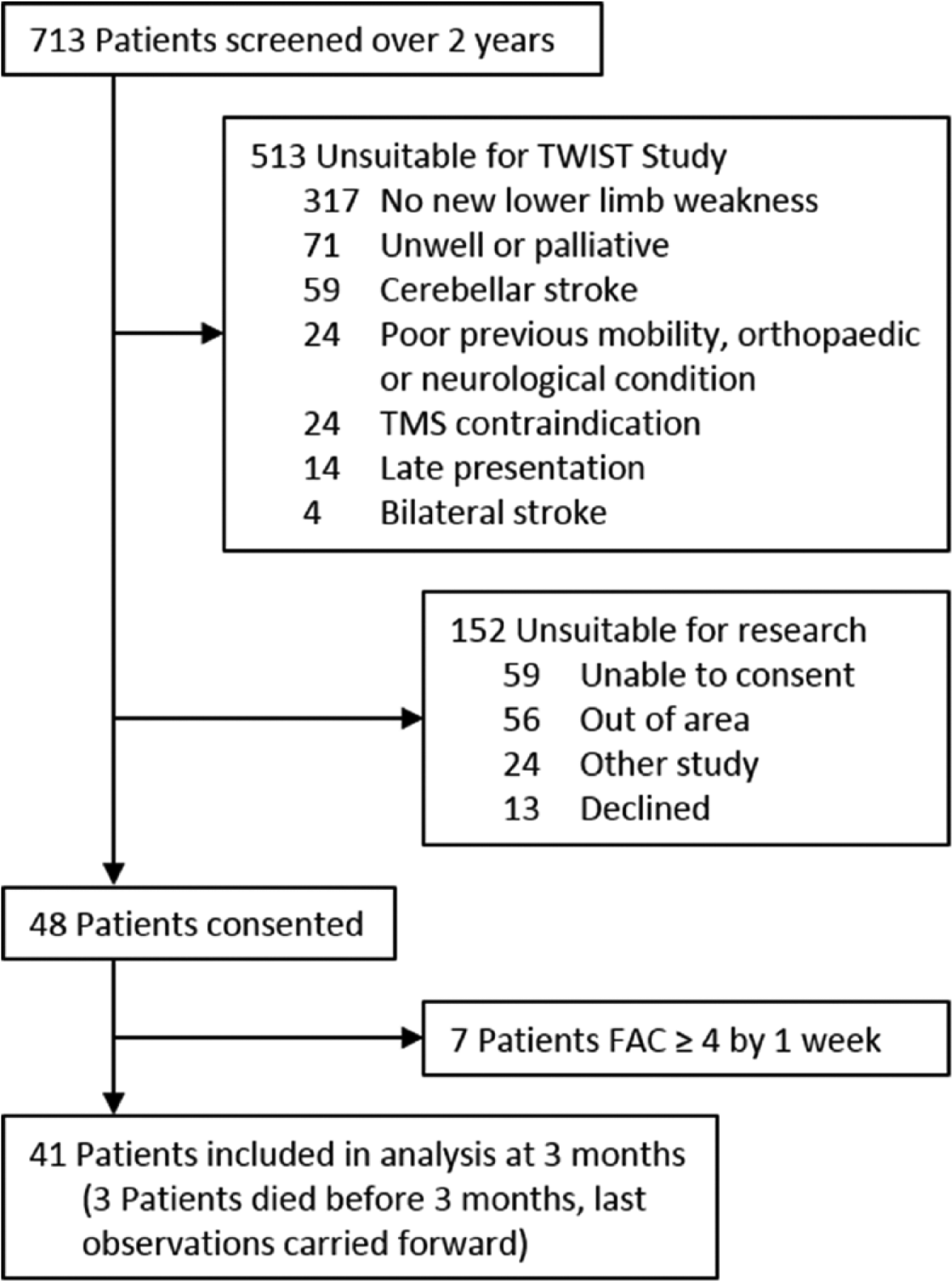
Study flowchart.
Only 5 participants achieved independent walking between 4 and 6 weeks, therefore these 2 categories were combined. Although the number of participants in the 12-week category was also small (n = 5, 12%), they form a functionally distinct group from those participants walking independently by 6 weeks (n = 21, 51%) or not walking independently at 12 weeks (n = 15, 37%).
The CART analysis is presented in Figure 2. Age, therapy intensity (minutes per day), therapy dose (total therapy time), FAC at 1 week (out of 5), TCT at 1 week (out of 100), and MRC grades at 1 week (out of 5) for hip flexion and extension, knee flexion and extension, and ankle plantarflexion were entered into the CART analysis. The only predictors of the time by which participants regained independent walking were the TCT score and MRC grade for hip extension strength. Most participants with TCT >37 (21/23, 91%) achieved independent walking within 6 weeks of stroke. All participants with TCT ≤37 achieved independent walking between 6 and 12 weeks poststroke provided hip extension MRC grade was ≥3 (n = 4); otherwise they remained dependent at 12 weeks (n = 14). The logistic regression and CART analyses did not identify any other predictors from age; sex; stroke classification; stroke severity; stroke type (M, MS, MSH); comorbidities; 1 week FAC; MRC grades for hip flexion and abduction, knee flexion and extension, ankle dorsiflexion and plantarflexion; lower limb Motricity Index; therapy dose and intensity.
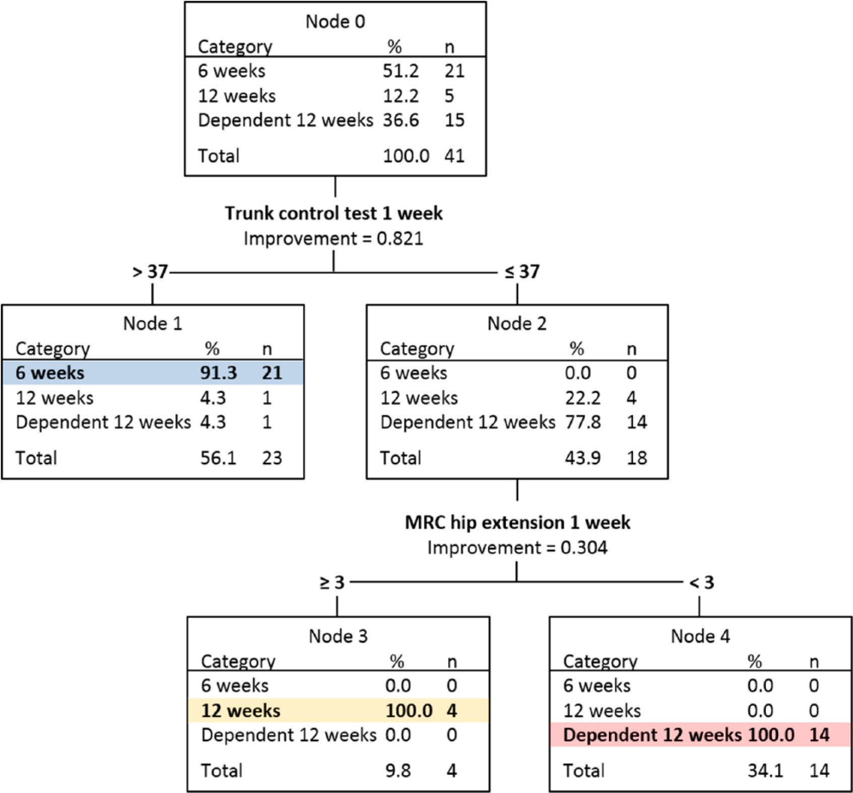
Classification and regression tree (CART) analysis. CART analysis identified factors that predict time taken to walk independently after stroke (6 weeks or 12 weeks, or dependent at 12 weeks). TCT, Trunk Control Test; MRC, Medical Research Council strength score.
Regression analyses were also conducted with the subsets of participants who had TMS and MRI. TMS and MRI measures were not found to have predictive value and therefore not included in CART analysis. Of the 25 participants with TMS data, 10 (40%) were MEP+ and 15 (60%) were MEP- (Table 2). MEP+ participants had better outcomes with 8 of 10 independently walking at 12 weeks compared with 7 of 15 MEP- participants; however, this finding was not significant (P = 0.10). Of the 13 participants who achieved independent walking by 6 weeks, only 7 (54%) were MEP+. MRI data for 30 participants were included in the regression analysis. No MRI measures were identified as predictors of outcome. There were no differences in MRI measures between participants who walked independently by 12 weeks and those who did not (all P >0.5).
The CART analysis results were used to create the Time to Walking Independently after STroke (TWIST) algorithm for use with patients who are not yet walking independently 1 week poststroke (Figure 3). For ease of clinical use, the dichotomized TCT score was rounded from 37 to 40 as this is easier to remember, and this change does not affect the decision tree as it is not possible to score between 37 and 40. The TWIST algorithm correctly predicted time to walk independently after stroke for 39/41 (95%) of participants. Accuracy, positive and negative predictive values, sensitivity, and specificity are reported in Table 3.

Time to Walking Independently after Stroke (TWIST) algorithm. The TWIST algorithm predicts time taken to walk independently after stroke (6 weeks, 12 weeks or dependent at 12 weeks). All assessments are at 1 week poststroke. Each outcome category is color-coded. The colored dots indicate the actual category outcome of patients as a proportion of the total patients predicted for each category, for example, 5% of patients predicted to walk independently by 6 weeks actually walked by 12 weeks and 5% were dependent at 12 weeks. All patients predicted to walk by 12 weeks or to be dependent at 12 weeks achieved their predicted outcome. FAC, Functional Ambulation Category; TCT, Trunk Control Test (out of 100); Hip extension, Medical Research Council (MRC) grade for hip extension strength.
Sensitivity and Specificity of Time to Walk Independently after Stroke (TWIST) Algorithm.
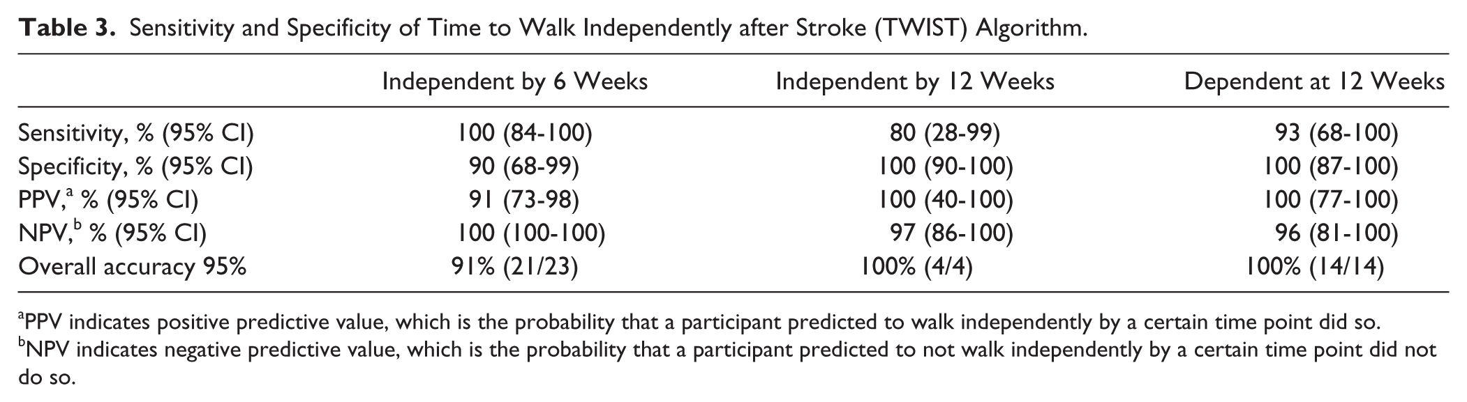
PPV indicates positive predictive value, which is the probability that a participant predicted to walk independently by a certain time point did so.
NPV indicates negative predictive value, which is the probability that a participant predicted to not walk independently by a certain time point did not do so.
Discussion
This is the first study to identify predictors of both whether and when an individual patient will walk independently after stroke. Using simple bedside assessments at 1 week following stroke, the TWIST algorithm based on the CART analysis predicted with 95% accuracy the time taken to walk independently. The first step of the algorithm is to assess trunk control. Most participants with good trunk control (TCT >40) at 1 week poststroke walked independently by 6 weeks poststroke. Participants with poor trunk control (TCT <40) only achieved independent walking by 12 weeks poststroke if they had hip extension of MRC grade 3 or more. Those with poor trunk control (TCT <40) and hip extension of MRC grade 2 or less at 1 week poststroke were predicted to be dependent at 12 weeks poststroke (Figure 2).
The ability to predict whether and when a patient will regain independent walking may give a patient and their family realistic expectations of the level and duration of support they will need after discharge and allow clinicians to begin discharge planning earlier. 39 The TWIST algorithm predicts walking independence, rather than the quality of walking, walking speed, or community ambulation. Walking speed is often the target of prediction studies due to the relationship between walking speed, community ambulation, and falls.2,39,40 However, the ability to walk indoors without another person present (FAC ≥4), is more likely to influence the timing and destination of discharge from hospital than walking speed or community ambulation. It may also influence the level of support required after discharge. It is important to note that while the TWIST algorithm predicts that some patients will not be walking independently by 12 weeks poststroke, they may be able to do so at a later time point.
These findings support previous work identifying trunk control and lower limb strength as predictors for the return of independent walking, 5 with trunk control being the stronger predictor of the two. 6 In contrast to previous studies which used the Motricity Index as the sole measure of lower limb strength,5,11 MRC grades for individual muscles were also included in this analysis. This allowed for muscles that are not included in the Motricity Index to be considered as potential predictors. The Motricity Index combines the scores of hip flexion, knee extension, and ankle dorsiflexion. These muscles, along with ankle plantarflexion, are important for walking performance.41-45 It may therefore seem counterintuitive that the strength of these muscles was not identified as a predictor in the present study. This may be because the main outcome of this study is the ability to walk independently, rather than walking performance in terms of quality or speed. Provided the patient has good trunk control, compensatory strategies involving the trunk, such as shifting weight away from the paretic side or using a walking aid, enable walking even with little voluntary control of the lower limb. 6 To achieve independent walking, it is possible to compensate for poor hip flexor strength with lateral flexion and rotation of the trunk combined with circumduction of the hip to initiate swing; compensate for poor knee extensor strength with hyperextension of the knee or the use of a stick during stance; and compensate for poor ankle dorsiflexor strength by either high-stepping or using an ankle splint. The findings from the CART analysis suggest that the presence of hip extension may provide compensatory proximal stability for those patients with poor trunk control.
Several potential clinical predictors were not included in this exploratory study and would benefit from further investigation in a larger study. These include the presence of increased tone, proprioception, visuospatial inattention, cognition, mood, and previous stroke. More sensitive measures of somatosensory and vision impairment than provided by the NIHSS could also be investigated.
Total therapy dose and therapy intensity (minutes per day) did not predict when a participant achieved independent walking. This may be due to the large separation between the outcome categories (6 and 12 weeks). Participants in this study were provided with standard care for this center, and lower limb therapy intensity was relatively low (median 18 minutes per day of inpatient stay, range 2-116 minutes), and could have been overestimated. 46 Further research is needed in different rehabilitation centers to establish whether therapy provided at higher intensities influences the time taken to walk independently.
Neurophysiological and imaging measures were not predictors of whether or when participants would regain independent walking, which is in contrast to studies of recovery in the upper limb.18,21,47 Participants with tibialis anterior MEPs generally had better walking outcomes than those without MEPs. However, almost half of the participants without MEPs still walked independently by 12 weeks, and MEP status did not predict when a participant would walk independently. In a study of 41 chronic stroke patients, more than half of the participants with absent tibialis anterior MEPs and disruption of CST on diffusion tensor imaging (complete corticospinal tract injury) were able to walk independently or with supervision. 48 This suggests that the role of the corticospinal tract may be less important for the lower limb than the upper limb.48-50 Neuroanatomical control of the lower limb and walking differs from the upper limb, with the presence of bilateral and alternative descending pathways providing more redundancy.49,50 The finding that trunk control strongly predicts recovery of independent walking may reflect the contribution of medial descending pathways such as the reticulospinal tract and vestibulospinal tract. 51 TMS specifically targeting the trunk muscles, 52 and MRI measures of the projections from the cerebellum to the reticular and vestibular nuclei, were not assessed in this study and could be considered in future studies.
These findings do not align with the few previous studies reporting that MEP status of lower limb muscles may be a predictor for recovery of walking.13,22-24 However, it is difficult to draw conclusions from these studies due to small sample sizes and variations in selected outcome measures, timing of assessment, and TMS technique. Although previous MRI studies report that patients with less corticospinal tract damage achieve better overall lower limb strength and gait outcomes, they do not specifically predict recovery of independent walking for an individual.12,26,40,53 The sample size in this study was small for the subsets of participants with TMS and MRI data; therefore, firm conclusions cannot be drawn from these neurophysiological and imaging results and further investigation is warranted.
This study has several strengths. The clinical measures used in the TWIST algorithm are simple and quick to administer at bedside. Clinicians routinely assess trunk control and lower limb strength at several time points after stroke, making this algorithm practical and straightforward to complete. Clinical experience identifies sitting balance and lower limb strength as important in walking recovery. The TWIST algorithm provides a structured approach to combining these assessments to make predictions for individual patients. These features support the potential translation of the TWIST algorithm to clinical practice.
This exploratory study has several limitations. The sample size is small and not all participants had TMS and MRI. The small number of participants who walked independently between 4-6 and 6-12 weeks reflects the nonlinear recovery timeline, with most participants recovering independent walking early after stroke or not at all. 3 This necessitated merging of the outcome categories for participants who achieved independent walking by 4 weeks and 6 weeks. The small sample size also has potential effects on the results of the CART analysis if a large number of variables are included. This was partly mitigated by the use of logistic regression analyses to select the variables for the CART analysis. It is possible that the TMS technique used here produced false negatives, due to the deep location of the lower limb motor cortex. Also, TMS and MRI measures did not target alternative or uncrossed descending pathways controlling the trunk. The study did not differentiate between participants who were nonambulatory at 12 weeks and those who could walk with assistance from one person. These 2 groups of patients may have quite different support needs on hospital discharge.
Larger independent samples are required to refine and validate the TWIST algorithm to ensure that results are generalizable, to further investigate the potential roles of TMS and MRI, and to assess patients at later time points.
Conclusions
In conclusion, this exploratory study has identified a simple algorithm based on 2 clinical measures made at the bedside 1 week poststroke that may predict whether and when a patient will walk independently within 12 weeks after stroke. Further work is needed to develop and validate the algorithm with a larger sample of patients and further investigate the role of TMS and MRI as predictors for independent walking.
Footnotes
Acknowledgements
The authors thank Suzanne Ackerley and Victor Borges.
Declaration of Conflicting Interests
The authors declared no potential conflicts of interest with respect to the research, authorship, and/or publication of this article.
Funding
The authors disclosed receipt of the following financial support for the research, authorship, and/or publication of this article: This study was supported by the Health Research Council New Zealand (11/270) and the Julius Brendel Trust.