
Abstract
Background. Improvement of postural control in persons with multiple sclerosis (PwMS) is an important target for neurorehabilitation. Although PwMS are able to improve postural performance with training, the neural underpinnings of these improvements are poorly understood. Objective. To understand the neural underpinnings of postural motor learning in PwMS. Methods. Supraspinal white matter structural connectivity in PwMS was correlated with improvements in postural performance (balancing on an oscillating surface over 25 trials) and retention of improvements (24 hours later). Results. Improvement in postural performance was directly correlated to microstructural integrity of white matter tracts, measured as radial diffusivity, in the corpus callosum, posterior parieto-sensorimotor fibers and the brainstem in PwMS. Within the corpus callosum, the genu and midbody (fibers connecting the prefrontal and primary motor cortices, respectively) were most strongly correlated to improvements in postural control. Twenty-four-hour retention was not correlated to radial diffusivity. Conclusion. PwMS who exhibited poorer white matter tract integrity connecting the cortical hemispheres via the corpus callosum showed the most difficulty learning to control balance on an unstable surface. Prediction of improvements in postural control through training (ie, motor learning) via structural imaging of the brain may allow for identification of individuals who are particularly well suited for postural rehabilitation interventions.
Introduction
Postural dyscontrol is a major source of disability in persons with multiple sclerosis (PwMS), leading to reduced walking speed, falls, and reduced quality of life.1-3 Given the well-established relationship between postural control and falls,2,4 improving balance is an important target for neurorehabilitation.
Recent studies demonstrate that PwMS can improve upper extremity (UE) motor performance through practice,5-7 although these improvements may be less pronounced compared to controls.8,9 Considerably less is known about the ability of PwMS to improve postural motor control. In fact, to our knowledge, only 2 studies have directly investigated postural motor learning in PwMS. Hatzitaki et al 10 showed that PwMS were able to learn a postural visuomotor task in which participants voluntarily leaned to the left and right. However, these improvements were less pronounced than in healthy adults. 10
Similarly, we recently showed that PwMS were able to improve postural responses through repeated exposure to continuous support surface translations. 11 PwMS maintained balance while the support surface continually slid forward and backward at a fixed frequency (see Gera et al 11 and Van Ooteghem et al 12 for details). We measured temporal performance, that is, ability to anticipate changes in direction, and spatial performance, that is, the ability to control the amplitude of sway, with repeated exposures to this moving support surface. Despite poorer performance than control subjects, PwMS exhibited improvements in temporal performance (over 1 day of practice) and retention (ability to maintain improvements 24 hours later) in a manner similar to the control group. Conversely, spatial performance improved to a lesser degree in PwMS than in control subjects and was not retained on the following day. We hypothesized that given the temporal consistency of platform movements, participants were able to improve temporal performance via a feedforward control strategy. However, given the relative inconsistency in amplitude of the forward and backward movements, participants were forced to rely on nonspecific learning, or improvements based on proprioceptive feedback control mechanisms to improve spatial performance. 11 Therefore, PwMS seemed to exhibit a specific deficit in nonspecific learning, but not feedforward learning, compared to people without MS.
The ability of PwMS to acquire and retain improvements in postural performance is promising for balance neurorehabilitation. However, the ability to learn varies widely in PwMS and the neural underpinnings of these motor learning processes are unknown. Previous UE research suggests that numerous structures, including the basal ganglia, cerebellum, and cortex play a role in motor learning. Not surprisingly, the white matter tracts connecting these structures also play an important role in this process.8,13-15 White matter tracts such as the corpus callosum (CC) are of particular interest as they are often disrupted in PwMS.16-18 Furthermore, PwMS exhibit considerable heterogeneity in the degree of degeneration of white matter tracts, which may contribute to the variability in learning ability in this population. Indeed, Bonzano et al 8 demonstrated that UE sequence learning was less pronounced in PwMS, and the degree of learning was directly related to structural connectivity of the CC, such that people with more pronounced worsening of CC structural integrity exhibited poorer learning. In a follow-up study, Bonzano et al 19 demonstrated that active UE motor rehabilitation resulted in retention of CC fibers and function, further underscoring the importance of the CC in motor learning and neurorehabilitation.
Based on these previous reports, it is possible that white matter degradation (particularly within the CC) affects motor learning in PwMS. However, research relating white matter disruption to motor learning in PwMS has been limited to UE learning tasks (eg, Bonzano et al 8 ); thus it is unknown whether these findings generalize to postural control. By investigating whether changes in the integrity of white matter predict deficits in postural learning, we may be able to identify PwMS most suitable for postural training, 20 thus improving the utilization of therapeutic resources.
To understand the neural underpinnings of postural motor learning in PwMS, we correlated the acquisition and retention of practice-related improvements of postural control to whole-brain structural connectivity using a tract-based spatial statistical (TBSS) approach. Given previous findings in UE literature, 8 we hypothesized that white matter connectivity of the CC, an area which is both important for motor learning, 13 and altered in PwMS, 16 would be related to improvement in postural control performance and retention of these improvements within PwMS.
Methods
Participants
We present the data for 29 PwMS and 15 age- and gender-matched healthy adults (healthy controls [HC]) used in our previous report. 11 Inclusion criteria for all participants were the following: ability to walk 500 m without assistance, ability to maintain balance independently by standing on toes for 3 seconds, and no known biomechanical conditions affecting balance. Exclusion criteria for PwMS and HC were the following: coexisting conditions that can mimic MS (eg, lupus or fibromyalgia), or additional conditions that may affect gait or balance (eg, arthritis, joint replacement).
Behavioral Protocol
Behavioral testing procedures have been described previously. 11 Briefly, participants underwent 1 day of balance training and 1 day of balance testing for retention. Participants stood on a hydraulically controlled platform that oscillated at a fixed, sinusoidal frequency (0.5 Hz) in the forward and backward directions (Figure 1). Participants were asked to maintain balance while keeping arms crossed across the chest to minimize contribution of the arms and looking straight ahead. Trials were 48-second long and the same sequence was repeated for each trial. On day 1, participants were trained with 5 blocks of 5 trials. On day 2 (24 hours later), participants completed 2 blocks of 5 trials to measure retention. As such, we were able to assess improvements in postural performance across day 1, as well as 24-hour retention of these improvements.
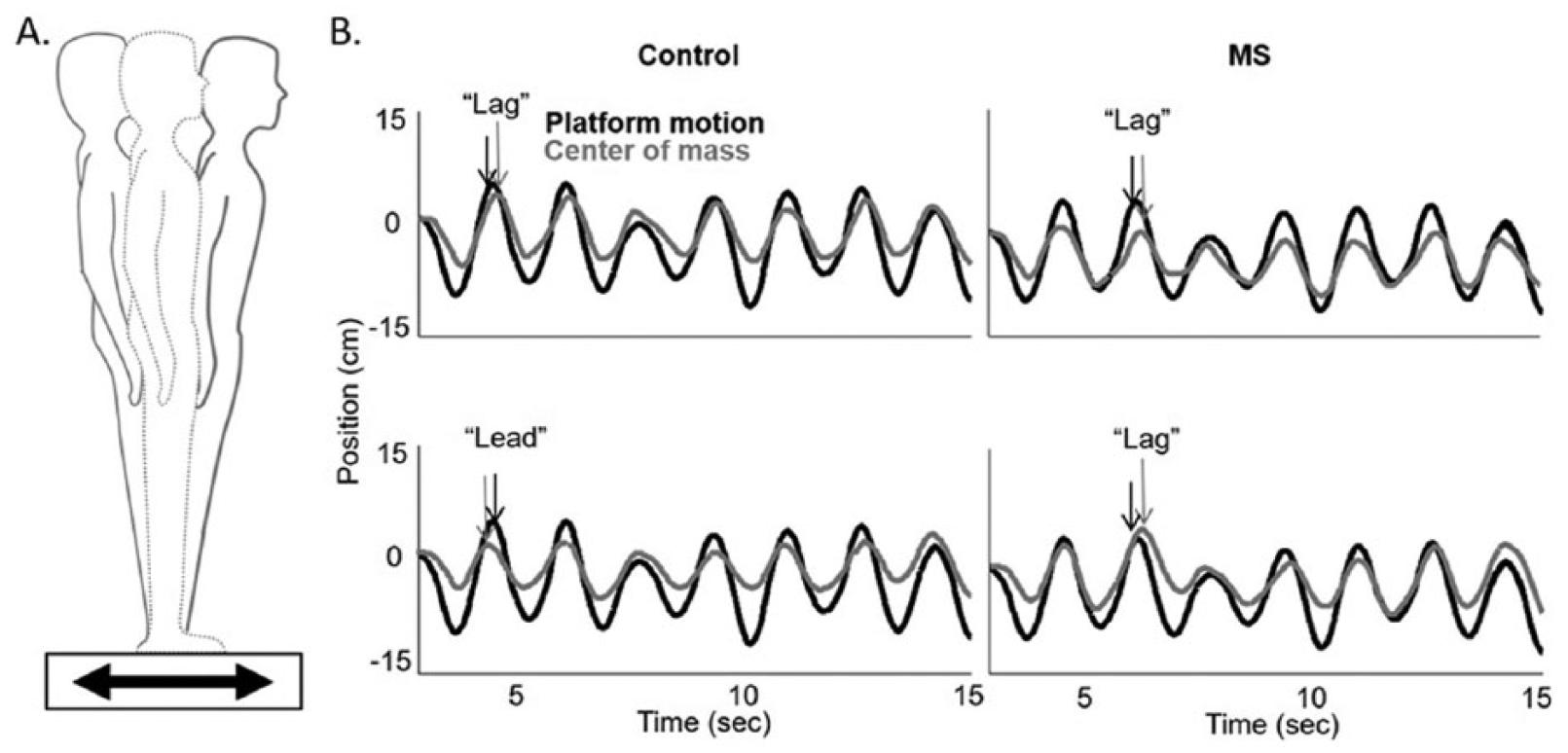
(A) Schematic of an individual while on the movable force plate. Forward motion (black) of the force plate results in backward sway, while backward motion of the plate (gray) results in forward sway. (B) Illustration of the position of the platform (black) and the center of mass (gray) for a control (left) and multiple sclerosis (MS; right) patient as it moved forward and backward during a trial. Temporal performance is noted as the difference in phase of the platform and center of mass. A “lag” in phase represents poorer performance, and “lead” represents improved performance. The top row shows the mean relative phase in the early training block, and the bottom row shows the late training block. With training, the phase “lag” relationship between the platform motion and center of mass changes to phase “lead” for the control participant but not so for the MS patient. Figure adapted from Gera et al. 11
Behavioral Data Analysis
Temporal and spatial control of body center of mass (COM) over the moving surface was used to measure postural performance. Whole-body COM was calculated from reflective markers placed on bony landmarks and measurement of body anthrophomorpic data. 21 Motion analysis was sampled at 60 Hz and low-pass filtered using a second-order, dual-pass Butterworth filter (5 Hz). COM position was tracked in the anteroposterior directions, and temporal and spatial COM outcome measures were calculated. The temporal COM measure was the mean relative phase of body COM position relative to the platform position at the instant of maximum platform displacement (peaks and valleys of sinusoidal platform movement). Negative values represent the COM “lagging” behind the platform, while positive values reflect the COM “leading” the platform in a predictive manner. Greater negative values represent worse performance. Thus, a phase lag moving toward zero (becoming less negative) represents an improved ability to predict platform perturbations, consistent with feedforward postural control. 11 Spatial COM performance was calculated as the position (relative to the starting position) of the COM at each peak and valley of the sinusoidal plate movement. COM displacements were normalized to the platform displacement. Thus, a mean gain of 1 would represent equal displacements of the platform and COM and would occur if participants’ COM followed platform motion exactly. Reduction of mean gain (toward zero), represents improved balance control with the body COM displacing less than the support surface and feet. Values at each peak and valley were averaged across each trial.
Improvement in temporal (phase lag) and spatial (mean gain) performance were calculated as the change in performance between block 1 and block 5 on Day 1. Retention of improvement was calculated as the change in performance between block 1 (day 1) and block 1 (day 2). Some previous investigations have shown retention as a lack of change between performance at the end of training and retention (ie, follow-up) periods. 22 We opted to calculate retention as the difference in performance between the beginning of training and the retention period.23-25 While both methods provide information regarding the retention of improved performance, we chose the latter option because it may provide more information about the degree to which improvements persist over the baseline value. 26 Improvements in postural motor learning and retention of these improvements were correlated to whole brain structural connectivity (see below). In addition, average temporal and spatial performance were calculated as the mean performance over all five blocks of practice on day 1. Group differences in average temporal and spatial performance were compared via independent sample t tests, and were also related to brain structural connectivity (see below).
Imaging Protocol
On a separate day, less than 2 weeks following behavioral testing, participants were scanned on a 3.0-T Siemens Magentom Tim Trio scanner with a 12-channel head coil at Oregon Health & Science University’s Advanced Imaging Research Center. One high-resolution T1-weighted MPRAGE sequence (orientation = sagittal, echo time [TE] = 3.58 ms, repetition time [TR] = 2300 ms, 256 × 256 matrix, resolution 1.0 × 1.0 × 1.1 mm, total scan time = 9 minutes 14 seconds) was acquired. A whole-brain echoplanar imaging sequence was used (TR = 9100 ms, TE = 88 ms, field of view = 240 mm2, b = 1000 s/mm2, isotropic voxel dimensions = 2 mm3); images were sensitized for diffusion along 90 different directions with a b value of 1000 s/mm2. For every 36 diffusion-weighted images, a non–diffusion weighted image (b = 0 s/mm2) was acquired (3 total). A static magnetic field map was also acquired using the same parameters as the diffusion weighted sequence. All neuroimaging testing occurred in the morning to maintain homogeneity of testing across participants. T1-weighted structural images were processed using the tools implemented in FMRIB Software Library (FSL; Version 5.0) to quantify metrics of brain volume. Briefly, using the FAST (FMRIB’s Automated Segmentation Tool) toolbox, 3-dimensional T1-weighted volumes were segmented to produce gray matter (GM), white matter (WM), and cerebrospinal fluid (CSF) images in standard space. 27 This technique is based on a hidden Markov random field model and an associated expectation-maximization algorithm, corrects for spatial intensity variations, and has been shown to be robust and reliable compared with most finite mixture model-based methods. 28 Images were modulated by multiplying each voxel intensity by the Jacobian determinant of the nonlinear transformation used for normalization. The volumes of each tissue were recorded and then used to calculate total brain volume as “GM + WM + CSF.”
Diffusion Tensor Imaging Data Analysis
Diffusion data were also processed using the tools implemented in FSL. The 3 raw data sets were first corrected for eddy current distortions and motion artifacts using FMRIB’s diffusion toolbox (FDT 1.0), then averaged to improve signal-to-noise ratio 29 and subsequently skull-stripped (using FSL’s brain extraction tool). The principal diffusion direction was estimated for each voxel as a probability density function, using Bayes’ rules in order to account for noise and uncertainty in the measured data. As described elsewhere, 30 the implicit modeling of noise in a probabilistic model enables a fiber tracking procedure without externally added constraints such as fractional anisotropy threshold or fiber angle. Thus, fiber-tracking in or near cortical areas becomes more sensitive. For each individual, the fractional anisotropy images were normalized into Montreal Neurological Institute (MNI) space by using a linear (affine) registration and Fourier interpolation through the FMRIB linear image registration tool. Using the averaged images with b = 0 and b = 1000 s/mm2, the diffusion tensor was calculated. Diagonalization of the diffusion tensor yields the eigenvalues λ1, λ2, and λ3 as well as the eigenvectors that define the predominant diffusion direction.
Radial diffusivity (RD) was chosen as our primary outcome measure to assess white matter microstructural integrity, as this measure is an indirect neural marker of myelination. 31 RD was calculated for each participant by taking the mean of the second and third eigenvalues: (λ2 + λ3)/2. In addition, we provide complementary analyses of both mean diffusivity (MD) and fractional anisotropy (FA). MD is a measure of the average molecular motion independent of any tissue directionality and is influenced by cellular size and integrity (λ1 + λ2 + λ3)/3. 32 For both RD and MD, lower values are interpreted as being indicative of better white matter tract microstructure. 33 FA is a normalized index ranging from 0 to 1 whereby higher values reflect increased alignment of cellular structures within fiber tracts and better microstructural integrity. 34
Tract-Based Spatial Statistics
We performed whole-brain, voxelwise analysis of RD, MD, and FA maps using tract-based spatial statistics (TBSS) within the FSL environment. TBSS provides analyses restricted to those white matter voxels that constitute the skeleton (core) of the brain’s connectional architecture and this skeleton can be matched more accurately (compared with whole-brain normalization) across subjects. 35 Each participant’s FA image was used as input for TBSS by registering all subjects’ FA maps to a common space (FMRIB_58 FA MNI template) via a nonlinear transform and then an affine transform to MNI152 space. The 2 transformations were combined before being applied, to avoid having to resample images twice. The above results in a standard-space version of each subject’s FA image, from which average group FA maps were created and skeletonized. The resulting alignment-invariant representation of the central trajectory of white matter pathways was used for a voxelwise statistical analysis (10 000 permutations). To identify group differences in white matter fiber tract microstructure, as assessed by RD, MD, and FA, the contrasts MS < HC and HC > MS were examined using threshold-free cluster enhancement, 36 with correction for multiple comparisons at α < .05 while controlling for age, gender, disease severity (Expanded Disability Status Scale [EDSS]), and total brain volume.
In addition, we used whole-brain TBSS skeleton regression analyses to identify relationships between white matter microstructure (RD, MD, and FA) and (1) temporal and spatial postural control for both improvement (over the course of day 1) and retention and (2) mean temporal and spatial average performance during day 1. All behavioral data were first demeaned across the entire sample and then regressed against the imaging metrics of interest using randomize within the FSL environment (10 000 permutations). All covariates described in the previous section were also controlled for in the TBSS regression models.
Post Hoc Region of Interest Analyses
White matter regions demonstrating significant correlations with postural motor control underwent post hoc region of interest (ROI) analyses to further describe these associations. To provide detailed information regarding interhemispheric callosal localization, we used a parcellation technique previously developed by our laboratory to differentiate interhemispheric connections between homologous left and right sensorimotor cortical regions, including the pre–supplementary motor area, supplementary motor area, primary motor cortex, and primary somatosensory motor cortex. 37 In addition, we use ROIs of the genu and splenium defined by the Johns Hopkins University white matter labels for both the genu and splenium. These callosal ROIs were identified on the mid-sagittal slice (X = 0) and extend ±4 slices in either direction.
Results
Of the 29 PwMS and 15 HC, data from 5 PwMS and 1 HC were excluded due to inability to complete the protocol (PwMS = 4; HC = 0), or technical issues during data collection (PwMS = 1; HC = 1). Of these 5 PwMS, 4 were relapsing remitting, and 1 was secondary progressive. Therefore, data presented in Table 1 represent 24 PwMS and 14 HC. Of the 24 PwMS included, 19 were relapsing remitting, 2 secondary progressive, 2 primary progressive, and 1 progressive relapsing.
Participant Characteristics. a
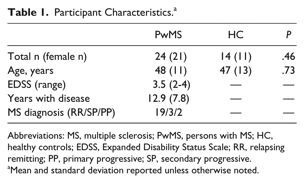
Abbreviations: MS, multiple sclerosis; PwMS, persons with MS; HC, healthy controls; EDSS, Expanded Disability Status Scale; RR, relapsing remitting; PP, primary progressive; SP, secondary progressive.
Mean and standard deviation reported unless otherwise noted.
Temporal Performance Was Worse in PwMS Than HC, but Improved Similarly With Training
PwMS exhibited worse temporal performance throughout the day 1 training period than HC (mean ± SD; PwMS, −8.97 ± 6.41; HC, −3.39±3.63; P = .001). Spatial performance trended toward worse in PwMS than HC, but this difference did not reach statistical significance (PwMS, 0.65 ± 0.09; HC, 0.62 ± 0.05; P = .208). As described in detail previously, 11 PwMS and HC improved similarly on both temporal and spatial performance on day 1; however, PwMS only retained improvements 24 hours later in temporal performance.
PwMS Exhibited Worse Structural Connectivity in the CC and Superior Cortical White Matter Tracts
Significantly smaller total brain volume as well as gray and white matter volume were observed within our cohort of PwMS compared with control subjects (Supplemental Figure 1). Furthermore, PwMS showed reduced supraspinal white matter integrity compared to HC. Specifically, we observed significantly increased RD (indicating poorer white matter integrity), principally within white matter tracts of the CC, corona radiata, and superior longitudinal fasciculi (SLF; Figure 2). PwMS also exhibited significantly reduced FA (indicating poorer white matter integrity) within similar, although fewer, neural regions as those white matter areas demonstrating increased RD (Supplemental Figure 2A). Finally, PwMS showed significantly increased MD (indicating poorer white matter integrity) compared to their age-matched counterparts within cortical white matter tracts including the SLF and corona radiata. It is worth noting that group differences in MD were lateralized to the right hemisphere (Supplemental Figure 2B).
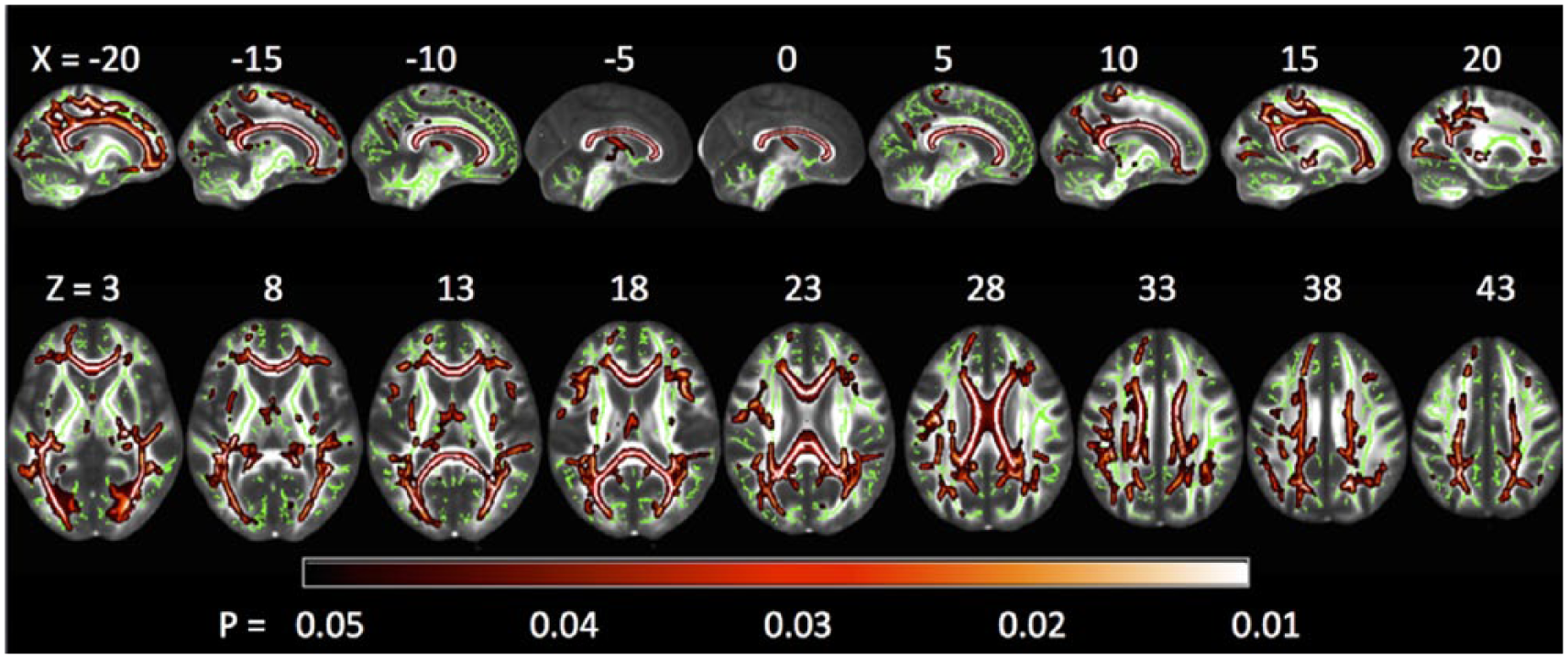
Tract-based spatial statistics (TBSS) whole-brain group comparisons of white matter microstructural integrity (assessed via radial diffusivity) showing the contrast of persons with multiple sclerosis (PwMS) > healthy controls (HC), with lighter colors indicating the largest differences between groups. Analysis is restricted to those white matter voxels that constitute the skeleton (green) of the brain’s connectional architecture, whereby this skeleton can be matched across subjects. PwMS had a large network of impaired white matter integrity (reflected by higher radial diffusivity values), most notably within interhemispheric callosal fibers. The reverse contrast of HC > PwMS yielded no significant differences. Results are multiple comparison–corrected and controlled for age, gender, brain volume, and Expanded Disability Status Scale (EDSS).
Temporal, but Not Spatial Improvements on Day 1 Were Correlated to Structural Connectivity in PwMS
Improved temporal performance over Day 1 was significantly correlated with the primary structural connectivity measure: RD, as well as secondary measures: FA and MD. Improved temporal performance for HC was not related to any of the measures of the white matter integrity. No significant correlations were observed between white matter integrity (RD, MD, or FA) and improvements in spatial performance for either HC or PwMS.
RD and Improved Temporal Performance
Statistically significant correlations were observed for improved temporal performance with RD measures of white matter integrity of the CC and white matter regions connecting the left posterior parietal cortex with the primary sensorimotor cortex of PwMS (Figure 3). Multiple comparison corrected post hoc analyses showed that correlations between improvement of temporal performance and structural connectivity (RD) were most pronounced in CC regions connecting prefrontal cortical regions (genu), and the region connecting the primary motor cortex (Table 2; Figure 3). No correlations were observed between temporal performance improvement and any cerebellar structural connectivity (RD) measures.
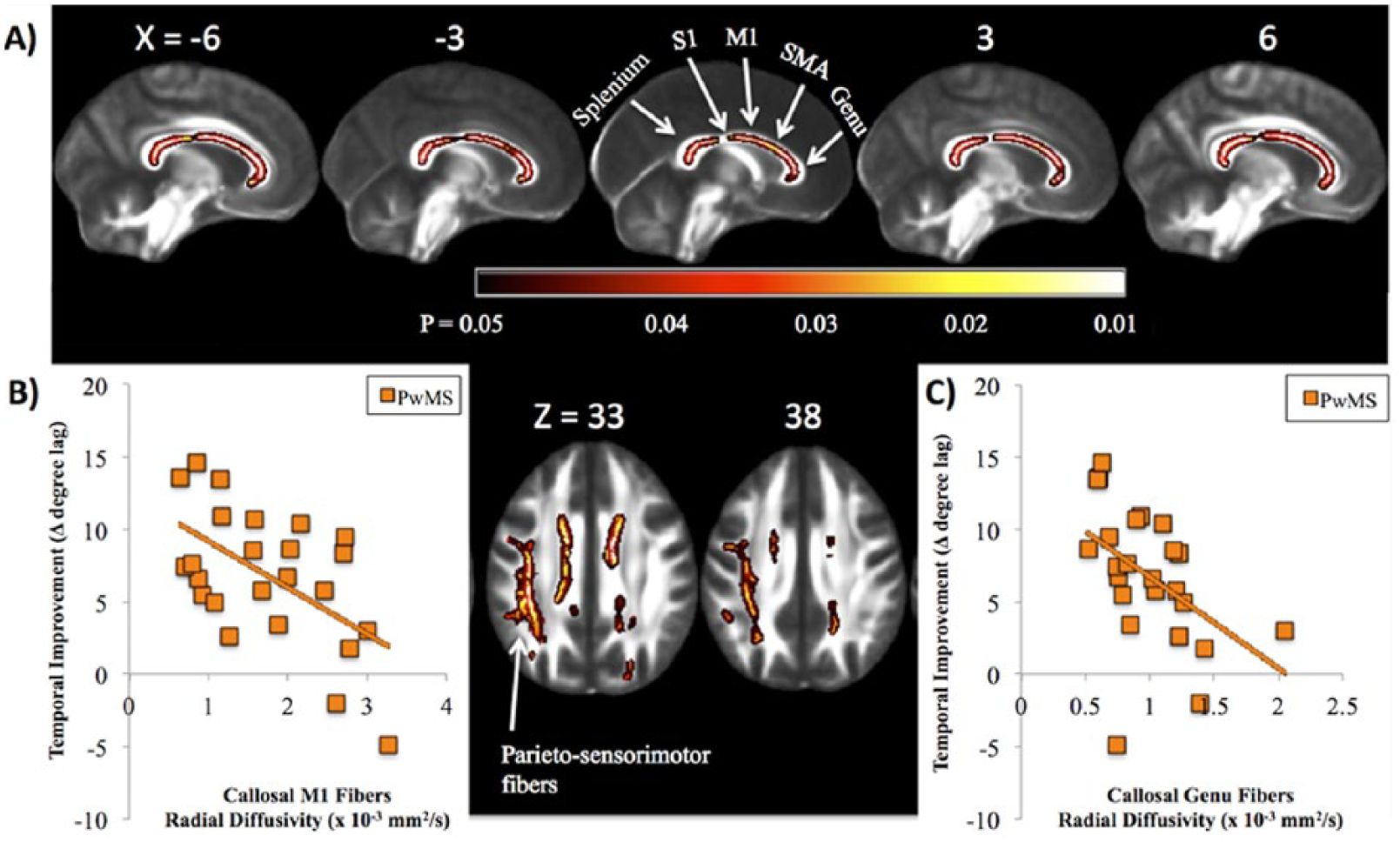
(A) Significant associations between temporal improvement and white matter microstructure are shown in people with multiple sclerosis (PwMS), with brighter colors (yellow, white) representing stronger correlation. Significant associations were localized to the corpus callosum (sagittal views: genu, body and splenium; top) and white matter connecting the posterior parietal cortices with the primary sensorimotor cortices within the left hemisphere (axial views; bottom). Results are multiple comparison–corrected and controlled for age, gender, brain volume, and Expanded Disability Status Scale (EDSS). Sections of the callosum connecting specific cortical structures (splenium, primary somatosensory cortex [S1]; primary motor cortex [M1]; supplementary motor area [SMA]), and genu are localized. Callosal locations are adapted from Fling et al. 37 Scatterplots represent individual values for PwMS displaying the significant association between temporal improvement in postural control and callosal fiber tracts connecting the (B) M1: r = −0.56; P = .004, as well as the (C) genu: r = −0.47; P = .01.
Post Hoc Analysis Correlating Fiber Tract Quality (Radial Diffusivity) of Specific Corpus Callosum Regions With Temporal Performance and Improvement in Temporal Performance in Persons With Multiple Sclerosis (PwMS). a
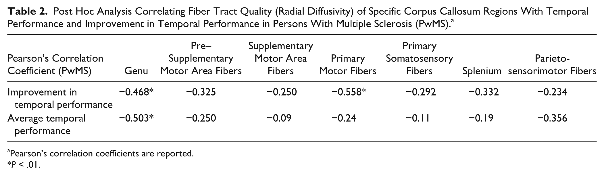
Pearson’s correlation coefficients are reported.
P < .01.
FA and Improved Temporal Performance
The relationship between FA and improved temporal performance was similar, albeit less pronounced, to RD (Supplemental Figure 3). Specifically, nonsignificant trends were observed between FA and improved temporal performance in the CC and left SLF.
MD and Improved Temporal Performance
A relationship between MD values and the improved temporal performance was observed in the left hemisphere’s SFL (posterior aspect) and arcuate fasciculus in PwMS (Supplemental Figure 4).
Retention of Improvements Tested on Day 2 Was Correlated to MD, but Not FA or RD Imaging Outcomes
No significant correlations were observed between RD or FA and retention of improvements of either temporal or spatial performance. However, MD values within the left hemisphere’s SLF (posterior aspect) and arcuate fasciculus were related to retention in PwMS, such that lower MD values were related to greater retention (Supplemental Figure 5).
Temporal Postural Performance Was Correlated to CC and Brainstem Structural Connectivity
Figure 4 shows the correlation between RD and mean temporal postural performance (averaged across day 1) in PwMS. CC (particularly within the genu of the CC—see Table 2) and brainstem white matter tracts were significantly correlated to temporal performance. Similar to RD, FA of the CC and brainstem was also related to mean temporal performance within PwMS (Supplemental Figure 6). No significant correlation was noted between MD and temporal performance.
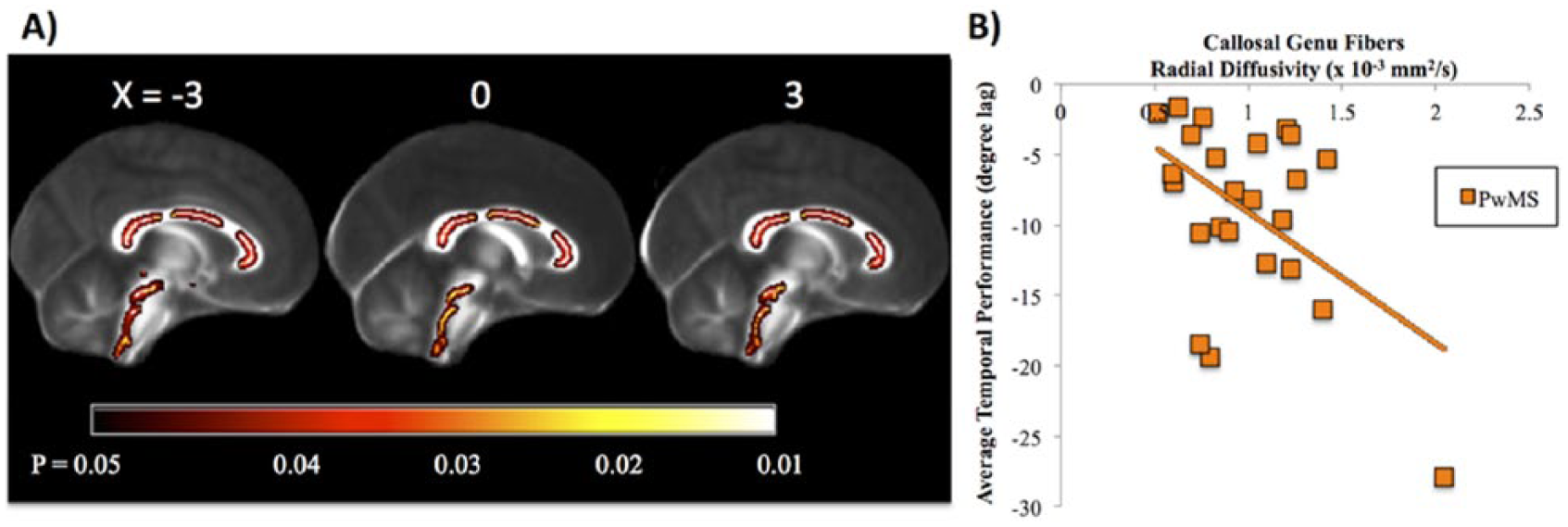
(A) In persons with multiple sclerosis (PwMS), significant associations between average temporal performance and white matter microstructure were localized to the corpus callosum (genu, body, and splenium) and within the brainstem. Results are multiple comparison–corrected and controlled for age, gender, brain volume, and Expanded Disability Status Scale (EDSS). (B) Scatterplot of individual participant values displaying the significant association between genu fiber tract integrity and average temporal performance in PwMS: r = −0.50; P = .006, but not health controls (HC): r = 0.01; P = .8 (HC data not shown).
Discussion
Our primary result is that integrity of CC white matter tracts was directly correlated to temporal postural motor performance as well as improvement in performance in PwMS, such that better connectivity was associated with larger improvements in postural control. In contrast, no significant correlations were observed between structural connectivity and spatial measures of postural control. These results are consistent with previous data suggesting the CC is related to improvements in UE motor learning in PwMS. 8 We extend this literature, showing that the CC, and interhemispheric communication, is also likely involved with postural motor learning in this population.
The observed relationship between improved temporal performance and CC integrity (measured via RD and FA) in PwMS is consistent with recent UE literature. Feedforward motor sequence learning is often measured by having participants tap individual fingers in response to repeated sequences of visual stimuli.7,38 Feedforward, sequence specific learning is measured by reduction in reaction time to these repeated sequences. Participants also improve reaction time when exposed to random sequences of visual stimuli. This improvement, characterized as “nonspecific” learning, is related to improved feedback response mechanisms. Bonzano et al 8 showed that the degree of UE feedforward learning, but not nonspecific learning, was strongly associated with the FA of the CC in PwMS. In the current study, CC integrity, measured both by RD and FA, was related to improvements in temporal, but not spatial, postural performance. We have shown previously that the constant sinusoidal frequency of support surface movements throughout trials allows for feedforward learning via prediction of the timing of these movements.11,12,39 However, given the relative inconsistency of movement amplitude forces, participants must rely on nonspecific learning using proprioceptive feedback for spatial improvements. 12 Therefore, our current finding that improvements in temporal performance (ie, feedforward learning), but not spatial performance (ie, nonspecific learning) is correlated to CC connectivity is consistent with the findings of Bonzano et al. 8 Together, these findings suggest that feedforward learning, but not nonspecific learning, is related specifically to CC structural connectivity.
Post hoc analyses showed that the strongest relationship between temporal performance improvement and CC fiber integrity occurred in the genu, connecting the bilateral prefrontal cortices, and the CC midbody, connecting bilateral primary motor cortices. This is consistent with reports showing regions of the CC connecting the prefrontal cortices to be critical for motor learning. For example, Sisti et al 13 found that upper extremity learning was directly correlated with anterior CC fiber tracts. Ours is the first study to suggest that connectivity between bilateral frontal and prefrontal cortices is also critical for learning to improve postural responses to surface displacements.
Similar to improvements in temporal performance, we also report that mean temporal performance was correlated with both RD and FA of the CC and the brain stem. The relationship between CC integrity and postural performance is consistent with previous research showing that the CC, and the genu specifically, is critical for UE motor performance (for review, see Gooijers and Swinner 40 ). In fact, the genu of the CC, connecting prefrontal cortices, has been shown to be particularly important for bilateral UE performance in PwMS. 41 Our finding relating postural performance to brainstem structural connectivity is also supported by previous research. Indeed, brainstem structures are critical for postural responses,42,43 as well as for maintenance of upright posture and tone in normal 44 and neurological 45 populations.
Although alterations in CC structure in PwMS are commonly reported,16-18 the mechanism by which CC structural deficits affect motor learning is unclear. However, recent work suggests reduced interhemispheric inhibition via CC structural deficits may affect learning. Considerable previous work suggests that learning (particularly bimanual learning) is reliant on the CC,8,13-15 and an important function of the CC is mediating interhemispheric inhibition. 46 Furthermore, intracortical inhibition contributes to motor learning,47-49 and PwMS demonstrate poor contralateral communication and intracortical inhibition.50,51 Perhaps unsurprisingly, recent results from our laboratory confirm that PwMS exhibit less specificity of interhemispheric network connectivity, likely related to altered CC structural connectivity. 52 Taken together, this work suggests that reduced interhemispheric inhibition may play a role in learning deficits in PwMS. However, more work is necessary to understand this possible relationship.
In addition to the interhemispheric connections between the prefrontal cortices, we also observed a significant correlation between improved postural control and the SLF, a tract that connects intrahemispheric posterior parietal cortex and motor regions. The posterior parietal cortex plays an integral role in voluntary movements by assessing the context in which movements are being made. Specifically, this region receives somatosensory, proprioceptive, and visual inputs, and uses this feedback to determine such things as the positions of the body and the target. 53 It thereby produces internal models of the movement to be made, prior to the involvement of the premotor and motor cortices. Therefore, alteration to the transmission of these models to the primary motor cortex via disrupted white matter tracts may have contributed to poorer improvement performance in PwMS. It is noteworthy that this association was lateralized to tracts connecting the left, but not right, posterior parietal cortex and sensorimotor regions. Recent work has shown these tracts, namely the dorsal SLF, to be asymmetric with the left hemisphere SLF exhibiting more pronounced connectivity between the dorsal precentral gyrus and the caudal middle frontal gyrus than the right. 54 As noted above, these regions play critical roles for the planning and execution of movement. Therefore, while replication of our finding is necessary, it is possible that left lateralized structural alterations may relate more closely to postural improvements than right-side dysfunction.
Interestingly, we did not observe correlations between white matter tracts to or from the cerebellum. While the cerebellum does play a critical role in learning, we note that no group differences were observed in cerebellar white matter integrity (Figure 2). Therefore, the lack of correlation in the current study may have been the result of relatively intact cerebellar white matter tracts in our group of participants.
Our primary variable of structural connectivity, RD, was not related to 24-hour retention, measured here as the degree to which improvements remained different from baseline. Although numerous cortical and subcortical structures have been shown to be related to retention of skill learning, to our knowledge, no previous studies have investigated the role of white matter integrity in retention. Late-stage learning is typically associated with reduced activity of cortical structures (ie, prefrontal cortex and dorsolateral prefrontal cortex), and increased deep brain activity (ie, basal ganglia and cerebellum).55,56 Therefore, it is possible that while acquisition of skill (ie, improvement through practice) relies on transcallosal fibers and cortical structures, later stage learning relies more on deep brain structures. It is therefore notable that white matter tracts emanating from the cerebellum were not related to retention in the current study. However, as discussed above, this may have been related to a lack of structural dysfunction of these tracts or potentially due to the short-term training protocol. Unlike RD, MD within the SLF and arcuate fasciculus of the left hemisphere were related to retention in PwMS. These tracts have previously been related primarily to language comprehension and production. 57 However, given the connectivity to motor regions, they may also play a role in motor function. The disparate findings in the current study regarding RD, FA, and MD are unclear. The lack of consistent coupling with FA is potentially the result of differences in how these measures are calculated, as MD represents the overall tissue diffusivity, whereas FA and RD represent the integrity of the primary and orthogonal directions, or tensors, respectively.
In addition to differences between RD and MD with respect to retention analyses, MS-related changes in MD were found to be somewhat distinct from FA and RD. Specifically, while FA and RD were widespread and consistent,58,59 with considerable alteration of the CC, MD was lateralized to the right hemisphere. The reason for these somewhat inconsistent and nonintuitive findings is unclear. However, previous studies have shown changes in MD to be both similar 60 and disparate 61 to FA in PwMS compared with controls. Given the lack of data relating different imaging measures to improvements in postural control with practice, additional work will be necessary to elucidate how each measure predicts postural control and improvement.
Several limitations should be noted. First, our population exhibited mild symptoms of MS. Furthermore, data were collected from a relatively small and somewhat homogeneous (primarily relapsing remitting) population of PwMS. Together, these limitations may reduce the confidence and generalizability of findings. In particular, results may not generalize to patients with more severe symptoms or those with secondary progressive MS. In addition, we were unable to calculate lesion load within this population. It is possible that lesions to grey or white matter also contribute to the heterogeneity of postural control or motor learning in this population. Finally, sensory loss was not accounted for in this analysis. Although sensory loss may have contributed to the reduced performance on the postural task, we find it unlikely to have affected the degree of learning in the MS population.
Conclusions
This study is the first to demonstrate that CC white matter tracts play a role in practice-related improvements in postural control in PwMS. Retention of these improvements was not related to structural connectivity of the CC white matter tracts. This information highlights the importance of interhemispheric white matter tracts for learning in PwMS. Furthermore, it has the potential to inform patient selection regarding ability to improve balance control over time. However, given the importance of retention of skills for neurorehabilitation, additional research is required to identify predictive factors of retention of skill acquisition in PwMS and how neuroimaging can best be used to predict one’s ability to learn.
Footnotes
Acknowledgements
We thank the volunteers for participating in this study. We are also grateful to Heather Schlueter and Jessica Nyugen for assistance in participant recruitment and data collection.
Authors’ Note
The contents do not represent the views of the US Department of Veterans Affairs or the US government.
Declaration of Conflicting Interests
The authors declared the following potential conflicts of interest with respect to the research, authorship, and/or publication of this article: FBH and Oregon Health & Science University (OHSU) have an equity/interest in APDM, a company that may have a commercial interest in the results of the study. This potential conflict of interest has been reviewed and managed by the Research & Development Committee at the Portland VA Medical Center and OHSU.
Funding
The authors disclosed receipt of the following financial support for the research, authorship, and/or publication of this article: This work was supported by grants from the US Department of Veteran’s Affairs Rehabilitation Research and Development Service (Career Development Award-1: #I01BX007080; PI: DSP) and VA Merit Award (E1075-R; PI: FBH) and the National Multiple Sclerosis Society (FG 2058-A-1 PI: GG; RG 5273A1/T PI: BWF; MB0011 PI: FBH). Additional support was provided by the Medical Research Foundation of Oregon (BWF, GG), and the NL Tartar Research Fund (BWF).
References
Supplementary Material
Please find the following supplemental material available below.
For Open Access articles published under a Creative Commons License, all supplemental material carries the same license as the article it is associated with.
For non-Open Access articles published, all supplemental material carries a non-exclusive license, and permission requests for re-use of supplemental material or any part of supplemental material shall be sent directly to the copyright owner as specified in the copyright notice associated with the article.