
Abstract
Objectives:
To use the superparamagnetic iron oxide (SPIO) contrast agent Resovist (±transfection agent) to label human melanoma cells and determine its effects on cellular viability, microstructure, iron quantity, and magnetic resonance imaging (MRI) detectability.
Materials and Methods:
Human SK-Mel28 melanoma cells were incubated with Resovist (±liposomal transfection agent DOSPER). The cellular iron content was measured, and labeled cells were examined at 1.5 T and 3.0 T. The intracellular and extracellular distributions of the contrast agent were assessed by light and electron microscopy.
Results:
The incubation of melanoma cells with SPIO does not interfere with cell viability or proliferation. The iron is located both intracellularly and extracellularly as iron clusters associated with the exterior of the cell membrane. Despite thorough washing, the extracellular SPIO remained associated with the cell membrane. The liposomal transfection agent does not change the maximum achievable cellular iron content but promotes a faster iron uptake. The MRI detectability persists for at least 7 days.
Conclusion:
The transfection agent DOSPER facilitates the efficient labeling of human metastatic melanoma cells with Resovist. Our findings raise the possibility that other Resovist-labeled cells may collect associated extracellular nanoparticles. The SPIO may be available to other iron-handling cells and not completely compartmentalized during the labeling procedure.
Introduction
Basic and clinical research investigating the anatomic and molecular pathways of tumor metastasis calls for labeling techniques that permit cell tracking while not altering tumor cell viability. Possible techniques include positron emission tomography (PET) imaging, 1,2 optical imaging, 3,4 and magnetic resonance (MR) imaging (MRI) using iron oxide MR contrast agents. 5,6 Despite its high sensitivity, the major drawbacks of PET imaging include poor resolution, patient exposure to radiation, and the limited half-life of radioactive tracers. Optical imaging is a nonradioactive and highly sensitive method. 7 However, due to its poor tissue penetration, it seems to be best suited for surface imaging (eg, wrist and breast) or for small animals in an experimental setup (eg, mice and rats). Magnetic resonance imaging using iron oxide particles, such as superparamagnetic iron oxide (SPIO), represents a good option in terms of sensitivity, resolution, and safety, and it enables the labeling and tracking of different cell types. 6,8,9
Hematopoietic and mesenchymal stem cells or human prostate cancer cells were efficiently labeled with SPIO-based MR contrast agents. 10 –12 Cells were visualized with clinical 1.5 T or 3.0 T MRI machines with a minimum detectable quantity of 1000 cells. In similar studies using human hematopoietic cells, the detection limit reported was 100 000 cells. 13
Melanoma cells can also be labeled with SPIO-based contrast agents and tracked with MRI. 14 After injection into an embryonic microenvironment, these cells migrate along neural crest pathways and can therefore be used to investigate migratory patterns during malignancy and embryonic development. 15 This finding is noteworthy, as the invasive behavior of melanocytes after their malignant transformation to melanoma cells may be linked to their developmental origins in the neural crest. 16 The noninvasive tracking of melanoma cells using MRI may therefore help to identify specific pathways promoting metastasis considering the neural crest origin of melanocytes.
Despite prior studies using SPIO labeling and MRI tracking of melanoma cells, an in-depth analysis of the effects of SPIO labeling conditions and transfection agents (TAs) on human melanoma cells has not yet been published. In contrast to epidermal tumor cell lines (such as TR131, SCLII, and FaDu) and melanoma cells derived from primary tumors (such as MelEi and MelHo), human skin SK-Mel28 melanoma cells are derived from a melanoma metastatic lesion and have high migratory and moderate invasive potentials. 17 As cell tracking is of an increased clinical relevance for metastatic disease, we chose the SK-Mel28 cell line for our study. Here we aimed to label human SK-Mel28 melanoma cells with the SPIO contrast agent Resovist and to examine the effects of the labeling conditions and TAs on cellular viability, ultrastructure, iron content, and MRI detectability.
Materials and Methods
Cell Culture and Labeling Protocols
Cell culture
Human skin melanoma cells (SK-Mel28) were obtained from the American Type Culture Collection (Rockville, Maryland). Routine cell cultures were grown in RPMI 1640 +
The SPIO contrast agent Resovist (SHU 555A, Schering, Berlin, Germany) 18,19,20 was used for all experiments. The SK-Mel28 cells were seeded at a density of 5 × 10⁵/cm2 in culture flasks and labeled with Resovist according to the protocol in Table 1. Additionally, to investigate whether Resovist SPIO clusters are firmly attached to the cells, the degree of Resovist detachment was evaluated with several washing steps, as described in Table 1.
Resovist Labeling and the Evaluation of Resovist Detachment.

The liposomal TA DOSPER (1,3-di-oleoyloxy-2-(6-carboxy-spermyl)-propylamid, Roche Diagnostics GmbH, Mannheim, Germany) was used as a carrier for the MR contrast agent to increase the labeling efficiency. For this purpose, 3 × 10⁵ SK-Mel28 cells/cm2 were seeded in culture flasks and labeled with Resovist and DOSPER according to the protocol in Table 2. Cell viability and proliferation were assessed using a CASY-TT cell counter according to the standard operating procedure (SOP) protocol (SOP CASY). Three 400 µL samples were used for each measurement. The cells were incubated for 24 hours with Resovist, after which the Resovist-containing medium was replaced with standard culture medium.
Enhancing Resovist Labeling With the Transfection Agent DOSPER.
Abbreviation: PBS, phosphate-buffered saline.
Magnetic Resonance Imaging
An agar matrix was used as a suitable environment for measuring SPIO-labeled cell cultures. 21 Briefly, a 1% agar solution (dissolved in PBS) was boiled and slowly cooled in a 37°C water bath to minimize air bubbles that would produce artifacts during MRI. Before congealing, the agar was embedded in specialized plastic boxes. A series of cone-shaped cavities were imprinted in the agar block with a specialized stamp (Figure 1).

A, Agar matrix with the embedded Resovist-labeled cells. The labeled cells were placed in the cone-shaped cavities (*) that were previously treated with a specialized stamp (#). Afterward, the cavities were occluded with agar. B, Wrist coil for the measurement with the 3.0 T Magnetom TRIO.
For MR measurement, 1000 to 1 000 000 SK-Mel28 cells were used. Cells were centrifuged (200 × g, 5 minutes), resuspended in a 4% gelatin-PBS solution (20 μL), and implanted into the previously created cavities within the agar matrix.
The cells were scanned with clinically used MR scanners at 3.0 T (Magnetom Trio, Siemens Medical Solutions, Erlangen, Germany) and 1.5 T (Magnetom Sonata, Siemens). High-resolution wrist coils were attached, and the 3D gradient-echo Fast-Low Angle Shot (FLASH), 22 and the Fast Imaging with Steady-State Free Precession (FISP) 23 sequences were used. The resolution can be changed at both field strengths. Pintaske et al 24 described that the use of high resolution improves the accuracy of geometric localization but decreases the MRI sensitivity. The resolution (0.25 to 0.50 mm) and cell number chosen for each experiment at the respective field strengths are specified in the figure legends. The TE varied from 5 to 25 ms, and the TR was 35 ms with a flip angle of 20°. The signal extinction was recorded by measuring the signal artifacts in all planes.
Quantification of Cellular Iron Content
Atomic Absorption Spectrometry
To examine the iron content with atomic absorption spectrometry (AAS), SK-Mel28 cells were incubated with SPIO-containing culture medium. Subsequently, the cells were washed 3 times with PBS, resuspended in Accutase (PAA-Laboratories, Pasching, Austria), centrifuged (5 minutes, 200 × g, RT), and resuspended. The cell numbers were measured with a CASY-TT cell counter, as described earlier. One million cells were centrifuged and sonicated in 3- to 10-second intervals (Branson Sonifier B-12, Branson Sonic Power Company, Danbury, Connecticut).
For the iron measurements, the cell samples and iron standards (Baker, Malinckrodt Baker Inc, Phillipsburg, New Jersey) were dissolved in a 0.3% HCl solution. The iron concentrations within the cell suspensions were assessed with an AAS system (Unicam SolaarM, Thermo Electron, Waltham, Massachusetts) using an air-acetylene flame. The baseline correction was performed with a deuterium lamp. The AAS settings were as follows: wave length, 248.3 nm; spectral bandwidth, 0.2 nm; lamp current, 7.5 mA; and gas volume, 0.9 L/min. The cell suspensions with low iron concentrations were diluted 1:2 to 1:5 (a detection threshold of 12 µg iron/L). The cell samples with an estimated higher iron content were diluted 1:10 or 1:20 (a detection threshold of 120 µg iron/L). The mean values of the diluted probes, which were measured 3 times, are shown.
Spectrophotometric Assay
The iron content of the labeled cells was assessed with a Ferrozine-based method (Eisen Ferene S Plus, Greiner Biochemica, Germany) using a ultraviolet (UV)-spectrophotometer (Ultrospec 3000, Amersham Pharmacia Biotech, Heidelberg, Germany), as described previously. 25 Fe3+ was reduced to Fe2+ with Ferene, forming a blue complex. The UV extinction of this complex, measured at a wavelength of 595 nm, is proportional to the iron concentration. The iron oxide of the contrast agent (Fe2+) was assessed without reduction. The iron concentration was measured using an internal standard solution. 26 After incubation with Resovist, the cells were washed twice with PBS, suspended in Accutase (PAA-Laboratories), centrifuged (5 minutes, 200 × g, RT), and resuspended in PBS. Next, the cells were counted, and 1 000 000 cells were centrifuged again. The cell pellet was dried for 12 hours at 70°C. Finally, the probes were treated overnight at room temperature and for 2 hours at 60°C in 200 µL of a perchloric acid/nitric acid (3:1) solution. The photometric measurements were repeated 3 times.
Electron Microscopy
The cells were fixed in 2.5% glutaraldehyde (Paesel-Lorei, Frankfurt, Germany) buffered with 0.1 mol/L cacodylate buffer (pH 7.4) and post fixed in 1% OsO4 in 0.1 mol/L cacodylate buffer. The pellet was then dehydrated with an ethanol series (50%, 70%, 96%, and 100%). The 70% ethanol was saturated with uranyl acetate for contrast enhancement. The dehydration was completed in propylene oxide. The specimens were embedded in Araldite (Serva, Heidelberg, Germany). Ultrathin sections were cut using an FCR Reichert Ultracut ultramicrotome (Leica, Bensheim, Germany), mounted on pioloform-coated copper grids, contrasted with lead citrate, and analyzed with an EM 10A electron microscope (Zeiss, Oberkochen, Germany).
Light Microscopy
The cells were seeded on specialized plates (Falcon CultureSlide, Becton Dickinson, New Jersey) and loaded with Resovist. For the Prussian blue staining, the cells were fixed with 4% glutaraldehyde, washed, incubated for 30 minutes in 2% potassium ferrocyanide in 6% hydrochloric acid, washed, and counterstained with nuclear fast red. The slides were analyzed using a Leica DM RBE microscope and an image analysis system (Quantimet 600, Wetzlar, Germany).
Statistical Analysis
Data are expressed as the means ± standard deviation of 3 to 9 independent data samples. The JMP5.1 software (SAS, Cary, North Carolina) was used for the statistical calculations. The viability data are expressed as the percentage of all cells. The absolute cell number and viability of labeled cells were examined with a multivariate analysis (the Tukey-Kramer test). The iron oxide concentrations were compared using Student t test. A P value less than .05 was considered statistically significant.
Results
Cell Viability and Microscopy
The growth of cultured SK-Mel28 cells was not altered by a 24-hour incubation in Resovist at concentrations ranging from 0 to 200 µg Fe/mL (Figure 2A). The fraction of living cells, which was between 80% and 90% during our experiments, was also not altered by a 24-hour incubation in the SPIO contrast agent Resovist at the indicated concentrations (Figure 2B). After 6 days, cell confluency was achieved (approximately 1 000 000 cells per culture flask), and the fraction of living cells within the culture dropped to 76% (no Resovist), 82% (50 µg Resovist-iron/mL), and 80% (200 µg Resovist-iron/mL). Transmission electron microscopy (TEM) did not reveal any structural changes to the labeled cells compared with the nonlabeled cells (Figure 3A-C). The intracellular accumulation of SPIO-containing vesicles appeared to increase as the amount of Resovist increased (Figure 3B). However, Resovist was also associated with the extracellular side of the plasma membrane (Figure 3C).

Growth of SK-Mel28 cells cultured in the presence or absence of Resovist. The cell numbers and viabilities were assessed with a CASY-TT cell counter. The experiments were performed in triplicate. A, No significant difference (P > .05) in cell proliferation was induced by Resovist labeling. The proliferation was inhibited by cell confluence after 5 to 6 days. B, No toxic influence of the superparamagnetic iron oxide (SPIO) labeling (iron concentration 0 to 200 µg/mL) was detectable, as no significant difference was observed with increasing iron concentrations (P > .05). The percentage of viable cells was not altered by the incubation with Resovist over a period of 7 days.

Analysis of the uptake of superparamagnetic iron oxide (SPIO) particles by transmission electron (A-C) and light (F-H) microscopy. A, Transmission electron microscopy (TEM) of an unstained melanoma cell. B, A cytoplasmic endosomal vesicle containing Resovist (arrow) and (C) an extracellular Resovist cluster associated with the cell membrane (arrow). Light micrographs show unstained (D-F) and Prussian-blue-stained (G-H) melanoma cells. D, The Resovist-labeled adherent melanoma cells are shown at 40× magnification. Light microscopy is not well suited to differentiate between extracellular and intracellular iron oxide aggregates. Nevertheless, in consideration of the TEM results, light microscopy indicates both (E) an extracellular association with the cell membrane and (F) an intracellular accumulation after detachment of the spheroidal shaped cells. G-H, The cellular association with iron (stained blue) was noticeably higher after 4 hours of incubation with both Resovist and DOSPER (H) than with Resovist alone (G). (E–H 100× magnification).
Using light microscopy, the iron of Resovist appears to be brown (Figure 3D-F). In an attempt to differentiate between the intracellular and the extracellular SPIO, the Resovist-loaded cells were detached with Accutase. Light microscopy is not well suited to differentiate extracellular and intracellular aggregates of iron oxide. Nevertheless, taking into account the electron microscopy results (Figure 3B-C), Figure 3E suggests an extracellular association with the cell membrane, whereas Figure 3F indicates a predominantly intracellular accumulation. Resovist is firmly associated with the cells: neither intense washing nor the TEM preparation procedures were able to remove it from the cell membrane.
Magnetic Resonance Imaging and the Measurement of the Cellular Iron Concentration
The quantitative assessment of the iron content of SPIO-labeled SK-Mel28 cells shows a correlation between the iron concentrations in the cells and in the culture medium (Figure 4A). With a concentration of 600 µg of iron per mL of culture medium, a maximum iron content of 68 pg/cell was measured via AAS. When using the Ferene-based photometric method, a maximum iron content of 84 pg/cell was measured (20% more than via AAS). The susceptibility artifacts of the iron-loaded cells in the agar phantoms at 3.0 and 1.5 T were also dependent on the cellular iron content (Figure 4B).

Correlation between the total cellular iron and increasing concentrations of Resovist in the culture medium. The iron concentration was between 50 µg and 600 µg per mL of culture medium, and the incubation time was 15 hours. The experiments were performed in triplicate. A, Quantification of the iron content of the Resovist-labeled cells with atomic absorption spectrometry (AAS) and a spectrophotometric assay. No transfection agent was added. The curve depicts the dependency of the uptake of magnetic resonance (MR) contrast agent on increasing iron concentration. B, Representative MR images of the Resovist-loaded cells with 3.0 T Trio Magnetom (FISP, 0.3 mm isotropic resolution, echo time (TE) 20 ms, 100 000 cells). C, Representative MR images of the Resovist-loaded cells with 1.5 T Sonata Magnetom (Flash 3D, 0.4 mm resolution, TE 20 ms, 100 000 cells). B and C, The MR signal enlarges due to blooming artifacts. Cell numbers were kept constant at 100 000 cells.
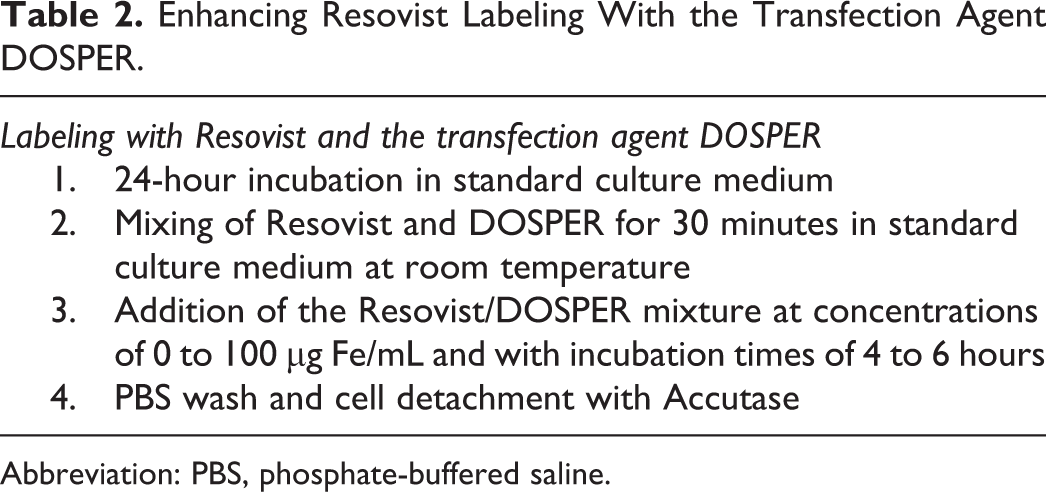
The degree of Resovist uptake correlates with the incubation time (Figure 5A). After 12 to 15 hours, the maximum iron uptake was achieved, and within the first 6 hours of incubation, approximately 90% of the maximum iron content was reached. An extended incubation with iron (72 hours) resulted in a decline in cellular iron (to only 70% of the maximum level). The susceptibility artifacts achieved at 3.0 T and 1.5 T paralleled this kinetic; nevertheless, the cells were effectively visualized after only 1 hour of incubation (Figure 5B).
Dependence of iron uptake on the incubation time. The iron concentration was 200 µg per mL of culture medium (analogous to Oppitz et al 14 ), and the incubation time varied from 5 minutes to 72 hours. The experiments were performed in triplicate. A, Quantification of the iron content of the Resovist-labeled cells with atomic absorption spectrometry. The curve depicts the dependence of magnetic resonance (MR) contrast agent uptake on incubation time. Within 6 hours, most of the iron was either absorbed by or adhered to the cells. An incubation time of more than 20 hours resulted in a decline in the cellular iron concentration. B, Representative MR images of Resovist-labeled cells with 1.5 T Sonata Magnetom (Flash 3D, 0.4 mm resolution, echo time (TE) 20 ms, 100 000 cells).
To evaluate the impact of cell division on the intracellular iron content and the MR susceptibility artifacts, iron-loaded cells were cultured in SPIO-free medium. A decline in the cellular iron content was observed (Figure 6A). Both measuring methods (Ferene-based photometry and AAS) documented a decline in the cellular iron content of 70% on day 7. However, the MR signal did not show a similar reduction, and the susceptibility artifact persisted.

Removal of the cell-associated iron oxide particles after incubation with Resovist. The cells were incubated with 200 µg iron/mL for 24 hours, washed with phosphate-buffered saline (PBS) and incubated in medium without Resovist. The experiments were performed in triplicate. A, At the indicated time points, the cells were washed and lysed, and the iron content was determined with atomic absorption spectroscopy and a spectrophotometric assay. The iron contents were expressed as a percentage of the initial iron concentration observed after Resovist incubation. After 7 days, only 30% of the initial cellular iron content could be measured. At later incubation times, no significant loss of cellular iron content was observed. B, Additionally, the cells were imaged by MR imaging at 3.0 T (500 000 cells) at the indicated time points.
Resovist tends to form clusters that bind to the outside of the cell membrane (Figure 3). To investigate whether these SPIO clusters are firmly attached to the cells, cultured iron-loaded SK-Mel28 cells were washed several times with PBS. Next, the cells were detached with Accutase, centrifuged, and washed again several times. After the second, fourth, and sixth cell washes, the iron content per cell was determined (Figure 7). Even after 6 cycles of washing and centrifugation, 81% and 88% (determined by AAS and Ferene-based photometry, respectively) of the iron remained attached to the cells. The iron particles could not be removed from the cells by the shearing forces induced by washing.

Removal of the cell-associated iron oxide particles after incubation with Resovist. The cells were incubated with 100 µg iron/mL for 24 hours, detached, and then washed several times with phosphate-buffered saline (PBS). The experiments were performed in triplicate. A, Quantification of the iron content of the Resovist-labeled cells with atomic absorption spectrometry and spectrophotometric assay. The iron contents are expressed as a percentage of the initial iron concentration observed after Resovist incubation. After 6 washing steps, the iron content remained at 81% (atomic absorption spectrometry [AAS]) and 88% (photometry). The iron particles could not be removed by the shearing forces induced by washing. B, Representative magnetic resonance (MR) images of the Resovist-loaded cells with 1.5 T Sonata Magnetom (Flash 3D, 0.4 mm resolution, TE 20 ms, 100 000 cells). Up to 6 washing steps did not produce an apparent discrepancy in the signal alteration.
Enhancing the Uptake of SPIO With the Liposomal TA DOSPER
The liposomal TA DOSPER did not change the absolute maximum cellular iron uptake (69 pg/cell with DOSPER compared with 68 pg/cell without DOSPER). However, the addition of DOSPER permitted the use of a lower amount of iron without compromising cellular uptake. In this way, the iron content of the culture medium could be reduced from 600 to only 100 µg/mL when the experiment was performed in the presence of TA. Furthermore, the incubation time was reduced from 16 hours to 4 hours (Figure 8). In the absence of DOSPER, the SK-Mel28 cells incorporated only 4.7 pg iron/cell when cultured in medium containing 100 µg/mL iron. Under the same conditions, the addition of DOSPER resulted in a 15-fold increase in the cellular iron concentration. In culture medium containing 50 µg/mL iron, DOSPER increased the cellular concentration of iron by a factor of 25 (64.5 pg iron/cell when using DOSPER compared to 2.5 pg iron/cell without DOSPER). The enhanced iron uptake mediated by DOSPER, despite lower Resovist concentrations in the culture medium was also demonstrated by microscopy (Figure 3G and H) and 3.0 T MRI in our agar phantom model (Figure 8B and C). Without DOSPER, the SK-Mel28 cells required incubation with at least 50 µg iron per mL of culture medium to become visible under the same experimental conditions.

Enhancing the iron uptake by the addition of the transfection agent DOSPER. The melanoma cells were incubated with increasing concentrations of Resovist. The experiments were performed in triplicate. A, Quantitative iron determination with atomic absorption spectroscopy. The graphs show cells incubated with Resovist in the presence or absence of the transfection agent (TA) for 4 hours. A dose-dependent iron uptake with an increasing load of Resovist in the incubation medium was observed. The incubation with the transfection agent resulted in a significant increase in iron oxide uptake (P < .05). B and C, Representative magnetic resonance (MR) images of the Resovist-loaded cells in the presence (B) and the absence (C) of the transfection agent were obtained with 3.0 T Trio Magnetom (FISP, 0.3 mm isotropic resolution, TE 20 ms, 500 000 cells, 4-hour incubation time). A dose-dependent increase in the susceptibility effects was observed. It is noteworthy that the cells incubated with the transfection agent display significantly higher susceptibility effects.
Discussion
Superparamagnetic iron oxide contrast agents are conglomerates of nanosized iron oxide crystals coated with dextran or carboxydextran that enable MRI with shortened T1 and T2 relaxation times. 27 Two SPIO MR contrast agents are clinically approved: ferumoxides (Feridex IV, Bayer Healthcare; Endorem, Guerbet) and Ferucarbotran (Resovist, Bayer Healthcare). Concentrations of Resovist up to 200 µg iron/mL did not have any effect on cell viability or cell proliferation in this study, which is in agreement with other reports using hematopoietic or mesenchymal stem cells. 28 –30
The susceptibility artifacts of labeled SK-Mel28 melanoma cells persisted for at least 7 days after the withdrawal of Resovist from the culture medium, despite a gradual decline in absolute iron content to only 30% of the initial level on day 7. Similarly, Sun et al observed decreased iron content after the culturing of SPIO-labeled cells over time and suggested a forced cellular iron elimination induced by incubation with SPIO. 28 In this regard, exocytosis is a possible mechanism for the disposal of excess SPIO and may explain the presence of SPIO at the extracellular surface of SK-Mel28 cells observed by TEM. Nevertheless, as SPIO-labeled cells grow in culture, the amount of cellular iron should decrease because the SPIO is not propagated with cell division. Although the transverse relaxation rates correlate with the amount of iron, we observed persistent MR signal alterations, despite a reduction in iron content. This result is consistent with the findings of Pintaske et al, 21 indicating that the diameters of the MRI signal extinction correspond to a logarithmic function that is dependent on the iron concentration and flatten out considerably with high iron concentrations. Furthermore, Pintaske et al 21 and Tanimoto et al 31 provided evidence that the extent of the signal extinction was dependent upon the geometric distribution of the iron particles.
In contrast to other studies, the efficiency of labeling melanoma cells by incubation with Resovist was satisfactory. Hematopoietic stem cells accumulated only 2.4 ± 0.7 pg iron/cell when incubated with Endorem (an SPIO particle with a mean diameter of 150 nm) or 1.1 ± 0.4 pg iron/cell when incubated with Sinerem (an ultrasmall SPIO particle with a mean diameter of 20 nm that is not approved for clinical applications). 29,32 The melanoma cells in our experiments accumulated 23 ± 3.8 pg iron/cell when incubated in culture medium containing 200 µg iron/mL. In our study, the maximum incubation period was 15 hours compared to 2 hours in other experiments. Nevertheless, the cellular iron content of melanoma cells was 9.35 pg/cell after only 1 hour of incubation. By increasing the iron concentration to 600 µg/mL, we were able to increase the cellular iron content by a factor of 3 (to 68 pg iron/cell).
Other reports in the literature describe an intracellular location for the iron. 29,33 Using electron microscopy, we observed that the majority of the iron was located on the outside the cells in the form of iron clusters associated with the cell membrane. Hence, the measurement of cellular iron does not reflect uptake alone but rather a combination of uptake and adherence. This finding raises questions regarding the fraction of SPIO adhering to cells with varying concentrations of Resovist in the culture medium and the fate of the extracellular SPIO adhering to labeled cells in long-term cultures and in vivo. We did not investigate the mechanism by which SPIO adheres to the cell membrane in detail and therefore cannot exclude that these extracellular nanoparticles could not dissociate under certain conditions. Nevertheless, we found that extensive washing does not result in iron detachment from the cell membrane. It is well known that increased levels of intracellular iron result in oxidative stress, which may lead to DNA fragmentation, apoptosis, and eventual cell death. 34 Therefore, an advantage of extracellular iron particles adhering to cells is the reduced intracellular radical formation induced by high iron concentrations. A drawback of extracellular adherent iron particles could be an alteration of cell mobility. However, the SPIO particles may translocate from transplanted cells to the surrounding tissue and label the extracellular surface of other cells, which could reduce the specificity of cell labeling. Indeed, Zacharovová et al observed considerable translocation of the SPIO particles after transplantation of pancreatic islet cells into the liver or kidney. Iron particles were found in macrophages, fibroblasts, and myofibroblasts surrounding the transplanted islet cells. 35 Similar results were reported by other groups investigating bone marrow stromal cells transplanted into an area of vascular inflammation, 36 neural precursor cells transplanted into the spinal cord 37 and stem cells transplanted into the heart. 38 Previously, the translocation of iron particles from SPIO-labeled cells to surrounding macrophages has been attributed to the viability of transplanted cells. 39 In contrast, our data suggest that when transplanting SPIO-labeled cells for the purpose of cell tracking, the SPIO particles may be readily available to other iron-handling cells and not completely compartmentalized during the labeling procedure.
Liposomes are used as carrier systems for cytotoxic drugs, DNA or gene therapy targets. Resovist is coated with carboxydextran and is usually phagocytosed by cells of the liver reticuloendothelial system. DOSPER binds to these negatively charged molecules resulting in the formation of polycationic complexes that are nonspecifically absorbed by negatively charged cell surfaces. 40 We used the liposomal TA DOSPER as a carrier for the MR contrast agent to increase labeling efficiency. In doing so, we significantly reduced the incubation time and the iron concentration within the culture medium. Nearly 70 pg of iron/cell was achieved with an incubation time of only 4 hours (25% of the time needed without DOSPER) and a Resovist iron concentration within the culture medium of only 50 µg (8% of the concentration needed without DOSPER). Additionally, Arbab and Janic et al reported that the labeling of hematopoietic stem cells or macrophages with ferumoxides using TAs did not alter the immunological properties of the cells. 41,42
There are several limitations of this study that necessitate discussion. First, the specific mechanisms resulting in the cellular uptake of the SPIO contrast agent were not investigated. Second, the cell viability was not re-evaluated after the addition of the TA DOSPER. However, in former studies, significant levels of cellular toxicity induced by liposomal TAs have not been observed, 43 and DOSPER did not result in cellular toxicity or gene rearrangements. 44 Third, we observed a minor difference between the AAS and photometric methods of measuring iron content. Although AAS should more specifically measure the absolute iron content, it is more expensive and time consuming. Nevertheless, the photometric measurements appear to be sufficient keeping in mind a possible measurement error, and as we did not observe consistent differences between the AAS and photometric measurements, a systematic correction of the photometric measurements was not feasible.
Conclusion
Although relatively long-term SPIO labeling for MR tracking of human melanoma cells is feasible by simply incubating cultured cells with Resovist, the efficiency of labeling can be improved by the addition of the TA DOSPER. SPIO incubation does not interfere with cell viability or proliferation. This study provides evidence that extracellular SPIO adheres to SK-Mel28 cells despite thorough washing procedures. Our data suggest that when transplanting SPIO-labeled cells for the purpose of cell tracking, the SPIO may be readily available to other iron-handling cells and not completely compartmentalized during the labeling procedure. Further studies are required to investigate resident and circulating macrophages that may dispose of the extracellular iron and confound cell tracking.
Footnotes
Declaration of Conflicting Interests
The author(s) declared no potential conflicts of interest with respect to the research, authorship, and/or publication of this article.
Funding
The author(s) received no financial support for the research, authorship, and/or publication of this article.