
Abstract
As a kind of nanometric lipidic vesicles, exosomes have been presumed to play a leading role in the regulation of tumor microenvironment through exosomes-mediated transfer of proteins and genetic materials. Tumor-derived exosomes are recognized as a critical determinant of the tumor progression. Intriguingly, some current observations have identified that exosomes are essential for several intercellular exchanges of proteins, messenger RNAs, noncoding RNAs (including long noncoding RNAs and microRNAs) as well as to the process of cancer metastasis and drug resistance. Herein, we review the role of exosomes and their molecular cargos in cancer invasion and metastasis, summarize how they interact with antitumor agents, and highlight their translational implications.
Introduction
Exosomes are spherical bilayered membrane vesicles with an average diameter of 30 to 100 nm and “saucer-shape” morphology. 1 They are the only type of extracellular vesicles formed from endosomal compartment invaginations, which are called multivesicular bodies (MVBs) and are released in the endosomal network. 2 –4 After the fusion of MVBs with plasma membrane, the internal contents are released into the extracellular space as the form of “exosomes” (Figure 1). Exosomes can be found in a range of fluids, including blood, plasma, saliva, urine, synovial fluid, amniotic fluid, malignant ascite, and pleural effusions. 5 –9 The biogenesis mechanisms of exosomes have not been fully elucidated, but they are secreted from nearly all cell types including normal cells and diseased cells. 10 –12 In particular, tumor cells possess more exosomes-releasing properties when compared to normal cells. 13 Exosomes from different cell phenotypes and body fluids contain various bioactive molecules such as proteins (including oncoproteins, tumor suppressor proteins, and transcriptional regulators), lipids (including phosphatidylcholines, phosphatidylethanolamines, and phosphatidylserines), 14 –16 DNA (including single-strand DNA, genomic DNA, XX DNA, and retrotransposon elements), 17 –20 and RNAs (including messenger RNAs [mRNAs], long noncoding RNAs [lncRNAs], microRNAs [miRNAs], and other non-coding RNAs [ncRNAs]). 21 –23 Circulating exosomes are complex molecular assemblies of these bioactive molecules, and these biomolecules permit horizontal transfer of oncogenic traits from the primary tumor to recipient target cells located in distant organs. Pioneering works call this phenomenon “genometastasis” 24 as an implication of the intercellular trafficking of oncogenic macromolecules via exosomes to explain cancer metastasis. 25 Since Abdouh et al 26 confirmed the validity of the genometastatic theory in human cells for the first time, recent studies provide more evidences to support this idea, and experimental data demonstrate the role of circulating factors carried by exosomes in conferring mesenchymal–epithelial transition (MET), 27 mediating oncogenic transformation 28 and contributing to cancer metastasis.

Generation and the structure of exosome. Exosomes are vesicles with a phospholipid bilayer membrane; they are considered to be secreted from intracellular multivesicular bodies (MVBs or late endosomes) into extracellular space. The exosomes contain a range of proteins, RNAs, mRNAs, and DNA molecular cargoes, with surface protein markers including CD9, CD63, CD81, TSG101, and ALIX, and there are equal or asymmetrical distributions of phospholipids between the 2 leaflets of exosome membrane. mRNAs indicates messenger RNAs; TBS, Tris-buffered saline; TSG101, tumor-susceptibility gene-101; ALG-2-interacting protein X (ALIX).
Although it is possible that horizontal transformation may require preexisting alterations in recipient cells, 29 Abdouh et al 30 also reported that human cells carrying a single oncosuppressor mutation would show an increased uptake of exosomes when exposed to blood-circulating cancer factors and were capable of integrating cancer factors at metastatic sites. This further demonstrates that oncogenic factors transferred via circulating cancer exosomes induce malignant transformation of target cells even at distant distance.
The constituent information about the exosomal molecular cargos can be obtained in public access databases, such as ExoCarta 31 and EVPedia. 32 Under the stimulation of external extreme conditions such as oxidative stress and serum starvation, most cells can produce exosomes containing altered RNA contents. 33,34
In addition, mutated oncogenes may also alter exosome secretion. 1,35,36 Tumor cells release exosomes into the circulation, leading to significant increase of exosomes in patients with cancer compared to the healthy controls, 13 and the abnormal exosomes may be associated with decreased overall survival. 37 Tumor-secreted exosomes can even influence organotropism. It is proved that tumor-derived exosomal molecules guide exosomes to specific organs and promote organ-specific metastasis because exosomes from different cancer recapitulate the organ specificity of their cell of origin and prepare premetastatic niches as well. 38,39
There are several approaches for the isolation of exosomes: Ultracentrifugation 1,40 –42 and commercial kits (ExoQuick kit, System Biosciences, USA; Total Exosome Isolation Reagent, Invitrogen, USA; Qiagen miRNeasy Kit, Qiagen, Germany) are usually used. Generally, traditional ultracentrifugation-based approach can provide cleaner exosomes, but this method is labor intensive and time consuming and in need of expensive laboratory equipment and larger sample volumes. For the commercial kit approach, the acquired exosomes contain additional molecular complexes that are unnecessary. Therefore, an improved isolation technology is required to obtain exosomes effectively. 39
In this article, we review the role of exosomes and its molecular cargos in tumor invasion and metastasis, summarize how they interact with antitumor drugs, and show the potential of clinical application.
Exosomes in Tumor Metastasis and Drug Resistance
Exosomes were considered “garbage bag” of unnecessary cellular materials when they were first discovered. 11,43 However, many important biological functions of exosomes have been convincingly demonstrated in the recent years. They are involved in intercellular communication, immune system modulation, cell growth enrichment, energy pathways maintenance, and propagation of viruses via releasing intracellular cargoes (eg, proteins, lipids, DNA, and RNAs including tumor-derived ncRNAs). 40,44 –46 One type of the exosomes, tumor-derived exosomes, have been found to apparently facilitate tumor growth and metastasis. 47 Rather than simple cellular debris, tumor-derived exosomes act as extracellular organelles with roles in remodeling tumor microenvironment by delivering messages for cell–cell communication. 48 –51 It is a well-built means of intercellular exchange via exosomes, and especially, the exosomes transport the nucleic acids and proteins from tumor cells to neighboring cells in tumor microenvironments. 52
First, tumor-derived exosomes induce proinflammatory phenomena. They interact with their proteins and miRNAs to result in an inflammatory behavior, and this is essential for recruiting inflammatory CCR6+CD4+ Th17+ into specific tumor sites 53 to contribute to proliferation, angiogenesis, and metastasis of malignant cells in tumor microenvironment. 54,55 For instance, when taken up by resident cells, tumor-derived exosomal integrins (ITG) increase proinflammatory S100 gene expression. 38 Second, tumor-derived exosomes mediate vascular leakiness as a key feature of premetastatic niche formation. 56 Similar to cytokines, tumor-derived exosomes can recruit bone marrow-derived cells to premetastatic tumor tissue, thus contributing to creating a permissive microenvironment for tumor metastases. 57 In this way, they could remodel the extracellular matrix (ECM) to support tumor growth and a prometastatic phenotype. 58,59 Third, tumor-derived exosomes are involved in immune response. They suppress the activation of effector T-cells, trigger the apoptosis of activated T-cells, and help metastatic cells to escape tumor immune surveillance. 60,61 Fourth, tumor-derived exosomes promote tumor progression via regulating drug resistance: (1) tumor cells can utilize the exosome secretion to extrude anticancer drugs; (2) exosomal molecular cargoes can compete with anticancer drugs, especially antibody-based drugs, to bind with oncogenic targets, thus lowering their therapeutic effect 62,63 ; and (3) exosomes contribute to the lateral transfer of drug resistance from drug-resistant cells to drug-sensitive cells. 64
Exosomes and Tumor Metastasis
Exosomes can mediate important cancer-related pathways to enhance tumor invasion and metastasis through different manners, including controlling angiogenesis, 65 –69 modulating stromal cells, 70 –72 remodeling the ECM, 73 transferring malignant traits, 30 and establishing the premetastatic niche. 74 –77 Exosomes containing ncRNAs and proteins play significant roles in these pathways (Table 1 and Figure 2). However, the biological function of exosomal ncRNAs and proteins in tumor invasion and metastasis remains unclear.
Functions of Exosomal Noncoding RNAs and Proteins in Tumor Metastasis and Invasion.

Abbreviations: EDIL-3, epidermal growth factor-like repeats and discoidin I-like domain 3; FN1, fibronectin-1; ECM, extracellular matrix; EMT, epithelial–mesenchymal transition; MMP9, matrix metalloproteinase-9; TGF-β, transforming growth factor β; ITG, integrins; miRNA, microRNA; MET, mesenchymal–epithelial transition; lncRNA, long noncoding RNA; MALAT1, metastasis-associated lung adenocarcinoma transcript-1; CRNDE-h, colorectal neoplasia differentially expressed-h.

Regulating network of exosomal proteins and ncRNAs in tumors. Molecular signaling of exosomal nc-RNAs (ie, exosomal miRNAs [exo-miRNAs] and exosomal lncRNAs [exo-lncRNAs]) and proteins in most frequent cancer-related pathways involved in cancer metastasis (ie, EMT, TGF-β, and Wnt). These bioactive molecules carried in exosomes take effects in metastasis promotion or repression and interact with others. EMT pathway is a process by which epithelial cells lose their cell–cell adhesion and acquire migratory property to become mesenchymal stem cells; TGF-β pathway is involved in cellular processes including cell growth, differentiation, and invasion; and Wnt pathway regulates crucial aspects of cell fate determination and cell migration during embryonic development. ncRNAs indicates noncoding RNAs; miRNA, microRNA; lncRNAs, long noncoding RNAs; EMT, epithelial–mesenchymal transition; TGF-β, transforming growth factor β.
Association between exosomal proteins and tumor metastasis
Exosomes contain numerous proteins, which may be associated with the growth and survival of metastatic tumor cells. Chen et al 40 revealed some upregulated exosomal proteins from patients with colorectal cancer (CRC) are involved in the modulation of pretumorigenic microenvironment for metastasis and invasiveness. These exosomal proteins, such as fibronectin-1, mediate cytoskeletal organization and actin dynamics to enable cell adhesion and motility. 80 Matrix metalloproteinase-9 (MMP9), a protein found to be overexpressed in exosomes of patients with CRC, plays a key role in ECM degradation via the activation of transforming growth factor β (TGF-β)/Smad signaling pathway. 81 Additionally, some exosomal protein markers could be used as predictive markers for organ-specific metastasis. For example, epidermal growth factor (EGF)-like repeats and discoidin I-like domain 3 protein from urinary exosomes of patients with bladder cancer can be used for predicting muscle metastasis. 78,79 Furthermore, surface proteins of exosomes are also involved in metastatic tropism. Hoshino et al revealed a close correlation between exosomal ITG and metastatic tropism. 38 They found that exosomal ITG could regulate local microenvironments within future metastatic organs. They also reveal that the expression of exosomal ITGα6 and its partners ITGβ4 and ITGβ1 was associated with lung metastasis; ITGαv and ITGβ5 were linked to liver metastasis, while ITGβ3 underlied organotropism to the brain. 38 Recent studies show that the metastatic tumor cells-derived exosomes are enriched with proteins, such as S100A8 and S100A9. 99 Exosomal S100A8/A9 proteins can be upregulated from tumor cells with high activity of Wnt/β-catenin pathway, and these proteins are able to recruit leukocytes and probably indirectly involved in the formation of premetastatic niches in distal metastatic organs. 82
Association between exosomal miRNAs and tumor metastasis
Micro-RNAs, a large class of ncRNAs, play pivotal roles in various cancers through the repression of downstream cancer-associated mRNAs. 100 Since miRNAs are detected in serum from patients with diffuse large B-cell lymphoma first, 101 different exosomal miRNAs have been reported in distinct types of malignancies, including tongue cancer, 102 breast cancer, 103 lung cancer, 104 ovarian cancer, 105 prostate cancer, 106 CRC, 107,108 and gastric cancer. 83 These findings support that these miRNAs are potential biomarkers for cancer diagnosis and prognosis. 109 There are abundant miRNAs in the exosomes. Notably, the expression and distribution exosomal miRNAs are often dysregulated in cancerous tissue. Cancer-derived exosomes transfer miRNAs into target cell cytoplasm and then result in downregulation of expression of specific target genes, thus affecting cell function. This process is possibly modulated by important signaling pathways in recipient cells, such as Wnt, Ras, TGF-β, and p53 signaling pathways. 110,111 Intriguingly, packaging of miRNAs into exosomes is not stochastic, miRNAs are preferentially selected into CD63-positive exosomes via a ceramide-dependent pathway, and some oncogenic signaling, such as Kirsten rat sarcoma viral oncogene homolog, may result in selective packaging of miRNA into exosomes, thus generating different cancer-specific exosome profiles. 112,113
Among those exosomal miRNAs mentioned earlier, some miRNAs are closely associated with tumor invasion and metastasis. For example, exosomal miR-31 from breast cancer cells influences metastasis by suppressing local invasion and metastatic colonization, and it is an onco-miR whose overexpression enhances cancer cell proliferation and migration. 41 Similarly, miR-130a and miR-328 are observed to be upregulated in exosomes derived from metastatic MDA-MB-231 cells. 85 Moreover, the let-7 miRNA family, including let-7a, let-7b, let-7c, let-7d, let-7e, and let-7i, is packaged into exosomes from metastatic AZ-P7a gastric cancer cell to maintain their oncogenesis and invasiveness. 83 Thus, this miRNA family has a vital role in the delivery of oncogenic signals to promote metastasis. Le et al 114 clearly demonstrated that exosomes containing miR-200 could transfer the metastatic capability between metastatic and nonmetastatic cancer cells. Bigagli and her group also found that exosomes containing miR-210 might be considered as epithelial–mesenchymal transition (EMT) promoting signal that guides metastatic cells to free new sites of dissemination with low level of vimentin and high level of E-cadherin. 87 Zhang et al 86 showed that exosomes from the human monocytic cells human acute monocytic leukemia cells (THP-1) with a high level of miR-150 could deliver miR-150 into the human endothelial cells human microvascular endothelial cells (HMEC-1) and then affect MYB proto-oncogene (c-Myb) expression and cell migration. Likewise, exosomes derived from MDA-MB-231 cells and Michigan Cancer Foundation-10 (MCF-10) cells harbor plentiful miR-105, which was responsible for promoting metastasis by suppressing the expression of tight junction protein zonula occludens 1 (ZO-1). 84 Abd Elmageed et al 27 revealed a new role for prostate cancer (PC) cell-derived exosomes in triggering neoplastic transformation and MET through trafficking of oncogenic factors (including onco-miRNAs miR-125b, miR-130b, and miR-155) into adipose-derived stem cells of patients with PC. In contrast, some exosomal miRNAs are negatively correlated with invasion ability. Shota et al 88 observed an increased expression of exosomal miR-200c and miR-141 in SW620 and SW620/OxR CRC cells after decitabine treatment, accompanied by the acquisition of epithelial cell-like characteristics. So exosomal miR-200c and miR-141 may be an indicator for MET and potentially associated with metastasis inhibition.
Association between exosomal lncRNAs and tumor metastasis
The lncRNAs from exosomes may serve as signaling molecules in various biological processes mediating intercellular communication. High levels of exosomal lncRNAs not only provide a novel diagnostic method for early prediction but also show predictive potential for tumor invasion and metastasis. 115,116 Notably, quantitative comparisons prove that exosomes lack mRNAs but contain abundant lncRNAs. 117,118 A subsequent attempt points out that lncRNAs occupy 3.36% of the total exosomal RNA content. 119 Liu et al 92 also indicated that exosomal colorectal neoplasia differentially expressed-h (CRNDE-h) levels were associated with distant metastasis and lymph node metastasis, accompanied by high expression of iroquois homeobox protein-5 in the metastatic sites. 120 Besides, this exosomal lncRNA decreased the sensitivity to the cytostatic effect of TGF-β and granted a growth performance to tumor cells in TGF-β/SMADS pathway. 120 Kogure and colleagues 97 suggested that the exosome-mediated transfer of lncRNA-TUC339 could improve the growth of cancer cells and facilitate the spread of hepatocellular carcinoma (HCC). Through the secretion of exosomes containing lncRNA-H19, CD90+ liver cancer cells can modulate endothelial–mesenchymal phenotype and involve in hepatic metastases by inducing an increase in vascular EGF and intercellular adhesion molecule-1 transcripts, 121 thus influencing tumor microenvironment in a prometastatic way. 122 Through the release of exosomes containing metastasis-associated lung adenocarcinoma transcript-1 (MALAT1), liver cancer cells can modify RNA alternate splicing by adjusting the levels of phosphorylated to dephosphorylated serine/arginine-rich (SR) proteins. 89 This process is connected with metastasis and recurrence of liver cancer as well as a diverse range of cancers. 90 It has also been suggested that MALAT1 could alter the protein expression of E-cadherin, ZO-1, β-catenin, vimentin, and snail, which are involved in EMT. 93
Through the release of exosomes containing lncRNA-ATB, the TGF-β signaling pathway can be modulated. In detail, lncRNA-ATB binds to interleukin 11 and triggers signal transducer and activator of transcription 3 (STAT3) signaling pathway to promote the invasion–metastasis cascade. 91 Analogously, exosomal lncRNA-p21 has been reported to regulate mRNA translation and to suppress the p53 to affect global gene expression by binding to the heterogeneous nuclear ribonucleoprotein K (hnRNP-K) complex. 94,117,123 Besides, exosomal lncRNA-HOTAIR in urothelial bladder cancer cells can regulate the expression of EMT genes, including SNAI1, LAMB3, TWIST1, MMP1, ZEB1, ZO-1, JAM2, ABL2, and LAMC2, thus inducing tumor migration and invasion. 124 Yan et al 125 also showed that HOTAIR was involved in migration and invasion through inhibiting the canonical Wnt pathway antagonist protein WIF-1 in vitro.
However, some other exosomal lncRNAs show a function to inhibit tumor metastasis. The lncRNA-LET binds to and destabilizes nuclear factor of activated T-cells 90 kDa (NF90), 126 thus suppressing cell growth and metastasis in HCC. 127 With these observations, exosomal lncRNAs may be useful for diagnosis and prevention of metastasis. Exosomal miRNAs have been extensively studied in various cancers, 98 whereas exosomal lncRNAs are being pursued for similar goals. Although the specific association of exosomes with cancer carcinogenesis and metastasis remains unclear, their stable property makes them to be widely used in cancer research. It is reported that exosomes remain stable even at a temperature range from 4°C to 42°C. 95
Exosomes and Drug Resistance
Exosomes-mediated drug resistance
Drug resistance is a major challenge for cancer therapy. Several studies reveal that exosomes are involved in the modulation of chemosensitivity by transferring the resistant phenotype to recipient cells. 64 Transport of ncRNAs, including miRNAs and lncRNAs, mediated by exosomes, is believed to be an effective mechanism for acquiring drug resistance in cancer cells (Table 2). For example, in ovarian cancer, exosomal transfer of miR-433 can promote paclitaxel resistance through the induction of cellular senescence. 126 In HCC, exosomal transfer of lncRNA-ROR can increase TGF-β-dependent chemoresistance. 127,98 In breast cancer, exosomal transfer of lncRNA urothelial cancer associated-1 (UCA1) can increase tamoxifen resistance in estrogen receptor-positive MCF-7 cells through mTOR signaling pathway. 96 Exosomal transfer of lncRNA UCA1 can also increase chemoresistance of bladder cancer cells via activating the Wnt signaling pathway. 128 Exosomes expressing full-length human epidermal growth factor 2 molecules enable them to bind and sequester Trastuzumab, thus lowering the therapeutic effectiveness of Trastuzumab in breast cancer. 62 In renal cancer, exosome-transmitted lncRNA Activated in RCC with Sunitinib Resistance (lncARSR) can disseminate Sunitinib resistance by acting as a competing endogenous RNA for miR-34 and miR-449, thus facilitating expression of receptor tyrosine kinase (AXL) and c-MET. 129 Exosomes are speculated as a novel target for cancer therapy because they can promote angiogenic phenotype and cell-to-cell adhesion in target cells. 130
Functions of Exosomal ncRNAs in Tumor Drug Resistance.
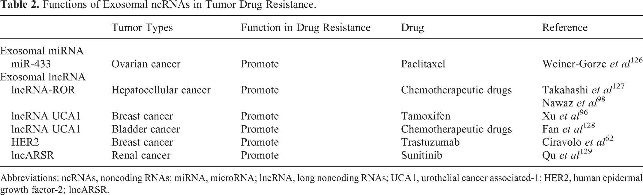
Abbreviations: ncRNAs, noncoding RNAs; miRNA, microRNA; lncRNA, long noncoding RNAs; UCA1, urothelial cancer associated-1; HER2, human epidermal growth factor-2; lncARSR.
A study suggests that the acid microenvironment of the tumor–host interface may stimulate the output of exosomes, 131 and such exosomes could release several contents that transport a resistance phenotype to sensitive tumor cells by changing cell cycle control and stimulating antiapoptosis programs. 132 It has also been proposed that EMT inducers can increase resistance to chemotherapy. 133 Bigagli et al 114 proved the chemosensitivity of metastatic cells undergoing EMT was diminished compared to adherent HCT-8 colon cancer cells, since exosomes secreted from HCT-8 cells might impact the EMT program and made the metastatic cells spontaneously insensitive to chemotherapeutic strategies. There is also a deduction that vesicles such as exosomes are able to move between cells using specific structures called nanotubes. 133 However, it is not clear which of these theories may be correct.
Therapeutic potential of exosomes in drug resistance of cancer
Treatment with locked nucleic acids targeting exosomal lncRNA and miRNAs can restore drug sensitivity. Through the untiring efforts of researchers, the lncRNAs and miRNAs are considered as putative targets for cancer therapy via exosome-mediated mechanism, such as lncRNA-H19 and lnc-ARSR9. 121,134 Exosomal ncRNAs-based therapeutic protocols, such as antisense oligonucleotides, have been proved to be capable of restraining pathological lncRNAs. Therefore, these exosomal ncRNAs are inspiring and appealing for cancer therapy. In addition, there is a speculation that exosomes can deliver drugs to selective targets as a novel platform in a drug delivery system. In particular, exosomes are capable of crossing the blood–brain barrier without inducing an immune response. 135 However, a set of troubles have been suffered during the clinical development of exosomes-based therapeutic technologies for cancers, including their indefinite functions in the complex networks, the difficulty in accurate quantification of exosomal lncRNAs and miRNAs, the hard transport of several lncRNAs and miRNAs antagonists or mimics as well as the unclear clinical pharmacokinetics and drug toxicity. 39 Therefore, more studies are needed to verify the potential of applying exosomes and their contents in cancer therapy. Exosomes-based drugs are expected to enter clinical trials, and their great potential may be revealed.
Prospects and Challenges
From the above, we conclude that tumor-derived exosomes not only discard cellular waste but also trigger signaling pathways and drug resistance in target cells and facilitate the growth of metastatic cells. It is encouraging that nanomaterials or microbodies, such as exosomes-mediated delivery system, provide more possibility. Exosomes containing MAGE family member A3 (MAGE-3) peptides show minimal toxic effects after treatment in patients with stages III/IV melanoma. 135 However, small RNA-based drugs, such as miRNAs and circRNAs, face different kinds of challenges due to the lack of targeted delivery strategies in cancer-targeted therapy. 136
Switching to a wider scientific horizon, exosomes are still on initial exploration at present, and only a few molecular mechanisms about exosomes in cancer progression are discovered. In addition, the strategies and methods used for investigating exosomes are still limited and challenging. The identification methods of exosomes are diverse; for instance, the size of pelleted particles can be measured using dynamic light scattering such as Brookhaven instruments BI-9000 digital correlator (Brookhaven instruments, USA), Zetasizer Nano ZS (Malvern, UK), LM10 NanoSight (Malvern, UK), or BI200-SM goniometer (Brookhaven instruments, USA), configured with a solid-state laser tuned at 532 nm. 122,124 The morphology of exosomes is suitable to be examined under a transmission electron microscope (TEM) at 80 keV, with a diameter of approximately 30 to 100 nm under the TEM ultimately. The typical electron micrographs can be captured by a megapixel digital camera, such as Erlangshen 11 digital camera (Gatan, USA). 124 Exosomes can also be identified by Western blot analysis with exosomal surface protein markers antibodies such as anti-CD9, anti-CD63, anti-CD81, anti-CD82, and so on. 137 However, from the reported studies, we can see that the identification of exosomes is not coherent and comprehensive, which cannot avoid the occurrence of false-positive or false-negative results.
Currently, the large-scale profiles of miRNAs or lncRNAs in exosomes are usually obtained using microarrays of next-generation sequencing followed by the extraction of exosomes, which can be customized and time reduced. 138 Besides, quantitative polymerase chain reaction (PCR), digital PCR, and NanoString nCounter Gene Expression Assay (NanoString nCounter system, USA) can be used for detection of miRNAs or lncRNAs, with features of expensive and uneffective but high sensitivity. 139 Exosome protein can be isolated by optimized lysis buffer 140 and quantitated by a microBCA Protein Assay kit (Pierce, USA). When the focus is on the number of total exosomal proteins in patients with cancer, a quantitative proteomic analysis can be used. 141
In conclusion, more details regarding the functional contents of exosomes involved in cancer progression is highly desired, and exploring their biological mechanism will improve the clinical applicability of exosomes in cancer diagnosis and therapy.
Footnotes
Authors’ Note
Chengcheng Zhang, Qing Ji, and Yue Yang contributed equally to this work.
Declaration of Conflicting Interests
The author(s) declared no potential conflicts of interest with respect to the research, authorship, and/or publication of this article.
Funding
The author(s) disclosed receipt of the following financial support for the research, authorship, and/or publication of this article: This work was supported by International Cooperation Key Project of National Natural Science Foundation of China (81520108031) and National Natural Science Foundation of China (81573749 and 81473478), Key Project of Shanghai Municipal Science and Technology Commission (16401970500), Program of Shanghai Academic Research Leader (16XD1403600), Program for Shanghai Outstanding Medical Academic Leader, Natural Science Foundation of Shanghai, China (16ZR1437700), and Research Project for Practice Development of National TCM Clinical Research Bases (JDZX2015067).