
Abstract
This study focused on fabrication of lead-free PVDF electrospun nanofibers with enhanced piezoelectric properties and reduced fiber diameter, achieved by incorporating ZnO nearly spherical nanoparticles (NSNs). Incorporation of ZnO NSNs at different concentrations (0–15 wt.%) progressively decreases fiber diameter from 705 nm to 200 nm and increases β-phase content from 58.90% to 90.07%, as confirmed by FE-SEM and FTIR analyses. XRD results also revealed that the addition of ZnO NSNs enhances the electroactive β-phase content and crystallinity of PVDF nanofibers, confirming successful ZnO incorporation. These structural enhancements transformed into a significant rise in piezoelectric performance, which was evaluated by measuring the generated output voltage by the nanofibers under applied pressure using a custom-built device, revealing a significant improvement reaching 1100 µV at 15 wt.% ZnO NSNs, over a 200% increase compared to pristine PVDF. The enhancements are attributed to inherent piezoelectricity of ZnO NSNs and their ability to modulate the dielectric properties of the solution, promoting enhanced jet stretching and β-phase nucleation during electrospinning. The resulting nanofibers demonstrate promising potential for applications in energy harvesting and wearable electronics.
Introduction
In recent years, advances in science and technology have increasingly focused on clean, sustainable, and renewable energy sources as alternatives to traditional energy production methods. Researchers are actively investigating various natural energy resources; to harness these resources effectively, it is essential to develop innovative tools and devices capable of generating such energy. Among the promising materials in this field are piezoelectric materials, which can convert mechanical energy into electrical energy and vice versa, making them valuable for energy harvesting applications.1–3
Piezoelectric materials, including ceramics, polymers, and their composite structures, play a crucial role in various advanced technological applications. Piezoelectric ceramics, such as lead zirconate titanate (PZT), are known for their exceptionally high piezoelectric coefficients, which make them particularly suited for high-performance sensors and actuators. Nevertheless, these ceramics exhibit brittleness and lack mechanical flexibility, limiting their scope in applications requiring flexible and durable materials. 4 On the other hand, piezoelectric polymers, such as polyvinylidene fluoride (PVDF), exhibit notable advantages, including flexibility, low density, and ease of fabrication, which make them highly suitable for applications in wearable electronics and biomedical devices. 5 Despite these advantages, the piezoelectric performance of polymers is generally inferior to that of ceramics. Piezoelectric composite materials merge the distinct strengths of both ceramics and polymers, resulting in materials that demonstrate improved electromechanical properties, enhanced mechanical flexibility, and the customizability of properties through diverse connectivity configurations. 6 These piezoelectric composites are advantageous as they achieve a balance between superior piezoelectric performance and mechanical durability, making them appropriate for a diverse array of applications, including medical imaging, energy harvesting, and beyond. 7
Fiber-shaped piezoelectric composites have observed significant attention in recent years due to their exceptional combination of flexibility, durability, and high piezoelectric performance. These materials can be woven into fabrics or embedded within flexible electronic systems, making them ideal for wearable devices and smart textiles. 8 Their high efficiency in converting mechanical energy into electrical energy has unlocked novel opportunities for energy harvesting applications, particularly in portable and wearable electronic devices. 9 There is a significant body of research on the fabrication of piezoelectric composite fibers for energy harvesting, as documented in the literature. For instance, Yuan et al. fabricated fiber films of PVDF embedded with PZT using the electrospinning process. 10 In another study, Chamankar et al. investigated PVDF nanocomposite fibers doped with PZT particles for energy harvesting applications. 11 Since lead is a component of PZT, there has been a global effort to develop lead-free piezoelectric materials to replace PZT in various applications, driven by environmental and health concerns.12,13 Thus, researchers have focused their efforts on developing lead-free piezoelectric composites by replacing PZT with alternative piezoelectric materials, such as barium titanate (BaTiO3), to fabricate PVDF-BaTiO3 piezoelectric composite fibers.14–16
Another of the few highly regarded lead-free piezoelectric ceramics is zinc oxide (ZnO), which has been demonstrated to be environmentally safe for use.17,18 ZnO is a multifunctional material known for its outstanding piezoelectric properties, making it suitable for applications in energy harvesting, sensors, and nanogenerators. 19 The enhanced piezoelectric properties of ZnO at the nanoscale, as opposed to its bulk form, establishes it as a promising candidate for lead-free piezoelectric materials. 18 PVDF is a semicrystalline polymer recognized for its excellent piezoelectric performance, thermal stability, and mechanical robustness. PVDF can be processed into various forms, such as films and fibers, enabling its use in applications including sensors, actuators, energy harvesting systems, and biomedical engineering. 20 Its remarkable piezoelectric characteristics make it particularly advantageous for self-powered devices and flexible wearable technologies. 21 As a matter of fact, recent research efforts have concentrated on PVDF-ZnO piezoelectric composite fibers, as their nontoxicity, breathability, and flexibility make them highly desirable for modern technological applications.22–24
Among various fiber fabrication techniques, electrospinning stands out as an innovative and highly adaptable method for producing fibers from polymer solutions, yielding fibers with diameters ranging from a few nanometers to several micrometers. The process begins when a high voltage is applied to the polymer solution, creating an electric field that surpasses the surface tension of the liquid, resulting in the formation of a structure known as a Taylor cone at the tip of a needle. As the charged polymer jet is ejected from this cone, it undergoes stretching and thinning, driven by electrostatic forces and whipping motions, ultimately depositing onto a collector surface. This versatile technique enables the fabrication of fibers with varied shapes and orientations, making it highly applicable across diverse applications, such as biomedical engineering—where it serves as a basis for tissue scaffolds—as well as in filtration systems and energy storage devices. Furthermore, electrospinning is capable to produce composite fibers by integrating functional materials, thereby expanding its potential in advanced technological applications.25,26 Moreover, electrospinning is a highly advantageous method for fabricating piezoelectric fibers, as the high-voltage electric field and mechanical tension induced by the stretching of the solution jet during fiber formation contribute to in-situ poling and the generation of piezoelectric properties in nanofibers. This process eliminates the need for additional polarization steps, which are typically required in conventional fiber fabrication techniques. 27
Several research articles have studied incorporating BaTiO3 and/or ZnO nanostructures into PVDF electrospun nanofibers, resulting in significant enhancements in the properties of nanofibers. Studies indicate that adding 5 to 15 wt.% of BaTiO3 nanoparticles leads to increase in β-phase content (e.g. from 59.38% in pristine PVDF nanofibers to 71.65% in PVDF-BaTiO3 nanofibers), a reduction in fiber diameter (e.g. from ∼181 nm at 5 wt.% to ∼161 nm at 15 wt.% of BaTiO3), and substantial improvement in piezoelectric output. FTIR and XRD analyses confirm that BaTiO3 promotes the α-phase to β-phase transformation effectively, attributed to strong interactions between BaTiO3 and PVDF chains.16,28 The incorporation of ZnO nanorods (NRs) has been shown to significantly facilitate the transformation of PVDF from the non-polar α-phase to the electroactive β-phase, primarily due to its one-dimensional, rod-like geometry that promotes orientational crystal interfaces and strong dipole interactions with the PVDF chains.29–31 For example, the β-phase content in PVDF/ZnO NR composite nanofibers can reach up to 90.7%, compared to 61.3% in pristine PVDF, and the piezoelectric output can increase dramatically. 31 However, in terms of morphology, the addition of ZnO NRs can reasonably increase the diameter of nanofibers. 31 Even when ZnO NRs are well-dispersed within the PVDF matrix, studies show that the resulting nanofibers maintain diameters comparable to or only slightly larger than pristine PVDF nanofibers.29,32
In this study, lead-free PVDF-ZnO composite piezoelectric electrospun nanofibers with ZnO nearly spherical nanoparticles (NSNs) have been fabricated. This study aims to investigate the potential effects of ZnO NSNs addition on decreasing the average diameter of PVDF nanofibers, leading to higher β-phase content and superior piezoelectric properties. ZnO NSNs tend to have lower piezoelectricity than ZnO nanorods, 33 thus our investigation focuses to enhance the piezoelectric performance of PVDF electrospun nanofibers through the incorporation of ZnO NSNs, utilizing the synergistic effects arising from the reduction in fiber diameter and the intrinsic piezoelectric properties of ZnO. Therefore, PVDF-ZnO(NSNs) piezoelectric composite nanofibers that can be used in energy harvesters and wearable electronic devices were fabricated in this study.
Materials and methods
Polyvinylidene fluoride powder (Kynar® 761, molecular weight ∼440,000 g/mol, ARKEMA, France) was selected as the polymeric matrix and ZnO nearly spherical nanoparticles (ZnO NSNs, average particle size: 10–30 nm, US Research Nanomaterials, Inc., United States) were used as the ceramic additive. N, N-dimethylformamide (DMF, Merck, Germany) was employed as the solvent.
Description and composition of each sample.

The morphological analysis of nanofibers performed using Field Emission Scanning Electron Microscopy (FE-SEM; MIRA3 TESCAN-XMU, Czechia Republic), and the diameter of nanofibers was measured by image analysis software Digimizer v6.4. Fourier Transform Infrared Spectroscopy (FTIR; Perkin-Elmer Spectrum RXI FT-IR Spectrometer, United States) and X-ray diffraction (XRD; STOE STAD IP, Germany, by Cu Kα radiation with λ = 1.54 Å) were conducted to evaluate the β-phase formation in the nanofibers. The piezoelectric properties of the samples were evaluated based on their voltage output in response to applied pressure, using a device known as piezo-tester (Advanced Materials and Technologies in Textiles Research Center, Department of Textile Engineering, Amirkabir University of Technology, Iran). The piezo-tester operates by applying mechanical loads to fibrous piezoelectric sensors, enabling the assessment of their sensitivity by measuring the electrical response generated under varying pressure intensities. The piezo-tester records the output voltage as the applied pressure increases at a consistent rate, allowing for the determination of sensor sensitivity and response time. This systematic approach provides an accurate characterization of the performance metrics of piezoelectric devices under diverse mechanical stimuli (Figure 1-f). A comprehensive explanation of the piezo-tester has been reported elsewhere.
34
Fabrication process and piezoelectric performance testing of nanofibers: (a) Solution preparation, (b) dispersion process, (c) ultrasonic bath, (d) electrospinning process, (e) real-life fabricated nanofibers, (f) piezo-tester device, and (g) design of the nanofiber energy harvester. “Created with BioRender.com”.
Results and discussions
FE-SEM images in Figure 2 show the well-interwoven structure of PVDF nanofibers and PVDF-ZnO(NSNs) composite nanofibers, fabricated in a felt-like configuration (Felt refers to a type of nonwoven fabric formed through stitching and needle-punching processes, widely used for various applications.
35
). Figure 2 and Table 2 demonstrate that incorporating ZnO NSNs and increasing their concentration in nanofibers decreases the average diameter of nanofibers. Specifically, the average diameters of PVDF (Figure 2-a), PZ5 (Figure 2-b), PZ10 (Figure 2-c), and PZ15 (Figure 2-d) nanofibers are 705 nm, 270 nm, 240 nm, and 200 nm, respectively. Since all electrospinning parameters remain constant across samples, this reduction in fiber diameter attributes solely to the addition of ZnO NSNs. Similar observations regarding fiber diameter reduction in polymer-based piezoelectric nanofibers following the incorporation of ceramic nanoparticles have been reported by other researchers.36,37 FE-SEM images of (a) PVDF, (b) PZ5, (c) PZ10, and (d) PZ15 indicating decrease of diameter of the PVDF-ZnO(NSNs) nanofibers compared to pristine PVDF nanofibers. Descriptive statistics (Avg. ± SD, Min, Max) of the diameter of nanofibers measured by Digimizer v6.4.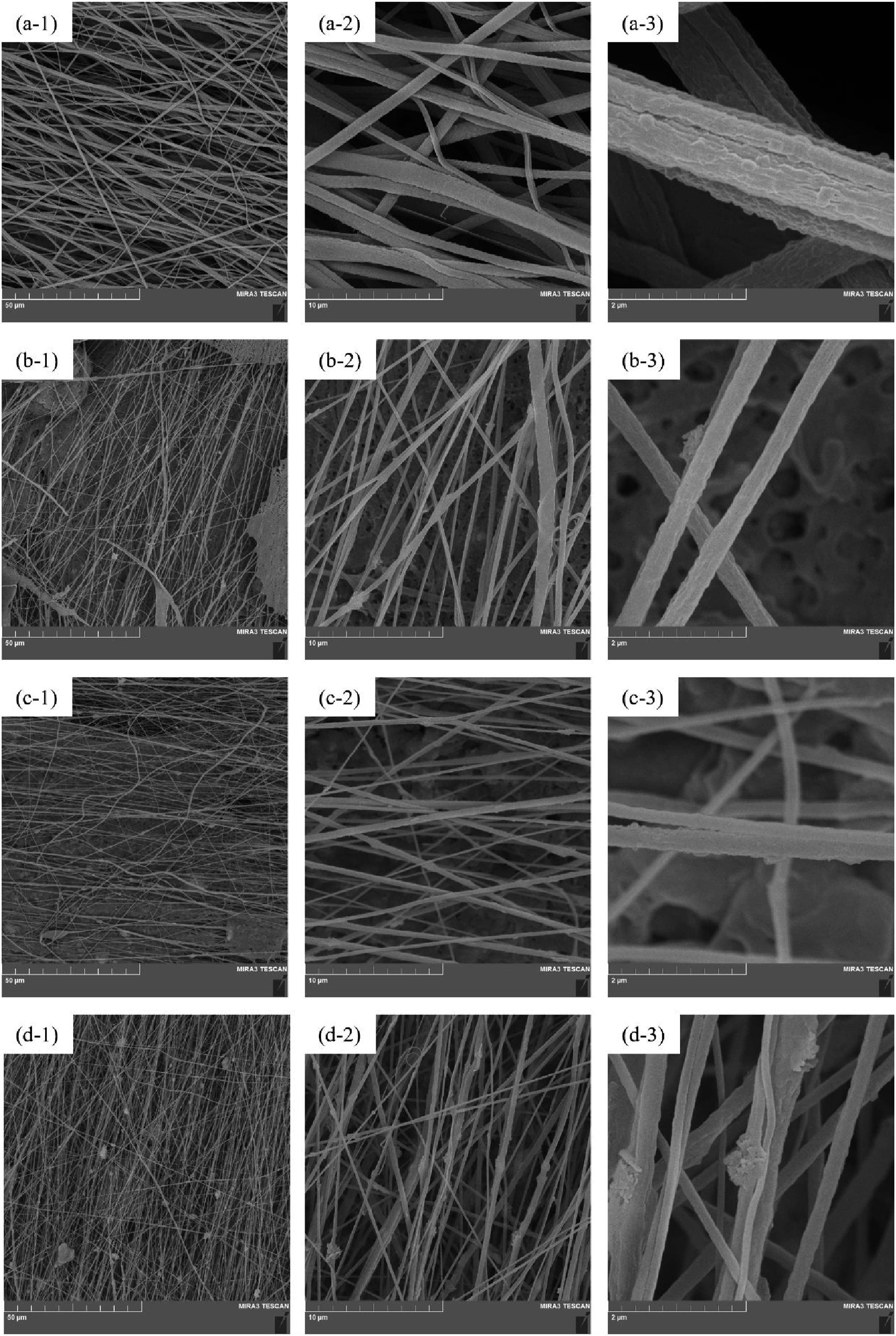

According to Mahboubizadeh et al. the incorporation of ceramic particles into a polymer solution can have a significant impact on the dispersion and viscosity of the solution. As the quantity of ceramic particles increases, the viscosity of the solution will also increase. This, in turn, can lead to the production of finer fibers due to enhanced stretching and thinning of the polymer jet through the process of electrospinning. 16 Furthermore, reports in literature claim that incorporating ceramic particles into the polymer solution can modify the chain alignment and stretching during electrospinning, ultimately resulting in fibers with a reduced diameter. 38
Electrospinning is a voltage-driven process in which a high electric field is applied between a polymer solution at the tip of a needle (nozzle) and a grounded collector, causing the solution to form a Taylor cone and eject a charged jet that stretches and solidifies into nanofibers as it travels toward the collector. If the conductivity of the electrospinning solution is excessively high, the rapid movement of charges can neutralize the potential difference between the nozzle and collector, thereby inhibiting the formation of a stable Taylor cone and preventing successful fiber formation, regardless of further increases in applied voltage. Conversely, when the solution exhibits lower conductivity, the charge accumulation and electrostatic repulsion at the nozzle are optimized, promoting efficient jet formation and stretching under the applied electric field.
The incorporation of ZnO NSNs, which are semiconducting in nature, into the PVDF solution increases the dielectric constant and effectively reduces the electrical conductivity of the solution. This modulation of dielectric properties enhances the electrostatic stretching forces acting on the polymer jet, facilitating greater stretching and thinning of the fibers as they travel toward the collector. As the concentration of ZnO NSNs increases, the dielectric constant of the solution further rises, resulting in even more jet stretching and the formation of finer nanofibers.
Some researchs indicated that a reduction in the average diameter of piezoelectric fibers is associated with an enhanced presence of the β-phase and increased piezoelectricity.16,27,39 Higher and better jet stretching during electrospinning, due to the incorporation of ZnO NSNs, aligns PVDF chains, transforming the non-polar α-phase into the polar β-phase, which enhances piezoelectric properties.
Moreover, as FE-SEM images depict in Figures 2-a-1, 2-b-1, 2-c-1, and 2-d-1, the presence of beads within the fibers noticeably increases. Higher concentrations of ZnO NSNs may disrupt the uniform flow of the electrospinning jet, leading to greater bead formation within the final nanofiber structure.24,40
It is well established that uniform fibers increase the piezoelectric properties of PVDF electrospun nanofibers,27,41 and bead formation in PVDF electrospun nanofibers can negatively affect their piezoelectric properties by disturbing the uniform alignment of polymer chains and reducing the effective area for dipole orientation, which are essential for efficient piezoelectric response.27,42 Beads may also act as structural defects, causing non-uniform stress distribution and localized charge trapping during mechanical deformation.27,30
Nevertheless, in PVDF-ZnO(NSNs) nanofibers, the addition of ZnO NSNs not only increases the β-phase content of PVDF due to higher stretching of the solution during electrospinning but also introduces additional piezoelectric active sites due to the intrinsic properties of ZnO.30,43 The synergistic effect of enhanced β-phase and interfacial polarization from ZnO NSNs can offset the negative impact of bead structures, resulting in a net improvement in piezoelectric performance. This suggests that, at optimal ZnO NSNs concentrations, the benefits of increased β-phase and ZnO-induced piezoelectricity outweigh the drawbacks associated with bead formation.
Energy dispersive spectroscopy (EDS) mapping was utilized to verify the incorporation of ZnO NSNs within the PVDF-ZnO(NSNs) electrospun nanofibers. The EDS maps in Figure 3 clearly show the presence and distribution of zinc (Zn) and oxygen (O) signals in the PVDF-ZnO(NSNs) composite nanofibers (Figures 3-b, 3-c, and 3-d), whereas no Zn or O signals are detected in the pristine PVDF nanofibers (Figure 3-a), confirming successful ZnO loading. However, it is important to note that EDS spatial resolution is limited by the interaction volume of the electron beam, which typically extends to approximately 1 μm under standard operating conditions.44,45 Given that the diameters of electrospun nanofibers are generally in the range of a few hundred nanometers, the EDS signal arises from multiple fibers and possibly the substrate within this interaction volume, limiting the accuracy of elemental mapping at the individual nanofiber scale.
46
Despite this limitation, the combined FE-SEM and EDS analyses provide qualitative confirmation of ZnO distribution in the composite nanofibers, with increasing ZnO content correlating to stronger Zn and O signals. EDS maps of (a) PVDF, (b) PZ5, (c) PZ10, and (d) PZ15 nanofibers indicating the detection and distribution of Zn and O signals within PVDF-ZnO(NSNs) nanofibers and no detection within pristine PVDF nanofibers.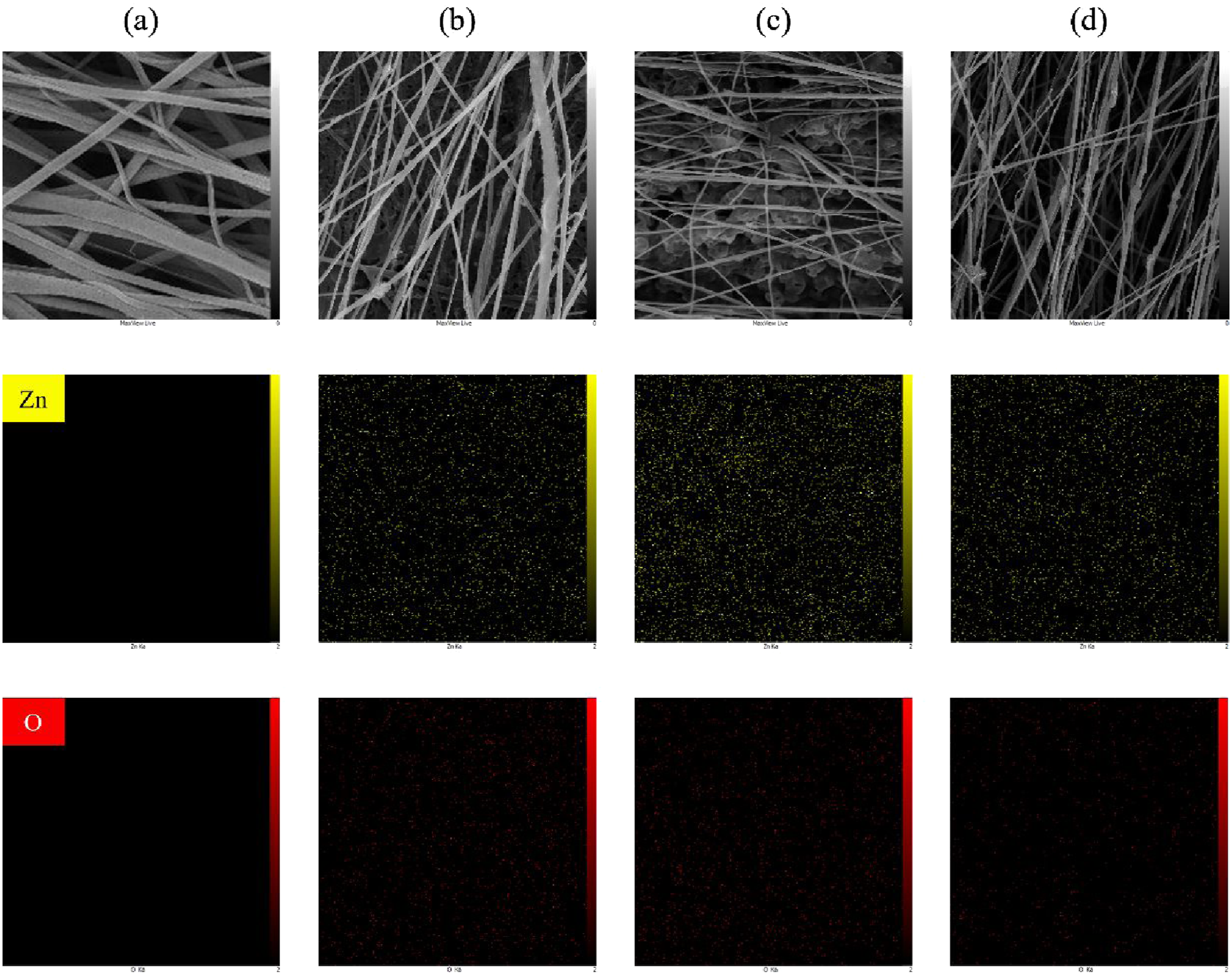
As Figure 4 demonstrates, FTIR was performed to study the content of β-phase created in the nanofibers. The α, β, and γ phases are three crystalline polymorphs exhibited by PVDF depending on the processing method of it.47,48 Table 3 lists the different absorption bands in α and β phases of PVDF in FTIR spectra. The β-phase has the maximum dipole moment per unit cell in PVDF, thus representing as the phase responsible for piezoelectricity of PVDF.1,51 Normalized FTIR spectra for PVDF, PZ5, PZ10, and PZ15 indicating the increase in β-phase content in PVDF-ZnO(NSNs) nanofibers compared to pristine PVDF nanofibers.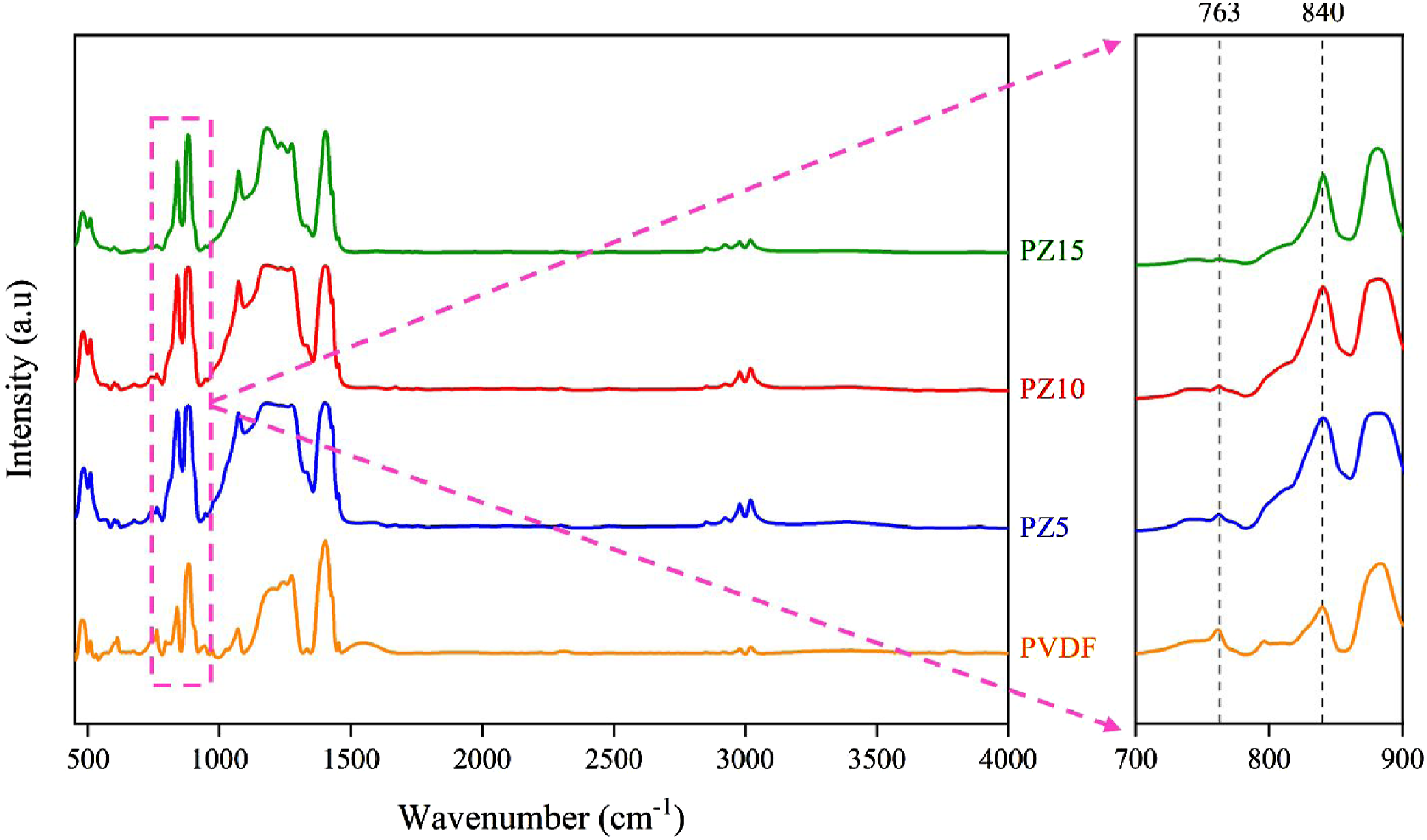

The β-phase of PVDF is a polar crystalline polymorph characterized by an all-trans planar zigzag conformation of the molecular chains, which results in a net dipole moment along the polymer chains. This molecular alignment imparts significant piezoelectric, pyroelectric, and ferroelectric properties to the β-phase of PVDF, making it highly favorable for applications in energy harvesting, sensors, and actuators. 52 In contrast, the α-phase of PVDF, which is the most commonly occurring polymorph, exhibits a non-polar trans-gauche conformation with antiparallel dipole arrangements that produce no net dipole moment, thereby resulting in negligible piezoelectric activity.52,53

Calculated relative fraction of β-phase in nanofibers admitting increase in β-phase content in PVDF-ZnO(NSNs) nanofibers compared to pristine PVDF nanofibers.
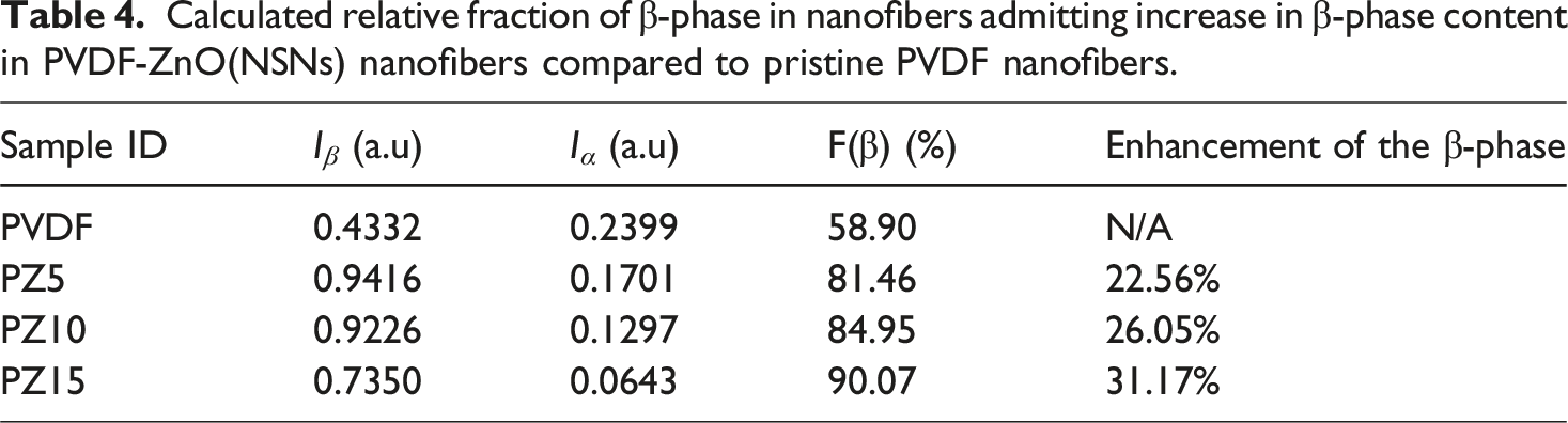
Studies have reported the increase in the β-phase content of PVDF electrospun nanofibers by incorporation of ZnO nanoparticles into them.24,29,40 For instance, Bafqi et al. enhanced the β-phase content of PVDF nanofibers, from 80% to 87%, achieved through incorporating ZnO nanoparticles. 23 In this study, as detailed in Table 4, the β-phase content of the PVDF, PZ5, PZ10, and PZ15 nanofibers are 58.90%, 81.46%, 84.95%, and 90.07%, respectively. Incorporating ZnO NSNs into PVDF nanofibers shows significant enhancement of β-phase by 22.56%, 26.05%, and 31.17%, for PZ5, PZ10, and PZ15 nanofibers, respectively.
This incorporation promotes the alignment and crystallization of PVDF nanofibers, thereby enabling a more efficient transition from the non-polar α-phase to the polar β-phase. The presence of ZnO NSNs within PVDF nanofibers enhances the molecular orientation of PVDF polymer chains, which is critical for the formation of the β-phase. ZnO NSNs serve as nucleating agents, improving the alignment and development of the β-phase during the crystallization process of PVDF nanofibers. At higher concentrations of ZnO NSNs, an increased number of nucleation sites is available, further enhancing the crystallization of PVDF nanofibers into the β-phase.
Furthermore, the higher stretching of PVDF solution during electrospinning significantly enhances β-phase formation through molecular chain alignment and electric field-induced polarization. When the polymer jet is stretched, the PVDF chains undergo reorientation, transforming from the non-polar α-phase to the electroactive β-phase, which is eventually responsible for piezoelectric behavior in the PVDF nanofibers. Stretching of the solution thins the nanofibers, which correlates with higher β-phase content. For instance, some researchers reported that increasing applied voltage of electrospinning process from 15 kV to 20 kV reduced fiber diameters while boosting β-phase content.54,55 Therefore, the thinner nanofibers resulting from the incorporation of semiconducting ZnO NSNs and increasing the concentration, contribute to the formation of a higher β-phase due to effective alignment of molecular chains, as a result of better stretching during the electrospinning process.
Rather than numerical comparison, Figure 5 demonstrates the decline of α-phase intensity peaks and rise of β-phase intensity peaks. In FTIR spectra of PVDF-ZnO(NSNs) nanofibers, no α-phase intensity peaks at 530 cm-1, 615 cm-1, and 795 cm-1 have been found, in contrast to PVDF nanofibers which show peaks in the mentioned absorption bands. The α-phase intensity peak at 975 cm-1 starts to decrease after incorporation of ZnO NSNs, until no peak is found in the PZ15 nanofibers. The decline of α-phase intensity peak at 763 cm-1 is also observable from Figure 5. Conversely, PVDF-ZnO(NSNs) nanofibers indicate higher β-phase intensity peaks at 510 cm-1, 840 cm-1, 1175 cm-1, and 1275 cm-1 in comparison to PVDF nanofibers. As a result of enhanced β-phase in PVDF nanofibers, it can be concluded that the piezoelectric performance of PVDF nanofibers will improve with increasing incorporation of ZnO NSNs. α-phase and β-phase intensity peaks in normalized FTIR spectra for PVDF, PZ5, PZ10, and PZ15 demonstrating the increase of β-phase and decrease of α-phase peaks in PVDF-ZnO(NSNs) nanofibers compared to pristine PVDF nanofibers.
The XRD patterns of electrospun nanofibers comprising pristine PVDF, PZ5, PZ10, and PZ15 are presented in Figure 6. The pristine PVDF nanofibers exhibit characteristic diffraction peaks at 2θ = 17.7°, 18.4°, and 19.9°, which correspond to the (100), (020), and (110) planes of the α-phase, respectively. Additionally, a prominent peak at 20.6° is observed, attributed to the (110)/(200) planes of the electroactive β-phase of PVDF.52,56,57 Upon incorporation of ZnO NSNs, new diffraction peaks emerge at 2θ = 31.7°, 34.2°, 36.2°, 47.2°, 56.1°, 62.2°, and 67°, which are indexed to the (100), (002), (101), (102), (110), (103), and (112) planes of hexagonal wurtzite ZnO, respectively (JCPDS Card No. 36-1451). The intensity of these ZnO-related peaks increases progressively from PZ5 to PZ15, confirming both the successful incorporation and the increasing crystallinity of ZnO NSNs within the PVDF matrix. Notably, the intensity of the β-phase peak at 20.6° increases with higher ZnO content, while the α-phase peaks at 17.7°, 18.4°, and 19.9° decrease in relative intensity. This trend indicates that ZnO acts as an effective nucleating agent for the β-phase, promoting the transformation from the non-polar α-phase to the electroactive β-phase as the ZnO concentration increases. The peak at 36.2°, present in all ZnO-containing samples, is primarily assigned to the (101) plane of ZnO; however, a minor contribution from a higher-order β-phase PVDF reflection cannot be excluded. The overall enhancement in peak sharpness and intensity with increasing ZnO content also suggests improved crystallinity and phase purity in the composite nanofibers. Collectively, these results demonstrate that the addition of ZnO NSNs into electrospun PVDF nanofibers not only introduces crystalline ZnO phases but also significantly enhances the electroactive β-phase content of PVDF, while suppressing the α-phase. This synergistic effect is most pronounced in PZ15, indicating that higher ZnO loading is particularly effective for tailoring the crystalline structure and functional properties of PVDF-based nanofibers for piezoelectric applications.58,59 XRD patterns of PVDF, PZ5, PZ10, and PZ15 electrospun nanofibers showing the evolution of α-phase and β-phase PVDF peaks, as well as the emergence and intensification of ZnO diffraction peaks with increasing ZnO content, demonstrating an increase in β-phase intensity and a decrease in α-phase peaks for PVDF-ZnO(NSNs) nanofibers compared to pristine PVDF, confirming enhanced β-phase formation and successful incorporation of crystalline ZnO NSNs.
The piezoelectric performance of the nanofibers is determined by measuring the output voltage signals in response to an external force. This measurement is conducted under stress conditions by applying a pressure force of 2.6 N at a frequency of 5 Hz, using a hand-made device. In addition to the piezoelectric response, the electrical sensitivity of nanofibers is calculated through equation (2).
Calculated piezoelectric properties of nanofibers.


(a) Piezoelectric performance of nanofibers showing higher piezoelectric properties for PVDF-ZnO(NSNs) nanofibers and (b) average diameter of nanofibers – piezoelectric performance diagram indicating higher piezoelectric properties as a result of decrease in the diameter of nanofibers.
Conclusions
In this study, PVDF nanofibers embedded with ZnO NSNs were successfully fabricated via electrospinning (PVDF-ZnO(NSNs) nanofibers), with the aim of enhancing piezoelectric performance of PVDF electrospun nanofibers through the incorporation of ZnO NSNs, through synergistical effects of the reduction in diameter of nanofibers and the inherent piezoelectric properties of ZnO. Unlike ZnO nanorods, which have been reported to increase the diameter of nanofibers due to their anisotropic structure and tendency to aggregate,61–63 the incorporation of ZnO NSNs significantly decreases the average diameter of nanofibers from 705 nm for pristine PVDF to 270 nm, 240 nm, and 200 nm for 5, 10, and 15 wt.% loading of ZnO NSNs, respectively. While the addition of 5, 10, and 15 wt.% ZnO NSNs reduces the average diameter of nanofibers due to increased dielectric constant and less electrically conductive of solution, creates better stretching of it during electrospinning, which leads to better chain alignment and the β-phase content increases from 58.90% to 81.46%, 84.95%, and 90.07%, respectively, observed from FTIR results. Moreover, XRD analysis confirms that the incorporation of ZnO NSNs into PVDF electrospun nanofibers significantly enhances the electroactive β-phase content while suppressing the α-phase, with increasing ZnO concentration promoting higher crystallinity and successful integration of crystalline ZnO within the polymer matrix. This morphological refinement and phase transformation results in a considerable enhancement of the piezoelectric response, with output voltage increasing by 45.63%, 159.56%, and 200.55% for 5, 10, and 15 wt.% PVDF-ZnO(NSNs) nanofibers compared to pristine PVDF nanofibers. Although PVDF-BaTiO3 composites and electrospun nanofibers are highly effective and reliable for piezoelectric applications, they often require high filler concentrations (above 20 wt.%) to achieve optimal performance, which can lead to agglomeration and processing challenges,17,64–66 while PVDF-ZnO(NSNs) nanofibers show significant improvement of piezoelectric response at lower than 20 wt.% concentration. For instance, in our previous work, 67 incorporating 5 wt.% of BaTiO3 particles increased the piezoelectric response of PVDF nanofibers from 366 µV to 383 µV, which is 1.39 times lower than incorporating 5 wt.% of ZnO NSNs according to the findings of current study. Rather than piezoelectric performance, ZnO NSNs are advantageous in biocompatibility, making them preferable for biomedical applications. It can be concluded from this study that PVDF-ZnO(NSNs) electrospun nanofibers perform as promising piezoelectric composites, which can be applicable in energy harvesting, sensors, and wearable electronics.
This study demonstrates that incorporation of ZnO NSNs concentrations up to 15 wt.% effectively enhances the properties of PVDF electrospun nanofibers. Although not exceeding 15 wt.% is to avoid nanoparticle agglomeration and maintain fiber uniformity, future work will investigate higher ZnO NSNs loadings facilitated by surfactant-assisted dispersion techniques to further improve the functionality of nanofibers. Moreover, exploring advanced fabrication techniques such as 3D-printing of wrap and weft nanostructures of PVDF-ZnO(NSNs) composites will be under investigation. To ensure the reliability of PVDF-ZnO(NSNs) electrospun nanofibers in real-world applications in energy harvesting and wearable electronics, assessing the long-term stability of them including mechanical durability under cyclic loading, environmental stability, and scalability will be evaluated.
Footnotes
Funding
The authors received no financial support for the research, authorship, and/or publication of this article.
Declaration of conflicting interests
The authors declared no potential conflicts of interest with respect to the research, authorship, and/or publication of this article.
Data Availability Statement
The datasets generated during and analyzed during the current study are available from the corresponding author upon reasonable request.