
Abstract
Electrospun nanofiber web were developed to examine the feasibility of developing chemical and warfare agent detector textile materials. Some parameters of nanofiber mats, including the polymer type, time of electrospinning and the polymer solution concentration, were designed to change and pH detection properties of specimens were compared. Multi-layered electrospun nanofiber mats equipped with pH sensitive dyes showed good performance in pH detecting property that opens up new possibilities for using in the protective garment application as the chemical and warfare agent detectors. It was also found that the developed electrospun fibers due to their porous structure and fiber diameter distribution are stable for chemical warfare detection in the form of gas.
Introduction
It is essential to be aware about different chemical substances, which are used in many contexts of industries and have considerable effect on environment and human's life. The first step to decrease destructive effects of being in contact with these types of chemical material is recognizing and knowing about their existence. Among several methods for recognizing these ingredients, the concept of colorimetric detection has gained much interest in academic world. In this method, the act of response between chemical ingredient and the used material in sensors cause the color change. Colorimetric detection is a wet chemistry technique formulated to indicate the presence of a chemical agent by a chemical reaction that causes a color change when agents come into contact with certain solutions or substrates [1].
There are many different triggers which can cause a color change in the fibrous materials. The most known chameleon fibrous materials have temperature and light as stimulus for the color change and are designated as thermochromic and photochromic systems [2,3]. The degree of acidity is, however, an important parameter in daily life and a pH-sensitive sensor could, therefore, be very useful. The most common colorimetric detectors come in the form of detection tubes, papers or tickets, which can detect nerve, blister and blood agents [1]. The application of pH-sensitive dyes to textile materials leads to flexible and easy-to-handle sensors that are able to give the first, immediately visible signal. Synthetic polymer systems with very similar attributes are often prepared for a broad range of applications, such as responsive bio interfaces that are functionally similar to natural surfaces; controlled drug-delivery and release systems; coatings that are capable of interacting with and responding to their environment; composite materials that actuate and mimic the action of muscles; and thin films and particles that are capable of sensing very small concentrations of analyses. In the last decade, smart textiles have become the subject of many studies all over the world. Within the group of smart textiles, a growing interest in color change materials, or so-called chameleon textiles, is recognized. There are several methods to identify these chemical ingredients. Every method has some advantages and disadvantages [1–5].
An effective method must have some parameters such as detection capability, selectivity, sensitivity, response time and portability. One of the approaches for improving the detection capability of pH sensors could be increasing the surface area of the sensor. Nanofibers, due to their fascinating properties such as porous structure, interconnectivity of pores between fibers and large surface area, is one of the interesting materials as a pH sensor. Among all approaches for producing the nanofiber mats, electrospinning have received large attention in many different fields [6–13]. Electrospinning as an effective and promising technique for the production of nanofibers provides a mat of extremely fine fibers with small pore size and high surface area. Electrospun nanofibers may have a broad application in composite nonwoven structures in traditional markets. The small fiber diameter and porous structure of the nanofiber mats gives rise to a large specific surface area. This is advantageous in a wide variety of applications such as high-performance filters, scaffold in tissue engineering, separation membranes, reinforcement in composite materials, templates for the preparation of functional nanotubes and many others [2,6–14]. One of the main attractive specifications of electrospun mats in the area of pH-sensitive materials could be the direct application of electrospun multi-layered mats to garments. Up to now, different types of pH detectors have introduced for different applications [15–18], but direct application of pH-sensitive electrospun webs to garment systems would eliminate costly manufacturing steps and solve seam-sealing problems that have been limiting factors in protective garments. The electrospun fibrous structure has shown some unique characteristics such as an extremely high surface area, high porosity and small pore size. These properties can be useful in a whole range of applications, including pH detector, since exchange of liquids and gases in the environment can be done in an effective way. The small fiber diameter and porous structure of the nanofiber mats gives rise to a large specific surface area which is the suitable option for physical contact of chemical warfare and the pH detector dye within the fiber structure in such a quicker way than the normal fibers. Meanwhile, the garment using the change of its color can easily detect changes in the environmental pH. Easy usage and a quick response time are the most important advantages of this method [19–22].
The electrospinning surface is produced to be sensitive to some characteristic components such as destructive chemical ingredients. The influences of temperature and light are unavoidable. Therefore, color changing must be introduced for the whole ranges of variation. New development of color changing of textile material is incorporation of a pH-indicator dye into a nanofibrous structure. It is possible to add dying indicators into structure of polymer solution before electrospinning.
In the present study, we report our attempt to incorporate pH indicator dyes in nanocomposite fibers using electrospinning technique in a way that structural parameters of the resulted material is suitable for detecting the chemical warfares in form of gas. This paper focuses on assessing color changing of the nanofibrous mats with different structural and chemical bases with their environment change of pH. Since different polymers can be used for different applications, we used two polymers in this research: Nylon 6 (polyamide 6) and Polycaprolatone (PCL). Nylon 6 is a proper polymer for military application since it can be placed or be stitched to NBC protection clothing for visual alarming and PCL, as a biocompatible polymer, can be used as a patch for biomedical application.
Characteristics of the fabricated sensor including large specific surface area, appropriate fiber diameter and porous structure of the nanofiber mats were designed to achieve the proper performances of the sensor compared to the other forms of the polymeric sensors such as film- and paper-based sensors. Another important characteristic of fabricated sensor is the ability of using them in the protective clothing sector, since they can be easily distort during the wearing without any compelling on the comfort properties of clothing. A comparison between nanofiber pH-sensitive and dye-coated conventional fabric also was carried out to show the differences of the above-mentioned characteristics.
Materials and methods
Materials
Polycaprolactone (PCL, 80000 MW, Sigma-Aldrich, St. Louis, MO) was dissolved in chloroform/methanol (3:1) (Sigma-Aldrich, St. Louis, MO) at a concentration ranged from 14 to 20 wt% polymer in solvent. Polyamide 6 (PA) was supplied by Sigma-Aldrich. Different polyamide solutions solved in formic acid were prepared with different polymer concentrations and the optimum solution was used for further analysis. The pH-indicator dyes (C18H14N4O, MW: 302.33 g/mol, Sigma-Aldrich, St. Louis, MO), formic acid and hydrochloric acid were also supplied by Merck.
Methods
Electrospinning of nanofibers was performed in a horizontal electrospinning setup (Fanavaran Nano-Meghyas Company, Iran, Figure 1). It consisted of a syringe positioned horizontally with its needle, a precisely-controlled syringe pump, a high voltage power supply capable of 0–25 kV and a grounded collector. Upon applying a high voltage (20 kV), a fluid jet was ejected from the tip of the needle. As the jet accelerated toward a target, which was placed at 15 cm from the syringe tip, the solvent evaporated and nanofibers were collected on an aluminum foil substrate. The mass flow rate of the solutions was 0.3 mL/h for PCL and 0.5 for polyamide solutions to produce uniform nanofiber mats. Descriptions of samples with different parameters produced in this research are given in Table 1.
Schematic of electrospinning setup used to producing nanofiber mats. Description of samples used in this research.

To make the fibers pH sensor within the fibrous mat, pH-indicator dyes are blended with polymer solution for 2 h before electrospinning with different concentrations. After that aqueous dye–PCL blend solutions were electrospun to make the fibrous mats, in which dyes were confined. After a pre-study of the best volume percent of dyes in the polymer solution, 8% v/v had chosen to make the fiber enough sensitive and uniform. To evaluate the effect of nanofibers and a comparison between the nanofibers and conventional fabrics, same appropriate fabric was used and the dye was coated on one side. The microstructure and the morphology of the nanofiber mats were characterized by using scanning electron microscope (SEM, XL30, Philips Co.). All samples were gold-coated (Bal-tec SCD50 sputter coater) and the images were taken at an acceleration voltage of 20 kV. The fiber diameter was measured using image processing software (ImageJ, National Institutes of Health, USA).
Light stability and fastness of sensors were evaluated according to 105-A03:1993 standard.
Results and discussions
Morphology study of the electrospun nanofiber mats
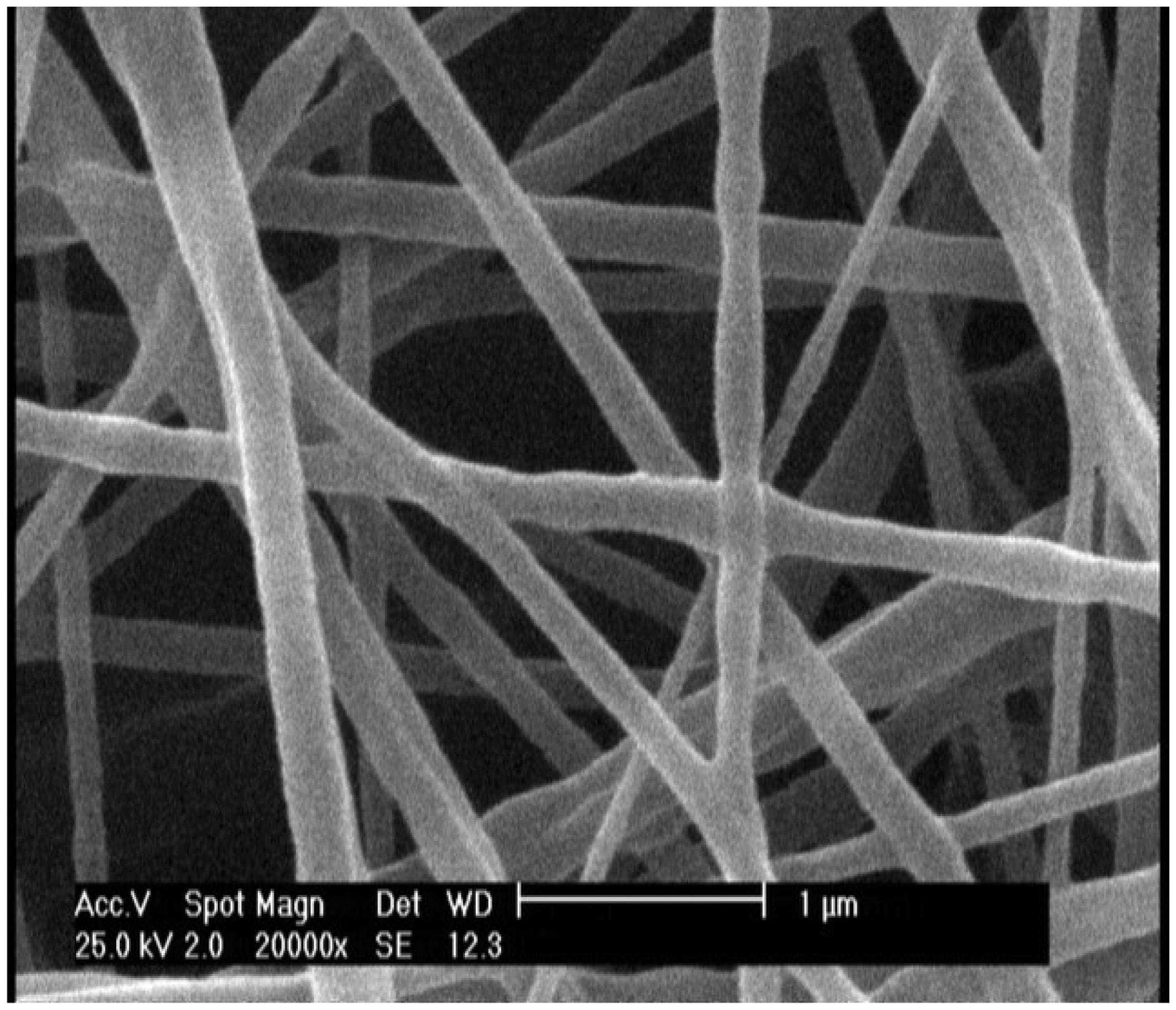
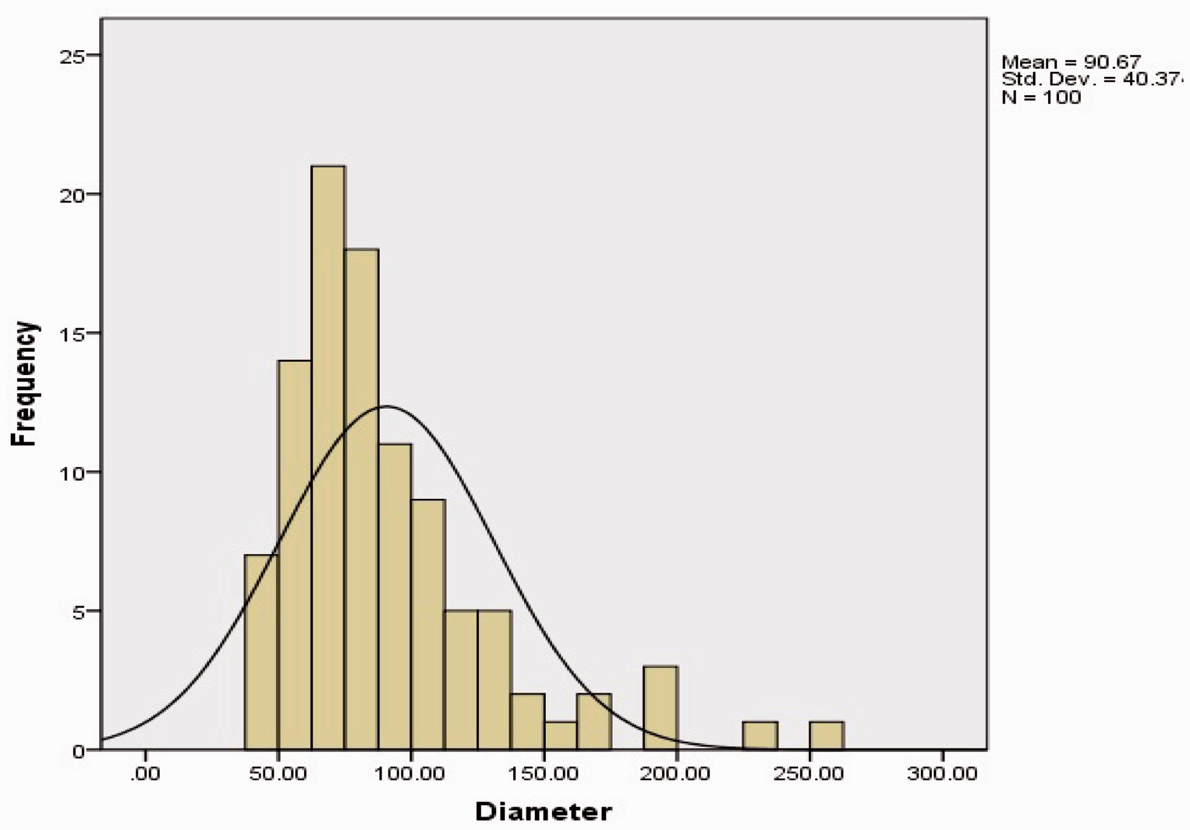
Diameters of the electrospun fibers were analyzed from the SEM images using ImageJ software. Typical SEM photographs of nanofiber mats with and without pH-indicator dyes are shown in Figures 2 and 3. Non-uniform nanofibers with different defects (such as beads and solution spray dots) were produced in some process conditions suggesting that solution or process parameters are not suited to make a uniform jet during the electrospinning, which after some parameter optimization of electrospinning process (including polymer in solution concentration, applied voltage, nozzle to collector distance) to fabricate a uniform fiber mats without any structural defects such as beads (Figure 2–9). To measure the average diameter of nanofibrous mats, 100 fibers in the SEM micrograph were selected randomly and the average value of them was considered as the average fiber diameter. The average fiber diameter of 91 nm and fiber diameter distribution shown in Figures 3 and 4 were measured for PA samples and the average fiber diameter of 87 nm and fiber diameter distribution shown in Figures 6 and 7 for PCL samples.
Typical SEM micrograph of PA nanofiber mats without pH indicator (SA3 sample without dye). Typical SEM micrograph of PA nanofiber mats with pH indicator (SA3 sample). Fiber diameter (nm) distribution for PA mats without pH indicator (SA3 sample). Fiber diameter (nm) distribution for PA mats with pH-indicator (SA3 sample). Typical SEM micrograph of PCL nanofiber mats without pH-indicator (SA7 sample). Typical SEM micrograph of PCL nanofiber mats with pH indicator (SA7 sample). Fiber diameter (nm) distribution for PCL mats without pH indicator (SA7 sample). Fiber diameter (nm) distribution for PCL mats with pH indicator (SA7 sample).


Although the pH-indicator dyes did not have much influence on the diameter of nanofibers, it could be asserted that the negative result of adding pH-indicator dyes in the solution are reduced considering the fact that by adding the pH-indicator dye the viscosity of solution was found to increase, consequently. In addition, the electrospinning process encountered some problems like having frequent droplets in front of the capillary. On the other hand, much incorporation of the pH-indicator dyes in the fiber structure expected to have more sensitivity to the change of the environmental pH. Therefore, different percentages of the pH-indicator dye were used in this study.
By comparing the results of fiber diameter distributions (Figures 4, 5, 8 and 9), it can be found that the pH-indicator dyes had the same result in two degradable and non-degradable polymers (PCL and PA polymers) and the differences are not considerable for all samples generally. Some variations were found for some samples which could be due to the changing in the electrospinning conditions because of environmental changes during the process.
Halochromic behavior of fabricated samples
The investigation of the halochromic behavior of pH indicator encapsulated nanofiber mats were performed by dropping the aqueous pH baths on the surface of the samples. Since the possibility of developing a fabric-based sensor with the same properties of normal fabric along with pH-sensory function was our main goal in this research, we only compared and optimized the structure and morphology of the nanofibrous mats with each other, although a comparison has been made with the results of conversional fabrics. Electrospun nanfibrous with incorporated pH-sensitive indicator are expected to show alternation in the environment with different pH due to their highly porous structure. This environment can be the aqua environment, liquid or a gas with high concentration which caused infection. In this research, we performed a dropping test of pH bath on the samples, which is the normal condition for testing the sensitivity of samples in comparing with the aqua and gas environment due to our limitation of the testing. Figure 10 shows the color transition observed almost instantaneously upon exposure to a drop of pH bath. Both PA and PCL samples showed a color transition with pH from orange to light blue.
Colorimetric transition of (a) PA, (b) PCL samples upon exposure to various pH bath and (c) pH-indicator dye-coated conventional fabric. (a) Electrospun PA nanofibers with pH-indicator. (b) Electrospun PCL nanofibers with pH-indicator. (c) pH-indicator dye-coated conventional fabric.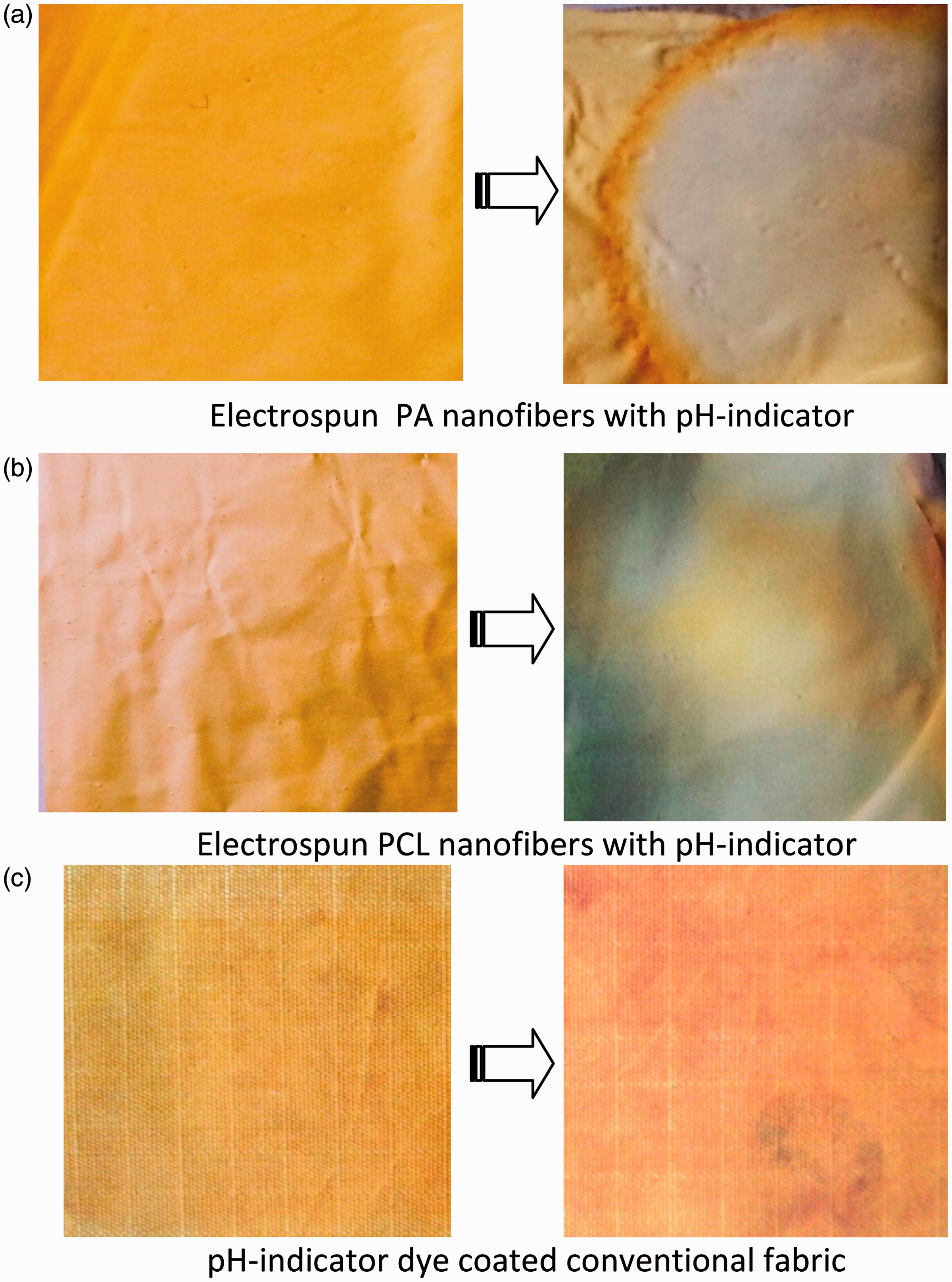
Assessment of the color fastness using a grey scale of Polyamide 6 (PA), Polycaprolatone (PCL) and conventional fabric (CF) samples.

Conclusion
The halochromic coloration of nanofibers was realized by directly incorporating the dyes into the fiber structure, which was shown to be highly effective. A clear and fast color shift was observed by changing the environmental pH which was attributed to the high porosity of the electrospun fibers and their nanoscale properties. Furthermore, by comparing the obtained results, it can be concluded that, generally no differences were observed between the pH sensing behavior of PA and PCL samples. Although the nature of the polymer type may have an influence on the halochromic properties, the effect of fibrous structure of both polymeric material used in this study had the dominant role. The promising results from the pH-detecting experiments demonstrated the possibility to develop a convenient and portable sensory system (e.g. as a strip in the clothing system) for gas and chemical agent detection which PA nanofiber detector has a better cumulative performance in terms of stability and sensitivity.
Footnotes
Declaration of Conflicting Interests
The author(s) declared no potential conflicts of interest with respect to the research, authorship, and/or publication of this article.
Funding
The author(s) disclosed receipt of the following financial support for the research, authorship, and/or publication of this article:
The ATMT Research Institute, Amirkabir University of Technology and INSF (Grant No. 91051749& 92039994).