
Abstract
Cellulose acetate nanofiber mats containing tetracycline hydrochloride were prepared by electrospinning. Incorporation of tetracycline hydrochloride (20 wt% based on the weight of cellulose acetate) in the cellulose acetate solution (17% wt/vol in 2:1 vol/vol ethanol-dimethyl sulfoxide) did not affect the morphology of the obtained fibers, as tetracycline hydrochloride-loaded cellulose acetate fibers were smooth. The average diameters of these fibers ranged between 50 and 90 nm. Determination of the release characteristics of tetracycline hydrochloride from the cellulose acetate nanofiber mats was carried out by total immersion in phosphate buffer solution, that is, in the total immersion method, the maximum amounts of the tetracycline hydrochloride released from the cellulose acetate nanofiber into the medium were 10 and 25% µg/100 ml. Antibacterial activity of prepared tetracycline hydrochloride-loaded as-spun cellulose acetate fiber mate was achieved using Escherichia coli and Staphylococcus aureus as a representative each of Gram-negative and Gram-positive bacteria, respectively. Antibacterial activity of tetracycline hydrochloride-loaded as-spun cellulose acetate fiber mat showed effectiveness with 77–88% bacteria reduction after 10 min–1 h for Escherichia coli and approximately 83–85% reduction after 10 min–1 h for Staphylococcus aureus.
Introduction
One of the significant achievements of nanotechnology today is the production of nanofiber. Electrospinning for nanofiber formation using a variety of materials has been the theme of a great deal of research activities. The principle of electrospinning is based on the extraction of a solution influenced by high electric field. Electrospun materials find widespread applications in tissue engineering, drug release, wound dressing, enzyme immobilization, and so forth. Emphasis is placed on types of materials that employ electrospinning apparatus with special reference to biomedical applications [1].
Cellulose is buildup of [1,4]-linked β-
Several solvents that directly dissolve cellulose have been investigated and used for electrospinning, including N-methylmorpholine N-oxide/water and lithium chloride/dimethyl-acetamide [3–7]. In addition, ionic liquids have recently been used to produce electrospun cellulose nanofibers [8]. Cellulose derivatives have been widely exploited to augment the solubility of cellulose and thus develop its electrospinnability. Cellulose derivatives can be easily electrospun into fibers and then converted to cellulose by aqueous or ethanolic hydrolysis. Cellulose derivatives used for electrospinning include cellulose acetate (CA) [9–12], cellulose triacetate [13], hydroxypropyl cellulose [14], ethyl cellulose [15,16] and methyl cellulose [17].
Recombinant human epidermal growth factor (EGF) was immobilized with electrospun block copolymers. The latter were composed of poly(epsilon-caprolactone) and poly(ethyleneglycol) nanofibers. They were exposed to in vivo wound-healing activities in diabetic ulcer mice. The results indicated that EGF-nanofibers displayed considerable in vivo wound-healing effects. EGF-conjugated nanofiber could be a potential candidate for management of wound complications [18].
Chitosan-ethylenediaminetetraacetic acid (CS–EDTA) solution was subjected to electrospinning process to produce nanofibrous mats with lysozyme. This lysozyme-loaded CS–EDTA nanofiber mats were subjected to in vivo wound-healing studies in Wistar rats. The results indicated that lysozyme release was observed from the nanofiber mats and improved the wound-healing effects [19].
Composite nanofibrous membranes (NFM) composed of collagen and CS showed wound healing and induced cell migration and proliferation in wound-induced rat model. These results signified that NFM was more suitable than gauze and commercial collagen sponge for wound healing [20].
This research is undertaken with a view to develop a novel drug delivery system. This involves synthesis of CA nanofiber mats loaded with hydrophilic antibiotic drug. Electrospinning technique has been used as an efficient method for this synthesis. The impact of electrospinning on the bioactivity of the drug and sustainability of the release behavior of the drug from the electrospun fibrous dressing are studied. Also, the antibacterial activity of the drug-loaded CA nanofiber mat vis-à-vis neat CA nanofiber mats is described.
Experimental methods
Materials
CA (white powder; Mw ∼30 kDa; acetyl content = 39.7 wt%; degree of acetyl substitution ∼2.4) was bought from Sigma-Aldrich, ethanol and dimethyl sulfoxide (DMSO) (analytical reagent grade) were used as solvents of the polymer. Tween 40× (analytical reagent grade; Fluka, Germany) was used as the non-ionic surfactant. Tetracycline hydrochloride (TC) was purchased from Sigma-Aldrich.
Preparation of polymer solutions for electrospinning
A weighed amount of CA powder was dissolved in 2:1 vol/vol ethanol/DMSO to acquire a CA solution at a concentration of 17% wt/vol. TC-loaded CA solutions were prepared by dissolving the same amount of CA powder and TC, the amount being 20 wt% based on the weight of CA powder dissolved in ethanol. These mixtures were stirred with an ultrasonic stirrer and were maintained under constant stirring for 1 h till clear solutions were obtained. Prior to electrospinning, the prepared solution was measured for its viscosity and conductivity using a Brookfield DV-III programmable viscometer and a Orion 160 conductivity meter, respectively. The measurements were carried out at 25℃ and average values for each solution were calculated from at least three measurements. Electrospinning of the prepared solutions was carried using NEU-010 Nanofiber Electorospinning Unit, Kes Kato Tech Co., Japan, connecting the emitting electrode of positive polarity from a gamma high-voltage research, high voltage DC power supply, to the solutions contained in a standard 20 ml syringe, the open end of which was attached to a blunt gauge-20 stainless steel needle, used as the nozzle, and the target drum, used as the fiber collection device. A fixed electrical potential of 20 kV was applied across a fixed distance of 15 cm between the tip of the nozzle and the outer surface of the target drum and the rotational speed of the rotating drum was 40 ± 5 r/min. The feed rate of the solutions was controlled at ∼1 ml h−1 by means of a syringe pump. Electrospinning was carried out under laboratory conditions (i.e., temperature = 25 ± 1℃; relative humidity = 71 ± 3%). For morphological observation of the electrospun products, the collection time was ∼5 min.
Characterization of TC-loaded CA nanofiber mats
Morphological appearance of TC-loaded CA electrospun nanofiber mats was observed using a JEOL JSM-5200 scanning electron microscope (SEM). The fiber mat sample was sputtered with a thin layer of gold prior to SEM observation. Diameters of the individual fibers in the electrospun fiber mats were measured directly from the SEM images The mechanical integrity in terms of the tensile strength and the strain at maximum of the neat similarly spun CA fiber mats control was investigated using a Lloyd LRX universal testing machine under laboratory conditions. Each specimen was cut into a rectangular shape (10 × 100 mm). The crosshead speed and the gauge length were 20 mm min−1 and 50 mm, respectively.
Release of tetracycline from TC-loaded CA electrospun nanofiber mats
Actual TC content
The actual amount of TC in the TC-loaded CA electrospun nanofiber mats was quantified by dissolving the sample in 10 ml of 2:1 vol/vol ethanol/DMSO. The obtained solution was measured for the amount of dissolved TC using a UV-visible spectrophotometer. The amount of TC originally present in the CA electrospun nanofiber mats was back-calculated from the obtained data against a predetermined calibration curve for TC.
Release of tetracycline from TC-loaded CA electrospun nanofiber mats
TC-loaded CA electrospun nanofiber mat was placed in separate test tubes. To this, 3 ml of phosphate buffer solution (PBS) pH 7.4 was added and kept in an orbital shaker with temperature maintained at 37 ± 0.1℃. The amount of tetracycline released from the fiber mat was estimated by collecting PBS from the test tube and replacing by adding fresh 3 ml buffer after 1 h. The same procedure was followed after 4, 24, 48 and 60 h. The concentration of tetracycline release was determined. The quantity of tetracycline released was calculated from the standard curve.
UV-visible spectrophotometeric analysis
UV-visible spectrophotometer using a UV-2200 made on Shimadzu double beam with matched 1 cm quartz cells in the range of 200–700 nm was used to quantify the amount of TC in a sample solution, according to a reported method [21].
Absorption spectra
The absorption spectrum of the colored species formed by mixing TC with p-N,N-dimethylphenylenediamine, sodium meta periodate (DMPD–IO4−), in appropriate pH medium exhibiting maximum absorbance was scanned in the wavelength region 400–700 nm against reagent blank. The λ max was 625 nm. IO4− with DMPD or tetracycline has practically no or low absorbance in this region.
Procedure
Into a series of 100 ml separating funnels, different volumes of the released solution (0.3–4.0 ml) were pipetted out. To each funnel 15 ml of phosphate buffer (pH 7.0), 1.0 ml of DMPD, 2.0 ml of NaIO4 and requisite volume of distilled water were added. After 20 min, 10.0 ml of n-butanol was added. The funnels were shaken gently for 2 min and the n-butanol layer was separated. A blank experiment was carried out omitting the TC. The absorbance of the colored extract was measured, λ max, at 625 nm during the stability period (30 min–2 h.) against a reagent blank. The mean value of three experiments was taken and the amount of the TC was obtained from a standard calibration curve prepared with the same TC under identical conditions.
Antibacterial activity
To test antibacterial properties [22] , 25 ml bacterial suspension, containing either Staphylococcus aureus or Escherichia coli, was placed between two pieces of electrospun CA nanofiber mats (10 mm × 10 mm) and held in place by a sterile weight. The bacterial suspensions contained 106–107 colony forming units (CFU). The actual number was determined by counting after spread-plating on trypticase soy agar plates. After 10, 30 and 60 min, the samples were placed in sterile conical centrifuge tubes, each containing 5.0 ml of sterile 0.01 M sodium thiosulfate and vortexed for 150 s to remove the bacteria. The plates were incubated at 37℃ for 24 h and then counted for viable CFU of bacteria. The reduction in bacterial count was calculated according to

Results and discussion
Morphology of neat and TC-loaded CA electrospun nanofiber mats
CA solution (17% wt/vol) in 2 : 1 vol/vol ethanol/DMSO was electrospun under an applied electrostatic field strength of 20.0 kV/15 cm, as detailed in the experimental section. A selected SEM image of the fibers thus obtained is shown in Figure 1. Obviously, the image of SEM of the electrospun nanofiber mats exhibits with much crossing among each other. These fibers are smooth with an average diameter 50 ± 45 nm. Due to the smoothness of the resulting fibers, the 17% wt/vol CA solution was used as the base solution, into which TC was added. After complete dissolution of TC, the obtained solution was measured for its viscosity and conductivity and compared with identical solution that was prepared without TC. The results obtained are shown in Table 1. The conductivity value of the CA solution containing TC is not much different from that of the neat solution. This could be attributed to the fact that TC contains no ionizable groups. On the other hand, the addition of TC despite its weight causes a decrease in the viscosity as compared with the neat solution. This could be associated with the drag-reducing effect of TC.
Scanning electron micrographs of a spun cellulose acetate nanofiber mats from 17% (wt/vol) ethanol/DMSO. Viscosity and conductivity of CA solution in the absence and presence of TC. CA: cellulose acetate; TC: tetracycline hydrochloride.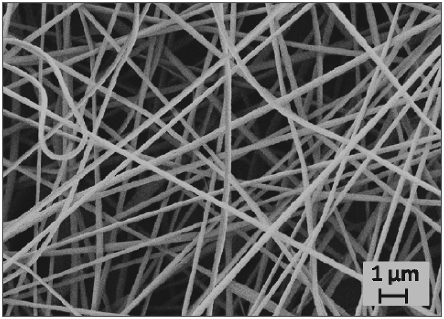

Figure 2 shows that the cross-sectionally round fibers with smooth surface of TC-loaded CA electrospun nanofiber mat are obtained. The average diameters for these TC-loaded CA electrospun nanofiber mats attained values of 75 ± 31 nm. Presence of TC in CA solution during the electrospinning process seems to cause varying levels of intimate association of the CA and in doing so, TC becomes an integral part of CA but at different contents. As a consequence, CA electrospun nanofiber mats containing TC display a wide range of diameter values compared with identical electrospun nanofiber without TC.
Scanning electron micrographs of TC-loaded as-spun CA fiber mat.
Tensile properties of TC-loaded CA electrospun nanofiber mats
The mechanical integrity in terms of the tensile strength and the strain at maximum of the neat and the TC-loaded CA electrospun nanofiber mats were investigated and the results are graphically shown in Figure 3. It is observed that the thicknesses of these fiber mats range from 20 to 30 µm. The tensile strength for all the CA electrospun nanofiber and TC-loaded electrospun nanofiber mats was in the range of 15.4–15.6 MPa, with the average value being 15.5 MPa. The strain at maximum for all of the electrospun nanofiber mats is in the range of 18.5–20.2%, with the average value being 19.6%. The results demonstrate that the presence of TC had no obvious effect on the tensile properties of CA electrospun nanofiber mats.
Mechanical properties of TC-loaded as-spun CA fiber mats: (a) tensile strength (MPa) and (b) strain at maximum (%).
Actual TC content in TC-loaded CA electrospun nanofiber mats
The actual amount of the TC incorporated in the TC-loaded CA electrospun nanofiber mats was determined prior to investigating their release characteristics. The actual amount of the TC in TC-loaded CA electrospun nanofiber (given as percentage of initial content of TC contained in spinning solution) mat sample amounts to 73%. The much less amount of TC incorporated in the fiber mat sample, in comparison with the relative amount of the spinning solution, could be ascribed to the poor stability of TC. The latter seems to lose its characteristics during the electrospinning under the high electrical potential applied to the solution. It may as well be emphasize that the smoothness of the surface of the TC-loaded CA electrospun nanofibers (see Figure 2) may be taken to indicate that tetracycline is encapsulated well within the fibers.
In vitro release study
Figure 4 shows the release patterns of TC from TC-loaded CA electrospun nanofiber mat during different periods of time: 1, 4, 24, 48 and 60 h. It is observed that in the initial 6 h, the drug release rate increases because of initial burst release, followed thereafter by a sustained release driven by diffusion of TC through CA electrospun nanofiber wall. The release of TC from the mat containing tetracycline occurs within a range of 10–20 µg/100 ml. This speaks of constant release of drug over the entire period of study (60 h).
Release of tetracycline from TC-loaded as-spun cellulose acetate nanofiber.
Antibacterial activity
Antibacterial activity of tetracycline enclosed in CA nanofiber against Staphylococcus aureus and Escherichia coli.

CA: cellulose acetate; TC: tetracycline hydrochloride.
Conclusion
Electrospinning technique was used as an efficient processing method to synthesize CA nanofiber mats containing TC and characterization of these synthesized products were undertaken. The synthesis involved preparation of CA solution (17% wt/vol) in DMSO/ethyl alcohol mixture (2:1). To this solution, TC (20% based on weight of CA) was added and the mixture was stirred to clarity using ultrasonic stirrer. Similarly, synthesis of CA electrospun nanofiber mats without TC was also performed. Thus, electrospun CA nanofiber mats were monitored for their morphology, tensile strength, actual TC content in TC-loaded CA electrospun nanofiber mats, in vitro release study and antibacterial activity. Also monitored were conductivity and viscosity of CA solution with and without TC. Major conclusion arrived at from these studies are as given under:
Image of ESM of the electrospun nanofiber mats shows well rounded fibers with much crossing among each other. These fibers are smooth and have average diameters of 50 ± 45 nm and 75 ± 31 nm in the absence and presence of TC, respectively. Incorporation of TC in CA solution before the electrospinning process causes no remarkable changes in the values of conductivity and viscosity. The tensile strength of the CA electrospun nanofiber mat and that of TC-loaded CA electrospun nanofiber mat are equal, with an average value of 15.5 MPa. The TC content in the TC-loaded CA electrospun nanofiber mats is much less than the TC content of the spinning solution. In vitro release study shows that the rate of TC release is characterized by an initial burst release in the first 6 h followed by sustainable release during the period of 10–60 h. TC-loaded CA electrospun nanofiber mats acquire antibacterial activity with effectiveness of 77–88% bacterial reduction in case of E. coli and 83–85% reduction in the case of S. aureus. This is in contrast to the neat CA electrospun nanofiber mat, which does not show significant effectiveness against both bacteria.
Footnotes
Funding
This research received no specific grant from any funding agency in the public, commercial, or not-for-profit sectors.