
Abstract
Background:
The incidence of intrahepatic cholangiocarcinoma (ICC) is increasing globally, and its prognosis has not improved substantially in recent years. Understanding the pathogenesis of ICC may provide a theoretical basis for its treatment. In this study, we investigated the effects and underlying mechanisms of fucosyltransferase 5 (FUT5) on the malignant progression of ICC.
Methods:
FUT5 expression in ICC samples and adjacent nontumor tissues was compared using quantitative real-time polymerase chain reaction and immunohistochemical assays. We performed cell counting kit-8, colony formation, and migration assays to determine whether FUT5 influenced the proliferation and mobility of ICC cells. Finally, mass spectrometry was performed to identify the glycoproteins regulated by FUT5.
Results:
FUT5 mRNA was significantly upregulated in most ICC samples compared with corresponding adjacent nontumor tissues. The ectopic expression of FUT5 promoted the proliferation and migration of ICC cells, whereas FUT5 knockdown significantly suppressed these cellular properties. Mechanistically, we demonstrated that FUT5 is essential for the synthesis and glycosylation of several proteins, including versican, β3 integrin, and cystatin 7, which may serve key roles in the precancer effects of FUT5.
Conclusions:
FUT5 is upregulated in ICC and promotes ICC development by promoting glycosylation of several proteins. Therefore, FUT5 may serve as a therapeutic target for the treatment of ICC.
Keywords
Introduction
Human primary liver cancers include hepatocellular carcinoma (HCC) and intrahepatic cholangiocarcinoma (ICC). The latter accounts for up to 20% of primary liver cancers, and its incidence is gradually increasing worldwide.1-4 Although surgery can contribute to a cure in a fraction of ICC cases, the 5-year survival rate for inoperable patients is only 5% to 10%. 5 Given the characteristic rapid development and high malignancy of ICC, patients are often already in the late stage of disease progression when diagnosed, thereby limiting the efficacy of treatment and disease prognosis, and leading to a lower quality of life and survival rate. Understanding the molecular mechanisms associated with the occurrence and progression of ICC will provide a theoretical basis for its diagnosis and treatment.
Glycosylation is an important post-translational protein modification that plays a key role in cancer cell growth, differentiation, adhesion, and tumor immune escape.6-9 Protein glycosylation mainly occurs in the endoplasmic reticulum and Golgi apparatus, and is catalyzed by a series of glycosyltransferases and glycosidases.6-8 Based on the properties of substrate sugar donors and catalytic glycosidic bonds, the glycosyltransferase superfamily can be divided into several functional subfamilies, including the fucosyltransferase, sialyltransferase, and N-acetylglucosamine transferase families. 10 Each of these catalyzes a group of specific substrates to generate unique glycosidic bonds. 10
Among the fucosyltransferases, 13 genes have been identified to date, namely, those encoding the protein fucosyltransferase 1-11 (FUT1-11), guanosine diphosphate-fucose (GDP-fucose) protein O-fucosyltransferase 1 (POFUT1), and GDP-fucose protein O-fucosyltransferase 2 (POFUT2).11-13 Recently, several studies have reported the role of glycosyltransferase 5 (FUT5) in tumorigenesis and cancer development. For example, Liang et al 14 showed that FUT5 promotes the proliferation, migration, and invasion of colorectal cancer cells by mediating the activation of the PI3K/Akt signaling pathway, whereas Padró et al 15 have demonstrated that knockdown of FUT5 promotes a reduction in the levels of sialyl Lewis antigens and reduces the adhesion capacities of gastric cancer cells.
ICC is a gastrointestinal cancer; therefore, we speculated that FUT5 may influence ICC development. To confirm this assumption, we compared the expression of FUT5 in ICC tissues and the corresponding adjacent nontumor tissues, and investigated the biological function and underlying molecular mechanisms of FUT5 in ICC. We believe that the findings of this study will help clarify the molecular mechanisms underlying ICC progression and provide a new perspective for the treatment of ICC.
Materials and Methods
Clinical specimens
In total, 63 pairs of paraffin-embedded ICC tissues and their corresponding adjacent nontumor tissues were collected at Peking University People’s Hospital. The pathological types of all samples were confirmed by pathologists. Sample collection was conducted in accordance with the Declaration of Helsinki.
Cell culture
In this study, we used 3 ICC cell lines: HCCC-9810, RBE, and HuCCT1. The HCCC-9810 and RBE cell lines were obtained from the National Experimental Cell Resource Sharing Platform (Chinese Academy of Medical Sciences, Beijing, China), and HuCCT1 cells were obtained from the National Collection of Authenticated Cell Cultures (Chinese Academy of Sciences, Shanghai, China). Notably, all 3 cell types were subjected to short tandem repeat authentication. Cells of each line were propagated in Roswell Park Memorial Institute Medium (RPMI) 1640 medium (Gibco, USA) supplemented with 10% fetal bovine serum (FBS) (Gibco), 100 U/mL penicillin, and 0.1 mg/mL streptomycin and cultured in a 5% CO2 atmosphere at 37°C.
RNA extraction and quantitative real-time polymerase chain reaction
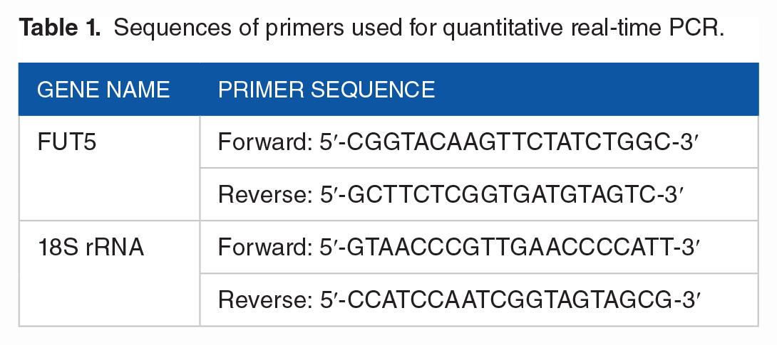
Total RNA was extracted from clinical samples or cultured cells using the TRIzol Reagent. The isolated RNA was used for reverse transcription, and subsequent first-strand cDNA synthesis was performed using the Transcriptor First Strand cDNA Synthesis kit (Takara, Japan). Polymerase chain reaction (PCR) was performed using an Agilent AriaMx Real-Time PCR System according to the manufacturer’s instructions, and 18S ribosomal RNA (18S rRNA) was used as an internal control gene for normalization. Sequences of the upstream and downstream primers are listed in Table 1.
Sequences of primers used for quantitative real-time PCR.
Immunohistochemistry
Paraffin-embedded tissues were deparaffinized, rehydrated, and rinsed with distilled water. Subsequently, the human FUT5 antigen was prepared. The paraffin-embedded slides were then incubated overnight with anti-FUT5 polyclonal Ab (1:200 dilution, Biorbyt, orb449427) at 4°C. The following day, slides were incubated with a secondary antibody (Solarbio) and stained with diaminobenzidine. The intensity of FUT5 staining was evaluated by randomly choosing 5 nonoverlapping fields at ×40 for each slide and then quantified by the percentage of the FUT5(+) biliary epithelial cells/field within all biliary epithelial cells per field. This immunohistochemical (IHC) quantification assay was based on a study by Lepore et al. 16
Knockdown of FUT5 in HuCCT1 cells
FUT5-specific shRNA sequences were initially cloned into GV344 vectors (GENECHEM Co., LTD, China). The shRNA sequences are listed in Table 2. Virus packaging experiments were performed on 293T cells following transfection with GV344-FUT5 shRNA#1 or GV344-FUT5 shRNA#2 plasmids. Thereafter, lentiviral particles expressing FUT5 shRNA were used to infect HuCCT1 cells, and stable cell lines expressing shRNA were selected using puromycin at 2 μg/mL. The knockdown efficiency of each shRNA sequence was assessed using quantitative real-time PCR and Western blotting.
Sequences of FUT5 shRNA and negative control.

Overexpression of FUT5 in HCCC-9810 and RBE cells
The coding sequence of FUT5 was cloned into CV146 vectors (GENECHEM Co., LTD) to obtain FUT5-overexpression plasmids. Virus packaging experiments were performed in 293T cells co-transfected with CV146-FUT5, pHelper 1.0, and pHelper 2.0, and lentiviral particles were used to infect HCCC-9810 and RBE cells. Cell lines stably overexpressing FUT5 were established based on selection with 2 g/mL puromycin and used for gene expression assays and further functional research.
Cell viability assay
Cell viability was evaluated using the cell counting kit-8 (CCK8) assay. Cells were initially plated in the wells of 96-well plates at an inoculation density of 1000 cells/well and thereafter cultured for 4 days in 100 μL of RPMI 1640 medium supplemented with 10% FBS. At daily intervals during culturing, 20 μL of CCK8 reagent (Sigma, China) was added to the designated wells, followed by incubation for 1 hour, after which the absorbance values (OD) were determined at 450 nm using a microplate reader (BioTek Instruments, USA). Cell proliferation curves were plotted for each time point.
Colony formation assay
Cells were seeded in the wells of 6-well plates at a density of 1000 cells/well. After mixing, the cells were cultured for 10 days in RPMI 1640 medium containing 10% FBS. To assess colony formation, we defined colonies as cell clusters containing 30 or more cells.
Transwell migration assay
To assess cell migration, we used Transwell chambers. We first seeded a suspension of 5 × 10⁴ cells in 200 μL of FBS-free RPMI 1640 medium into the top compartment. As a chemoattractant, RPMI 1640 medium supplemented with 10% FBS was then added to the lower compartment. After 24 hours of incubation, the cells in the upper chamber were removed with a cotton swab. Cell migration capacity was evaluated by counting the penetrating cells under a microscope (40 × 20), with 5 random fields of view assessed for each chamber. The cell numbers in all 5 fields were added for analysis.
Western blotting
Total protein was extracted using RIPA lysis buffer containing a protease inhibitor mixture. The protein samples were separated by SDS-PAGE and transferred onto polyvinylidene difluoride (PVDF) membranes. The membranes were blocked for 1 hour with 5% bovine serum albumin and incubated with primary antibody overnight at 4°C. The following day, the membranes were washed with Tris-buffered saline containing Tween-20, followed by incubation with a secondary antibody (1:1000 dilution for both anti-mouse and anti-rabbit antibodies) for 1 hour. Protein bands were visualized using a chemiluminescence detection system, with vinculin serving as a loading control.
As primary antibodies, we used anti-FUT5 (1:1000 dilution) (Biorbyt, orb449427), anti-vinculin (1:1000 dilution) (ABclonal, A2752), anti-ITGB3 (1:3000 dilution) (Abcam, ab197662), anti-Ki-67 (1:1000 dilution) (Abcam, ab16667), anti-E-cadherin (1:20 000 dilution) (Proteintech, 20874-1-1AP), and anti-GAPDH (1:2000 dilution) (Abcam, ab8245). Goat anti-rabbit IgG and goat anti-mouse IgG secondary antibodies were purchased from Beijing Solarbio Science and Technology Co., Ltd.
Lectin enrichment assay
Next, 100 μL of agarose-bound Aleuria Aurantia Lectin (AL-1393-2, Vector Laboratories) was added to 400 μg of total cell lysate and incubated at 4°C overnight on a rotator. α1,3-fucosylated glycoproteins were enriched by centrifugation and washed thrice with HBS buffer. The enriched glycoproteins were eluted directly with 2X Laemmli SDS buffer and heated at 100°C for 5 minutes before being subjected to SDS-PAGE and Western blot analysis.
Glycosylation/protein mass spectrometry
The sample was sonicated thrice on ice using a high-intensity ultrasonic processor (Scientz) in lysis buffer (8 M urea and 1% protease inhibitor cocktail). The remaining debris was removed by centrifugation at 12 000 g at 4°C for 10 minutes. For digestion, the protein solution was reduced with 5 mM dithiothreitol for 30 minutes at 56°C and alkylated with 11 mM iodoacetamide for 15 minutes at room temperature in darkness. Protein samples were diluted with 100 mM triethylammonium bicarbonate buffer (TEAB). Finally, trypsin was added at a 1:50 trypsin-to-protein mass ratio for the first digestion overnight and at a 1:100 trypsin-to-protein mass ratio for a second digestion (4 hours). The peptides were desalted using a C18 solid phase extraction column and then dissolved in 0.5 M TEAB. Each channel of the peptide was labeled with its respective Tandem Mass Tag reagent (based on the manufacturer’s protocol, Thermo Fisher Scientific) and incubated for 2 hours at room temperature. Quantitative glycosylation/proteomic analysis using LC-MS/MS was performed for each fraction. The resulting MS/MS data were processed using the MaxQuant search engine (v.1.6.15.0). All MS analyses were performed with the support of PTM-Biolabs Co. Ltd. (310018; HangZhou, Zhejiang, China).
Statistical analysis
Data are presented as means ± standard deviation of 3 independent biological replicate analyses of each group. The Student’s t-test was used to compare the values of treated and control groups. Statistical significance was defined as P < .05 and *P < .01. All calculations were performed using the SPSS software (version 13.0; SPSS Inc., Chicago, IL, USA).
Ethics approval and consent to participate
Written informed consent was obtained from all study participants. This study was approved by the Ethics Committee of the Peking University People’s Hospital (Approval No. 2014KT98).
Results
Expression of FUT5 is significantly upregulated in ICC and correlates with poor prognosis
FUT5 is a proto-oncogene that has been demonstrated to be somatically amplified in gastric and colon cancers.14,15 Given that ICC is a gastrointestinal cancer, we sought to determine whether FUT5 is overexpressed in ICC by analyzing the mRNA levels of FUT5 in 6 ICC tissues and corresponding noncancerous tissues. We found that FUT5 mRNA levels in ICC tissues were higher than those in the corresponding adjacent nontumor tissues (Figure 1A). In addition, mRNA levels of the remaining 12 FUTs were assessed (Supplemental Figure S1). To gain better insight into the clinical significance of FUT5 protein in ICC, we performed IHC analyses to assess the expression of FUT5 protein in 85 paraffin-embedded ICC specimens (Figure 1B and C). As previously studied, 17 FUT5 positive staining was mainly localized in the cytoplasm of ICC tumor cells. Interestingly, positive staining was observed in the extracellular matrix (ECM) of some samples, indicating that FUT5 may also act as a secretory protein. In addition, all 6 patients enrolled for the FUT5 mRNA level test also shows FUT5 high expression in the IHC assay. Consistent with the data obtained from real-time PCR analyses, we found that FUT5 protein was upregulated in ICC tissues compared with the levels detected in the corresponding nontumor tissues.

FUT5 was significantly upregulated in ICC on mRNA and protein level. (A) Tumor/nontumor ratio of the FUT5 mRNA levels in 6 ICC patients’ tumor samples compared with their nontumor samples. (B) Several examples of the immunochemistry results. (C) Protein level of FUT5 measured by staining percentage, Ca: Cancer samples, Pa: Para-cancer samples. (D) Survival analysis of the patients involved.
To determine whether the expression level of FUT5 in the tumor had any clinical impact, we performed survival analysis using clinical data from 85 patients. Patients in the FUT5 high expression group had shorter overall survival times (median survival time, 21 vs 34 months) (Figure 1D).
Overexpression of FUT5 in ICC cells clearly accelerates their proliferation
To examine the potential function of FUT5 in ICC, we determined the expression of FUT5 expression in 3 cell lines (HCCC-9810, RBE, and HuCCT1). Our data revealed that FUT5 protein levels were higher in HUCCT1 cells than in HCCC-9810 and RBE cells (Figure 2A). We subsequently constructed a stable cell line (HCCC-9810-FUT5) using a lentivirus packaging system and found that compared with HCCC-9810-control cells, the mRNA and protein levels of FUT5 were markedly upregulated in the HCCC-9810-FUT5 cells (Figure 2B and C). CCK8 and colony formation assays were used to examine the colony-forming efficiency of HCCC-9810-FUT5 and HCCC-9810-control cells (Figure 2D to F), and markers of cell proliferation and migration were also examined (Figure 2G). Accordingly, we established that the ectopic expression of FUT5 enhanced the proliferation and colony formation ability of HCCC-9810 cells.

Overexpression of FUT5 in ICC cells accelerated cell proliferation. (A) FUT5 protein levels in 3 ICC cell lines. (B) and (C) FUT5 expression is upregulated in HCCC-9810-FUT5 cells. (D) to (F) The effect of FUT5 overexpression in HCCC-9810-FUT5 cells compared with negative control cells. (G) Change of cell proliferation marker Ki-67 and migration marker E-cadherin.
FUT5 knockdown in ICC cells significantly suppresses their proliferation
To determine the effect of FUT5 on ICC cell growth, we generated 2 stable cell lines containing HuCCT1-FUT5 shRNA#1 and HuCCT1-FUT5 shRNA#2. Compared with HuCCT1-control cells, we found that the relative expression levels of FUT5 were significantly reduced in cells harboring HuCCT1-FUT5 shRNA#1 and HuCCT1-FUT5 shRNA#2 constructs (Figure 3A and B). The CCK8 assay revealed that FUT5 knockdown inhibited the proliferation of HCCC-9810 cells (Figure 3C), and the colony formation assay results indicated that FUT5 knockdown reduced the colony-forming efficiency of HuCCT1 cells (Figure 3D and E). Cell proliferation and migration markers were also examined (Figure 3F).

Depletion of FUT5 in ICC cells significantly suppressed their proliferation. (A) and (B) FUT5 expression is downregulated in HuCCT1-FUT5 shRNA#1 and HuCCT1-FUT5 shRNA#2 cells. (C) to (E) The effects of FUT5 knockdown on cell proliferation in HuCCT1-FUT5 shRNA#1 and HuCCT1-FUT5 shRNA#2 cells compared with negative control cells. (F) Change of cell proliferation marker Ki-67 and migration marker E-cadherin.
Using a Transwell migration assay, we examined whether FUT5 affects the migration of ICC cells. Our data revealed that the migratory properties of HCCC-9810-FUT5 cells were substantially higher than those of HCCC-9810-control cells (Figure 4A and B). In contrast, compared with the HuCCT1-control cells, the migratory capacity of the HuCCT1-FUT5 shRNA#1 and HuCCT1-FUT5 shRNA#2 cells was significantly impaired (Figure 4C and D). These observations provided evidence that FUT5 enhanced the migration of HCC cells.

The effect of FUT5 to the migration of ICC cells. (A) and (B) Overexpression of FUT5 substantially promoted the migration of ICC cells. (C) and (D) Knockdown of FUT5 markedly inhibited the migration of ICC cells.
FUT5 can regulate protein glycosylation to promote ICC progression
To gain a better understanding of the mechanisms underlying the effects of FUT5 on ICC development, we performed glycosylation mass spectrometry analysis to assess the effects of FUT5 on protein glycosylation. We detected 97 downregulated glycosylation sites in 63 glycoproteins following the knockdown of FUT5 (Figure 5A and B). Having identified these glycosylation sites, we subsequently divided them into 4 groups based on the KD/NC ratio of their glycosylation levels and conducted Gene Ontology (GO) category analysis. GO annotation indicated that the glycoproteins with the lowest glycosylation levels and KD/NC ratios mainly function in the interaction of cells with the ECM (including Integrin Binding) (Figure 5C and D). For example, versican (VCAN), which is a major component of the ECM and has been reported to promote the proliferation, invasion, and migration of various tumors, such as HCC, was found to have the most significantly downregulated glycosylation site among the glycoproteins assessed (Figure 5E).

FUT5 can regulate protein glycosylation to promote ICC progression. (A) and (B) Glycosylation sites found by the MS. (C) and (D) Grouped Gene Ontology of the differential glycosylation sites. (E) Several glycosylation sites which downregulate most when FUT5 was knockdown. (F) Volcano plot of protein expression measured by the MS. (G) Protein expression of those in (E) (Several proteins are not identified by MS may because of its low abundance). (H) Lectin enrichment assay shows change of the fucosylation level of ITGB3.
To exclude the effect of changes in protein expression on measured glycosylation levels, we conducted protein mass spectrometry analysis. Among the proteins showing the most obvious reduction in glycosylation level, we found that the expression level of ITGB3 did not change significantly after FUT5 was knocked down, while VCAN and ASAH1 showed a reduction in the expression level nearly equivalent to the reduction ratio of the glycosylation level (Figure 5F and G). ITGB3 is the most affected protein in the glycosylation process, in which FUT5 participates. We also conducted a lectin enrichment assay and validated the conclusions of mass spectrometry by Western blotting (Figure 5H).
These results may provide clues as to how FUT5 regulates the protein glycosylation profiles of ICC cancer cells and thereby indicate several potentially key factors contributing to the pro-cancer effects of FUT5.
Discussion
Fucosylation of cancer proteins has been observed in many tumors. The expression levels of FUT3-7 and FUT9 are increased in colorectal cancer, which promotes the expression of cancer-related Lewis antigens (such as sialic acid Lewisx and Lewisa), and are related to tumor metastasis.18-20 In breast cancer, FUT5 and FUT6 as well as their products, sialic acids Lewisa and Lewisx, are overexpressed and associated with both tumor metastasis and shorter overall survival. 21 The expression level of FUT2 in lung adenocarcinoma was higher than that in the adjacent tissues. In the lung adenocarcinoma cell lines A549 and H1299, FUT2 knockdown was found to inhibit tumor cell migration and invasion, and promote tumor cell apoptosis. 22
Many abnormal fucosylation processes have been found in HCC, which is also a type of malignant liver tumor, similar to ICC. The expression levels of FUT6 were significantly increased in HCC and were associated with disease progression. A study conducted in HCC cell lines showed that overexpression of FUT6 can increase the proliferative ability of tumor cells in vitro and their ability to form tumors in vivo.23,24 The overexpression of FUT4, FUT6, and FUT8 can cause HCC cells to acquire resistance to chemotherapeutic drugs. 25 In addition, GDP-fucose, the only substrate and energy source in the fucosylation process, is upregulated in HCC tissues. 26 FUT8 increases the glycosylation of core-focused alpha-fetoprotein (AFP-L3), which has been used as a tumor marker of HCC in the United States.27,28 However, few studies have examined the role of glycosylation in ICC.
Although the function of FUT5 has been studied in gastric and colon cancers, the association between FUT5 and ICC malignancy remains unclear. In this study, we sought to determine whether FUT5 can influence the proliferation and motility of ICC cells and to assess the clinical significance of FUT5 expression in ICC and the signaling pathway regulated by FUT5.
We initially examined the expression of FUT5 in clinical samples using IHC analysis. Previously, Liang et al 14 have shown that the expression of FUT5 was significantly increased in colorectal cancer tissues compared with that in normal tissues; consistently, we found that the average expression of FUT5 in 63 ICC tissues was significantly higher than that in the corresponding adjacent nontumor tissues. Survival analysis showed that high FUT5 expression was associated with a poor prognosis.
Previous studies have shown that FUT4 is not only highly expressed in breast cancer tissues but also in serum and could serve as a novel biomarker. 29 Our data revealed that FUT5 is not only localized in the cytoplasm and membranes of ICC cells but is also highly expressed in the ECM. Based on these findings, we speculated that FUT5 not only affects the characteristics of tumor cells but also functions as a secreted protein that contributes to the regulation of the tumor microenvironment. However, these conjectured roles need to be examined further in future studies.
Nevertheless, given the high expression of FUT5 in ICC tissues, we suspect that FUT5 may play a key role in facilitating the malignant progression of ICC. For verification, we conducted CCK8 and colony formation experiments to determine whether FUT5 influenced ICC cell growth. Our findings revealed that the overexpression of FUT5 facilitated the significant proliferation of HCCC-9810 cells, whereas FUT5 knockdown strongly suppressed the proliferative properties of these cells. Collectively, these findings indicate that FUT5 functions as a positive regulator of ICC proliferation.
Lymph node metastasis is found in 15% to 45% of patients with ICC, severely affecting their prognosis.30-33 Therefore, we examined whether FUT5 influenced the motility of ICC cells and found that FUT5 overexpression accelerated HCCC-9810 cell migration, whereas FUT5 knockdown repressed HuCCT1 cell migration. These findings indicated that FUT5 plays a role in enhancing the migratory ability of ICC cells.
To elucidate the mechanisms by which FUT5 promotes ICC proliferation and migration, we sought to identify the glycoproteins most affected by changes in FUT5 expression. Our observations indicated that the knockdown of FUT5 notably inhibits the glycosylation of a range of proteins in HuCCT1 cells, including VCAN, CST7, and ITGB3. After excluding the effect of changes in protein expression, ITGB3 was the protein most affected by FUT5 during glycosylation. However, whether FUT5 and ITGB3 directly interact with ICC cells remains to be established. Consequently, further studies are necessary to identify key proteins or proteins implicated in the FUT5-mediated promotion of ICC progression.
However, this study had some limitations. For clinical sample analysis, it would be better to include more cases to make the conclusions more convincing. Tumor cells may act differently in vitro and in vivo; therefore, animal experiments, such as subcutaneous tumor formation in nude mice, could provide more direct evidence. FUT5 may affect cell function through multiple pathways; therefore, further studies are warranted.
Conclusions
In this study, we demonstrated that compared with the corresponding para-tumor tissues, the expression of both FUT5 mRNA and protein was high in most of the assessed ICC tissues. We established that FUT5 is localized in both the nucleoplasm and ECM of ICC tissues; moreover, we found that knockdown of FUT5 resulted in the suppression of the malignant behavior of HuCCT1 cells, whereas its overexpression promoted HCCC-9810 cell proliferation and migration. We provide evidence that FUT5 is a potential mechanism of action in ICC as it mediates malignant progression by regulating protein glycosylation profiles. Consequently, these downstream proteins could serve as ideal targets for therapeutic interventions in patients with ICC. In summary, FUT5 may function as a positive regulator of ICC proliferation. The findings of this study provide a theoretical basis for future precision ICC therapies.
Research Data
sj-jpg-1-onc-10.1177_11795549231181189 – for Fucosyltransferase 5 Promotes the Proliferative and Migratory Properties of Intrahepatic Cholangiocarcinoma Cells via Regulating Protein Glycosylation Profiles
sj-jpg-1-onc-10.1177_11795549231181189 for Fucosyltransferase 5 Promotes the Proliferative and Migratory Properties of Intrahepatic Cholangiocarcinoma Cells via Regulating Protein Glycosylation Profiles by Jingheng Guo, Qian Cheng, Yongjian Li, Lingyu Tian, Delin Ma, Zhao Li, Jie Gao and Jiye Zhu in Clinical Medicine Insights: Oncology
Footnotes
Author Contributions
JG and QC made a substantial contribution to the concept or design of the work, drafted the article, or revised it critically for important intellectual content; YL, LT, DM, and ZL participated in acquisition, analysis, or interpretation of data; JGa and JZ made a substantial contribution to the concept or design of the work and approved the version to be published.
Ethics approval and consent to participate
Written informed consent was obtained from all study participants. This study was approved by the Ethics Committee of the Peking University People’s Hospital (Approval No. 2014KT98).
Declaration of Conflicting Interests:
The author(s) declared no potential conflicts of interest with respect to the research, authorship, and/or publication of this article.
Funding:
The author(s) disclosed receipt of the following financial support for the research, authorship, and/or publication of this article: This work was supported by the National Natural Science Foundation of China [grant nos. 81871291, 82172693 and 81872508].
Supplemental Material
Supplemental material for this article is available online.
References
Supplementary Material
Please find the following supplemental material available below.
For Open Access articles published under a Creative Commons License, all supplemental material carries the same license as the article it is associated with.
For non-Open Access articles published, all supplemental material carries a non-exclusive license, and permission requests for re-use of supplemental material or any part of supplemental material shall be sent directly to the copyright owner as specified in the copyright notice associated with the article.