
Abstract
Several fields in neuroscience have been revolutionized by the advent of optogenetics, a technique that offers the possibility to modulate neuronal physiology in response to light stimulation. This innovative and far-reaching tool provided unprecedented spatial and temporal resolution to explore the activity of neural circuits underlying cognition and behaviour. With an exponential growth in the discovery and synthesis of new photosensitive actuators capable of modulating neuronal networks function, other fields in biology are experiencing a similar re-evolution. Here, we review the various optogenetic toolboxes developed to influence cellular physiology as well as the diverse ways in which these can be engineered to precisely modulate intracellular signalling and transcription. We also explore the processes required to successfully express and stimulate these photo-actuators in vivo before discussing how such tools can enlighten our understanding of neuronal plasticity at the systems level.
Keywords
Introduction
External milieu and neuronal plasticity
In mammals, the ontogeny of the central nervous system (CNS) is achieved through different genetic programmes in prenatal life, which cause the diverse types of neuronal cells that build up the brain to be produced in the right number and at the right place. Modalities of sensory experience in post-natal life, however, markedly shape the CNS architecture by modulating the structure and function of synaptic connectivity. 1 The experience-induced modulation of neural circuit functionality leads to the formation of short-term and long-term forms of neuronal plasticity that alter the computational properties of sensory systems and ultimately have an impact at the level of higher brain functions and behaviour.
Environmental influences on brain development and function are mediated by the release of neurotransmitters at specific synapses between neural cells, which, following the binding to appropriate receptors on the post-synaptic neuron, set in motion a number of intracellular signalling pathways that arrive to the nucleus and regulate gene transcription. Different events can be drawn along these physiological mechanisms. First, a transient increase in calcium concentration within the post-synaptic cell promotes synapse-specific modifications that lead to changes in synaptic transmission.2,3 Second, the activation of intracellular signal transduction pathways results in the initiation of gene programmes that alter dendritic growth, 4 synapse development, 5 and neuronal connections. 6 Thus, modifications in the structure and function of synaptic connectivity within neuronal networks arise from intracellular transduction pathways driven by electrical signals associated with neuronal activity, which in turn control the expression of plasticity genes. This underlies the capability of the brain to adjust in response to changing environmental conditions.
Aside from identifying transcription factors that regulate gene transcription in response to external stimuli, epigenetic mechanisms that exert a long-lasting control of gene expression by altering chromatin structure, rather than changing the DNA sequence itself, have recently emerged as conserved processes by which the CNS accomplishes the induction of plasticity.7,8 A hot spot in the field of neuroscience is the identification of physiological mechanisms associated with experience that promote alterations in the pattern of DNA methylation9–11 and/or posttranslational modifications of histones12–14 that control the expression of plasticity genes in the brain. A new set of optogenetic tools capable of affecting both epigenetics and transcription is emerging (detailed below), offering new ways to study intracellular mechanisms underlying neuronal plasticity.
Many mechanisms have been implicated in the occurrence of activity-dependent plasticity.
15
According to Hebb’s rule,
16
neurons that fire together wire together, whereas neurons that fire out of synchrony lose their link. Decades of effort in the field have elucidated the involvement of different post-synaptic receptors in this mechanism. Glutamate (Glu) receptors of the α-amino-3-hydroxy-5-methyl-4-isoxazolepropionic acid (AMPA) and N-methyl-
Activity-dependent plasticity plays an important role in processes of learning and memory. Hence, it is responsible for adaptation of an individual to the environment. Until recently, optogenetic tools have been used to extrinsically modify neuronal activity using type-1 rhodopsin derivatives (described below), enabling a primary impact on the electrical state of the neuron and only a secondary impact on intracellular signalling and transcription (Figure 1). Novel optogenetic toolboxes described here have now the potential to affect both those processes directly.

Rhodopsin transmembrane photo-actuator. (A) Pictorial representation of rhodopsin structure in a membrane environment: the protein is represented as green cartoons and the retinal chromophore as red sticks. Membrane lipids are represented as grey sticks (hydrophobic region) or surface (hydrophilic region). Generated in the Visual Molecular Dynamics software. 23 (B and C) Examples of (B) type I and (C) type II rhodopsins, with their activation spectra depicted as coloured arrows and retinal cycle along with their influence on intracellular function and its impact on transcription and neuronal circuitries’ connectivity.
Optogenetics and neuronal plasticity
One major challenge for modern neuroscience is to control the activity of a single type of neuron in the mammalian brain while leaving others unaltered. 24 The application of molecular engineering to the field of optogenetics provided precious tools to control specific groups of neurons that underlie behaviour.24–28 Optogenetics has had a major impact in the unravelling of issues that include cross-modal plasticity, 29 information processing by neuronal circuitries,30,31 hippocampal memory formation,32,33 anxiety and depression,34,35 fear conditioning, 36 aggression, 37 feeding behaviour,38–40 restoration of visual functions in blind animals,41–43 the Parkinson disease,44,45 and epilepsy.46,47
Interpreting the occurrence of plastic phenomena under physiological conditions requires a well-defined correlation between the activity of specific neuronal subtypes and the computational properties of neuronal networks within sensory systems. Unfortunately, the lack of a clear relationship between morphological and functional characteristics of different cell types precludes a wide view of neuronal connectivity patterns within neuronal networks. This, together with the absence of experimental approaches with high temporal and spatial resolution to address the role of specific cellular subtypes in phenomena of plasticity, is one of the major reasons by which the screening of neuronal populations underlying brain plasticity has remained elusive. Only recently, the development and application of optogenetics in the field of neuroscience has made it possible to investigate the relationship between sensory perception, plasticity phenomena, and the activity of specific neuronal subpopulations in primary sensory areas.
The subcellular targeting of optogenetic tools has opened up new avenues in neuroscience as it enables researchers to address the function of well-defined intracellular domains.48,49 High-precision spatiotemporal control of gene transcription has been recently accomplished, 14 which allowed to modulate endogenous gene expression and epigenetic chromatin modifications that underlie behaviour (for review, see Moglich and Hegemann 50 ). The fast advances in the diverse optogenetic toolboxes described in this review provide novel ways to establish causal relationships between the activity of specific neuronal networks and the related functional outcomes.
Photoactivatable Actuators
Photoreceptor protein domains respond to light by absorbing photons at a prosthetic group or chromophore. Interaction with light induces a cascade of events called the photocycle, comprising molecular transformations including chemical changes at the chromophore, and conformational and functional changes of the protein scaffold. The photocycle usually connects 2 states, the dark and lit states, separated by intermediates and is in general reversible because the photoreceptor is able to recover its dark state by thermal fluctuations. Isomerization or establishment of a covalent bond with the protein chain is among the most common chemical changes observed at the chromophores.
Photoreceptors are usually classified by their chromophore cofactor: rhodopsins with retinal, light-oxygen-voltage (LOV) sensors with a flavin mononucleotide (FMN), blue-light sensors (BLUF), and cryptochromes (CRYs) with a flavin adenine dinucleotide (FAD) and phytochromes (PHYs) with tetrapyrrole molecules. The UV-B receptor UV-B resistance 8 (UVR8) constitutes a separate class of photoreceptors as it does not employ a cofactor but instead absorbs UV radiation through tryptophan (Trp) residues. Although rhodopsins (Table 1) are membrane bound, the other photoreceptors (Table 2) are cytosolic, allowing for photo-transduction within any cellular compartment.
Intracellular signalling from membrane photo-actuators.

Abbreviations: Arch, archaerhodopsin; ChR2s, channelrhodopsins; NpHRs, halorhodopsin; SFOs, step-function opsins.
Rhodopsins are activated by lights spanning the entire visual spectrum and bound to the cell membrane, giving them control over both membrane potential and intracellular signalling. Short Wavelength (SW), Medium Wavelength (MW), Long Wavelength (LW).
Intracellular signalling from cytosolic photo-actuators.
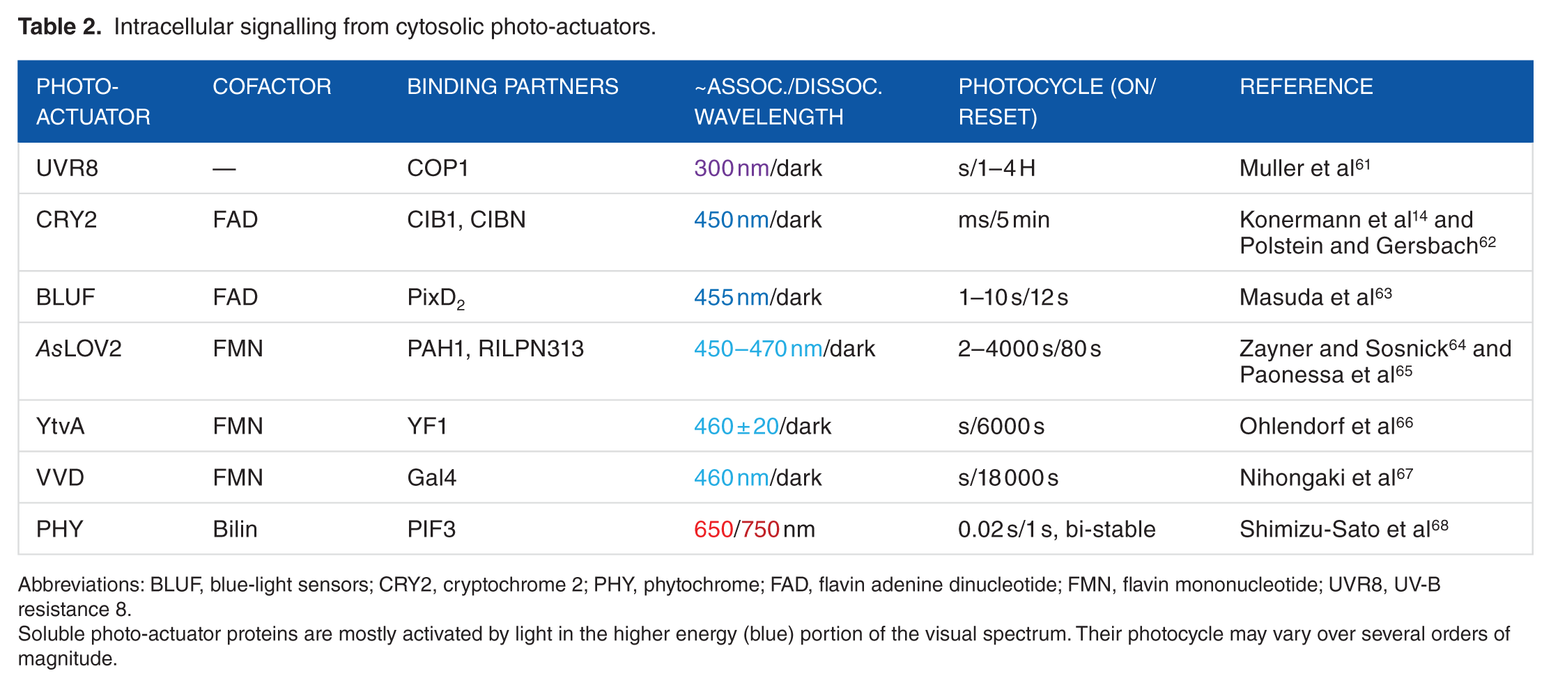
Abbreviations: BLUF, blue-light sensors; CRY2, cryptochrome 2; PHY, phytochrome; FAD, flavin adenine dinucleotide; FMN, flavin mononucleotide; UVR8, UV-B resistance 8.
Soluble photo-actuator proteins are mostly activated by light in the higher energy (blue) portion of the visual spectrum. Their photocycle may vary over several orders of magnitude.
Rhodopsins were the first proteins to be used as optogenetic actuators in neuroscience applications.69,70 This class of light-sensitive protein is composed of 7 transmembrane α helixes (Figure 1A) combined to the photo-isomerizable chromophore retinal. 71 The engineering of innovative opsins with diverse characteristics and functions originating from the two types of rhodopsins expressed in the microbial (type I) and animal (type II) kingdoms, have been extensively described.28,72,73
Type I rhodopsins
In type I rhodopsins, the transmembrane domain is generally an ionotropic effector (channel or pump) that is covalently bound to the chromophore allowing for dynamic modulation of the voltage across biological membranes at the sub-millisecond timescale 69 (Figure 1B). On illumination, all-trans retinal isomerizes to 13-cis retinal, leading to a conformational change in the transmembrane domain. The most cited examples of such optogenetic tools are channelrhodopsin (ChR2), 74 halorhodopsin from Natronomonas pharaonis (NpHR), 75 and archaerhodopsin (Arch). 76 Channelrhodopsin is permeable to sodium, potassium, protons, and, to a lesser extent, calcium on illumination with blue light (~470 nm) generating an inward current that leads to membrane depolarization and action potential firing. However, pump rhodopsins, such as NpHR, which drive chloride in the cytosol, and Arch, which pump protons out of the cell, lead to membrane hyperpolarization and a decrease in action potential firing on illumination with orange light (~590 nm). Photoactive chloride and proton pumps require constant illumination to go through their photocycle as opposed to ChR2, which only need channel opening, and bi-stable rhodopsins, which require a single pulse to move from one state to the other. Sensory rhodopsins trigger enzyme activity such as histidine kinase, 77 providing the opportunity to influence intracellular signalling. Type I rhodopsins and their uses as optogenetic tools are extensively reviewed elsewhere. 72
One of the most exciting features of the optogenetic revolution is the steady development of toolboxes of opsins exhibiting increasingly diverse spectra, excitation, and temporal kinetics. The NpHRs have been optimized to the point of being used in mammalian tissue 58 and having red/far red sensitivity. 48 Step-function opsins (SFOs) are a family of bi-stable ChR2 mutants 54 designed to stabilize the active retinal isomer. They display longer inactivation time constants, allowing the activation of populations of neurons for long times (from tens of seconds 54 to several 55 or even tens of minutes 56 ), together with the possibility of switching them off with a single pulse of yellow light. 54 These specifications enable a myriad of uses including calcium imaging or un-tethered behavioural experiments, in which the consequences of optogenetic activation can be investigated following a single pulse, without cumbersome stimulating hardware. 28 In addition to the development of such bi-stable optogenetic tools, multiplexing or even cooperative strategies are now emerging. The former allows various opsins sensitive to different wavelengths to be simultaneously but independently activated, such as the expression of ChR2 in the dendrites and the expression of NpHR in the soma of retinal ganglion cells to mimic the effect of centre-surround antagonism in the retina. 43 The recently developed ‘Cl-out’ strategy uses the outward current generated by the proton pump Arch to propel the extrusion of chloride ions through the concomitant activation of a co-expressed optogenetic chloride channel. 78
Type II rhodopsins
In type II rhodopsins, the 7 transmembrane domains form a G protein−coupled metabotropic receptor (GPCR), offering a broad influence over cellular physiology by tapping into secondary messenger systems. 11-cis-retinal dissociates on photo-isomerization to all-trans retinal, making the resetting kinetics considerably slower than type I rhodopsins, as well as requiring the exogenous application of 9 or 11-cis retinal anywhere other than in the retina. A large diversity of photo-transduction cascades are initiated following activation of type II rhodopsins including Gt-, Gq-, Go-, Gs-, Gi-, and Gi/o-coupled pathways. We discuss here the 3 most exploited tools (Figure 1C); for a more exhaustive review of type II rhodopsins, the reader is directed to the study by Koyanagi and Terakita. 73
Cone opsins and rod opsins are expressed in the outer segment of mammalian photoreceptors, coupling to Gα transducin and activating the Gi/o pathway 51 on illumination. Both cone and rod opsins present advantages, in that rod opsins are highly sensitive to light and cone opsins exist in various versions, sensitive to different wavelengths, 79 thus offering the potential for polychromatic multiplexing toolboxes. A major drawback in using these opsins is the high level of bleaching due to chromophore dissociation on activation. However, this feature was successfully exploited in vivo as a light adaptation mechanism to restore vision in genetically engineered blind mice. 80
Jellyfish opsins (JellyOp) have been found to signal through Gs proteins, 57 inducing translocation of adenylyl cyclase, leading to an increased production of cyclic adenosine monophosphate (cAMP) followed by activation of the protein kinases, PKA and ERK pathways. 81 The activation of all these proteins often leads to membrane voltage depolarization. Jellyfish opsin has a peak sensitivity of ~500 nm, and unlike visual opsins, does not bleach but is instead converted to a stable photoproduct that does not revert to its original dark state on subsequent illumination. 57
Melanopsin is a photopigment expressed in intrinsically photosensitive retinal ganglion cells (ipRGCs) and is responsible for the photo-entrainment of the circadian rhythm. 82 With recent evidence pointing to tri-stability, 53 melanopsin activation by blue light activates Gq signalling. 83 In ipRGCs, this leads to sustained firing of action potentials along axons, 80% of which (M1 ipRGCs) project to the suprachiasmatic nucleus (SCN, master clock of the CNS) leading to the daily plastic regulation of the sleep/wake cycle.82,84 Recent evidence suggests that the SCN integrates both irradiance from inner retinal ipRGCs and colour from outer retinal photoreceptors to determine the time of day. 85
To target signalling pathways that do not rely solely on generic Gα subunit coupling dynamics, recent effort has been made in the development of chimeric optogenetic tools (OptoXRs) composed of extracellular loops of type II rhodopsin and the intracellular loops/C-terminal tail of GPCRs endogenously expressed in the CNS. 86 The advantage of this approach consists in coupling the multiple inherent possibilities offered by type II opsins (high sensitivity, multi-chromaticity, bleach adaptation) with the quasi-infinite variety offered by endogenous GPCR systems. OptoXRs have been implemented successfully in vivo with the adrenergic, 87 adenosinergic, 88 opioid, 89 and serotonergic 90 receptor systems.
Although opsins represent powerful tools to modulate the physiology of entire cell populations, inducing or preventing plasticity in vivo through modulation of neural activity,33,51,88 they lack the spatial resolution to directly impact intracellular functions such as protein interactions or gene expression.
Soluble photoactivatable proteins
Light-oxygen-voltage (LOV) and phytochrome (PHY) protein receptors have a modular structure; ie, they are made of several adjacent globular domains originating from the same, continuous amino acid chain. Each module performs a different function. One domain is responsible for light absorption, whereas a separate one is responsible for biological activity, which can be protein binding, DNA binding, or enzymatic action. The transmission of the signal from the photo-sensor domain to the activity domain is usually a combination of structural and dynamic effects. The same principle is at the basis of engineered optogenetic probes, where the photo-sensor domain from a receptor is linked to a different protein domain exerting the biological function specifically required by the designer.
LOV domains
Light-oxygen-voltage sensor domains are found in proteins from bacteria, plants, and fungi, and they are the most widely used type of photoreceptors in optogenetic applications to influence intracellular signalling. All LOV domains show a fold typical of the so-called Per-ARNT-Sim (PAS) family, 91 with a central anti-parallel 5-stranded β-sheet surrounded by α-helices, and harbour an FMN cofactor that in the dark state is non-covalently bound to the protein chain. After blue-light absorption by the flavin nucleotide, a covalent bond is established between a conserved Cys residue of the LOV domain and the isoalloxazine ring of the cofactor. 92 Different LOV domains reveal different downstream effects such as dimerization Fungal photoreceptor Vivid (VVD93,94) or conformational changes Avena Sativa Light-Oxygen-Voltage domains 2 (AsLOV2 95 ). In all LOV domains, the lit state is metastable, and the dark state is recovered by thermal fluctuations in tens to thousands of seconds (Table 2). Different LOV proteins display distinct kinetics of adduct decay, 96 fast (τ < 1000 seconds; AsLOV2), intermediate (1000 seconds < τ < 10 000 seconds; YtvA), and slow (10 000 seconds < τ; VVD) cycling. Several studies have shown that LOV photocycles can be tuned by acting on the photochemistry of the transition process, and several mutations have been developed to influence the time scale of the cycle, by stabilizing or destabilizing 1 of the 2 states. 64
AsLOV2 is probably the most studied and employed LOV domain. Light absorption results in displacement and unfolding of the 20 residues C-terminal helix termed Jα, which in the dark state is folded and docked against the protein domain. To control the chemistry of the AsLOV2 photocycle, chemical events were investigated using a battery of mutations designed to alter side chain interactions with the FMN and the surrounding water molecules. 64 For this, the authors established a library of mutations located near the chromophore able to alter photocycle times from 2 seconds to more than 2000 seconds.
The mechanism of LOV domains photoexcitation has been widely used to engineer chimeric proteins whose action can be finely modulated by light. Remarkable examples realized using AsLOV2 include the control of gene transcription by modulating the action of the Trp repressor, 97 the binding of tetracycline to Tet repressor, 98 the activity of endogenous transcription factors, 65 the control of small GTPases’ activity and actin cytoskeletal dynamics, 99 and the caging of peptides. 100 Other LOV-based optogenetic probes successfully provided control over light-repressed or light-induced gene expression using YtvA, 66 caspase-9 activation to regulate apoptosis with VVD, 67 and gene activation by engineered zinc finger transcription factors. 101 Recently, LOV sensors were employed to modulate the action of RNA-guided programmable DNA endonuclease Cas9 102 and the structural disorder of diverse proteins, thus generating robust allosteric switches. 103
BLUF proteins
The Blue-light sensor (BLUF) proteins are found in bacteria, and they are unique among photoreceptors because the light-induced structural changes at the chromophore are very limited. 104 The BLUF structures that have been determined so far show 5 anti-parallel β-strands faced by 2 α-helices running parallel to the β-sheet that dock the FAD chromophore between them.105,106 Although the complete structural features of BLUF photocycle and signal cascade have not been clarified yet, it is known from spectroscopic and structural studies that, after light absorption, new hydrogen bonds are formed between the chromophore and a conserved Met or Trp residue in the protein. These changes trigger modifications in one of the strands (β5) that, in turn, induce structural modifications in the C-terminal α-helices. 104 Optogenetic applications include use of BLUF proteins to cluster transcription factors, thus regulating their activity, 63 and to control the conversion of adenylyl cyclases into guanylyl cyclases. 107
Cryptochromes
Cryptochromes (CRY) are blue light–absorbing photoreceptors found in all kingdoms of life as components of circadian clocks. 108 Several crystal structures of CRYs are available, which show binding of a FAD cofactor in a conserved α-helical domain. Although the fine details of CRYs’ photochemistry are still under debate, 109 it is known that after interaction with light, Arabidopsis thaliana cryptochrome 2 (CRY2) undergoes both homo- and hetero-dimerization, the latter occurring via association with its interacting partner calcium and integrin–binding protein 1 (CIB1). 110 The association with CIB1 takes place within milliseconds after illumination in mammalian cells, and the dimer shows a half-life of about 5 minutes in the dark. Although homo-dimerization has been observed only in plant cells, the property has recently started to be used also in applications to modulate functions of mammalian cells. Indeed, recent analysis revealed that the 2 processes can happen concomitantly in such cells, and that the use of specific CIB1 fusion proteins can suppress homo-oligomerization. Examples of CRY uses in optogenetics include the regulation of gene transcription in cultured neurons and in vivo 14 or the activation of the Raf/MEK/ERK pathway to induce neurite outgrowth. 111
Phytochromes
Phytochromes (PHYs) are found in fungi, plants, and bacteria and they bind covalently the red light– and far-red light–absorbing bilin tetrapyrrole chromophores. 112 They are made of 3 individual domains called PAS, cGMP phosphodiesterase/adenylcyclase/FhlA (GAF), and PHY, the last two being also structurally similar to PAS. The chromophore makes the larger number of contacts with residues in the GAF domain, but the presence of the PAS and PHY domains is also required for photoactivity, in all but a few limited exceptions. 113 In PHYs, the photocycle consists of conversions between spectral states that absorb far-red and red light. The 2 states are associated with dark and light conditions differently in diverse PHYs. Hence, apart from thermal fluctuations, the dark state can also be recovered by photon absorption. In both cases, the transition involves isomerization of the bilin cofactor. The fundamental details of signal transduction in PHYs are not yet known. Some crystal structures show parallel dimers with the individual domains linearly disposed around a central bundle of α-helices, but the effect of light signal propagation is still to be uncovered. 114 Bacterial PHYs have been engineered into several types of probes including fluorescent proteins,115,116 protein-protein interaction reporters, 117 and regulators of cAMP levels in zebra fish and mammalian cells. 118
UVR8
The UVR8 photoreceptor, mostly found in plants, forms a homodimer stabilized by salt bridges between facing Arg and Glu/Asp residues. 119 UV light absorption occurs at Trp residues clustered in proximity to the salt bridges and induces the dissociation of the dimer, most likely because of perturbations in the interaction pattern between monomers. The UVR8 receptor reverts back to its ground state by homo-dimerization and restores light responsiveness when UV light is switched off. 120 The UVR8 dimer restoration appears to be faster in vivo (1-2 hours) than in vitro (24-48 hours).120,121 It has been used to control gene expression, 61 protein recruitment to chromatin, 122 and protein secretion from mammalian cells. 123
Photo-labile protecting groups
The concept of caging bio-active compounds with a photo-labile construct to render them inactive until ‘un-caged’ by light was first developed to improve the spatial resolution of protein synthesis at the transcriptional level. 124 The technology rapidly evolved to enable control of both gene promotion and repression. 125 Systems neuroscience then greatly benefited from the technique, which allowed the spatiotemporally precise ‘uncaging’ of the amino acid Glu with light 126 to map out glutamatergic circuitry. One of the major drawbacks of this approach was the need to exogenously apply the caged compounds. However, recent advances have made it possible to genetically encode caging groups for lysines, tyrosines, and cysteines inside the protein sequences, enabling photo-control over protein localization, 127 signal transduction, 128 and gene expression. 129
Chemical optogenetics
Photoswitchable tethered ligands undergo conformational changes on illumination at a specific wavelength, therefore activating or antagonizing receptor activity on demand while preserving their physiological function. This approach was pioneered by the Isacoff group, who created a light-sensitive ionotropic Glu receptor (LiGluR) by tethering an agonist to its receptor via the azobenzene-photosensitive linker MAG (cysteine-reactive Maleimide/Azobenzene photoswitch/Glu head group). The conjugation of azobenzene to the receptor was made possible by a single cysteine substitution in the receptor sequence.130,131 The MAG linker isomerizes from trans to cis on illumination at 380 nm, presenting the agonist to the receptor, and reverts back to the trans conformation on 500 nm illumination. The photoswitch is extremely rapid, on the time scale of milliseconds, and could efficiently modulate firing and synaptic transmission in cultured neurons,132,133 allowing the activation of plasticity with single-synapse specificity. 134
Besides cultured neurons, this system was successfully used in entire organisms such as zebra fish132,135 and Drosophila larvae. 136 By following a similar approach, light-agonized and light-antagonized metabotropic Glu receptors (LimGluRs), 137 a light-regulated GABAA receptor (LiGABAR), 138 and a chimera of LiGluR and K+ channel (Hylighter) 139 were created. The 380 nm, near-UV wavelength required to activate the switch, however, was not convenient for biological experiments, which prompted further optimization of the initial LiGluR molecule 140 to achieve a red-shifted photoswitch (L-MAG0460), activated by 400 to 520 nm light, which undergoes spontaneous relaxation in the dark, thus displaying an interesting single-wavelength behaviour. 141 More recently, a wider panel of LiGlu/MAG variants was applied in vivo to control the activity of mouse brain when injected in the visual cortex. 142 Some of the abovementioned engineered variants have been used in mouse and canine models of blindness: when expressed by adeno-associated virus (AAV) injection and activated by subsequent MAG administration, they were able to restore responses to visible light in mouse and canine retinal explants and to restore light-dependent behaviour in rodents.143,144 Thus, despite the main disadvantage of being a 2-component, non-entirely genetically coded system, chemical optogenetic constructs hold a high potential in biomedical sciences.
Photo-actuated Transcription
Photo-transcription
The modulation of gene expression is crucial for long-term synaptic plasticity, and thus, the possibility to control protein levels by optogenetics is attracting increasing attention. The control of protein expression has been achieved through several strategies acting both at transcriptional and translational levels. Among the several available light-sensitive proteins, researchers have mostly exploited few plant-derived, light-dependent systems, such as (1) the blue light−dependent interaction between the photoreceptor CRY2 and the basic helix-loop-helix (bHLH) transcription factor CRY-interacting bHLH 1 (CIB1), 145 (2) the red/far-red–dependent association of Phy with the PHY interaction factor, 146 (3) the UVR8, 147 all derived from A thaliana, and (4) the LOV domain from Avena sativa and its variants (ie, the fungal circadian clock photoreceptor VVD or the 222 amino acid LOV-transcription factor from Erythrobacter litoralis EL222), which all undergo conformational changes in response to blue light 96 (Table 3).
Optogenetically mediated transcription.

Abbreviations: CRY2, cryptochrome 2; PHY, phytochrome; REST, RE1-silencing transcription factor; TALE, transcription activator–like effector; UVR8, UV-B resistance 8.
Examples of photo-actuated transcription, along with the actuators and a description of the biochemical pathways adopted.
Using various combinations of the abovementioned tools, modulation of transcription of exogenous genes has been achieved in lower organisms such as yeast and bacteria.68,151–154 Some of these tools have been successfully applied to the modulation of exogenous genes containing the appropriate consensus binding sites also in live organisms such as zebra fish155,156 and Drosophila melanogaster. 157 Two of these opto-tools have been tested in living rodents: the VVD-GAL4 construct, which can upregulate the expression of a reporter gene in the mouse liver, 148 and the ‘LITE’ system, which combined the customizable transcription activator–like effector (TALE) DNA-binding domain with the light-sensitive CRY2-CIB1 dimerizing system, 14 and could efficiently boost gene expression in the mouse brain. Among all these strategies, the LITE system was actually the only one that could target endogenous genes, rather than exogenous constructs artificially introduced into the animal.
Another challenging task, which goes beyond the ‘simple’ switching on and off of gene transcription, is to modify the epigenetic landscape of cells, to achieve a more physiologic variation of gene expression. 158 To our knowledge, only 2 attempts have been made to address this point. The above-mentioned ‘LITE’ system was coupled with several histone effectors to induce light-dependent epigenetic modification in primary neurons. 14 Following a different approach, the activity of the RE1-silencing transcription factor (REST), a transcriptional repressor that assembles a chromatin-modifying complex on the promoter of its target genes, could undergo light-dependent inhibition by the expression of inhibitory peptides fused to the LOV domain (Figure 2). Interestingly, when such probes were expressed in primary neurons, illumination triggered upregulation of selected neuronal genes and the consequent alteration of cell excitability. 65 On the same line, the inhibition of the activity of another pleiotropic transcription factor, cAMP response element binding protein (CREB), was achieved, in this case by the expression of a dominant negative CREB protein fused to the photoactive yellow protein. 159

The AsLOV2 domain and AsLOV2-PAH1 chimera. (A) Pictorial representation of the structure of LOV2 domain from Avena sativa phototropin 1 in the dark (left) and lit state (right); the protein is represented as cyan cartoons. The C-terminal Jα helix is coloured in red, and the FMN chromophore is represented as sticks. On illumination, the Jα helix undocks from the protein core and unfolds. (B) Pictorial representation of the AsLOV2-PAH1 chimera engineered in Paonessa et al 65 and obtained by attaching the LOV2 domain of A sativa to the REST-binding PAH1 domain of the mSin3a cofactor. The backbone of AsLOV2 and PAH1 domains is represented as cyan cartoons and the FMN chromophore as sticks. The amino acids linking the 2 domains are highlighted in orange. Generated in the Visual Molecular Dynamics software. 23
Opto-CRE/CRISPR
Besides targeting the genome, today’s molecular biology makes routine use of tools capable of modifying gene sequences, ie, the lox/Cre recombinase and the CRISPR/Cas9 systems. Both systems have been rendered light-sensitive through various optogenetic strategies. Photoactivatable Cre recombinases based on the CRY2-CIB1 system 149 and on the ‘magnet’ interacting proteins 160 were tested in primary neurons and live animals. Alternatively, a photo-caged Cre-ONBY has also been created, although its use has been so far limited to cells. 161 Similar to what has been described for Cre recombinase, also the CRISPR/Cas9 system has been combined with CRY2-CIBN 62 and with the magnet proteins 150 to achieve light-dependent genome editing.
A more subtle control on protein expression can be achieved by manipulating endogenous messenger RNA (mRNA) levels. This has proved to be a more complex issue, and only few attempts have been made to engineer mRNA-specific opto-probes. The CRY2-CIBN system was used to boost translation, by exploiting its ability to bind to specific sequences on target RNAs. 162 In addition, the cis-trans photo-isomerization of the photoresponsive 7-methyl-8-styrylguanosine (8ST) cap was exploited to engineer an 8ST-capped translation initiator. 163 However, despite these attempts, the optogenetic manipulation of RNA levels is still far from being satisfactorily achieved, and efficient RNA-directed optogenetic systems are still missing. 164 In this context, proteins of the PUF (Pumilio and FBF) family of RNA-binding proteins have been the tools of choice to make sequence-specific probes 165 and represent promising candidates to build mRNA-targeted, light-sensitive optogenetic proteins.
Applying Optogenetic Tools to In Vivo Systems
Targeted expression of photo-actuators
The design of effective optogenetic experiments relies heavily on the targeted expression of the photoactive actuators. As described above, these may be expressed in specific compartments (membrane, cytosol, or nucleus) of distinct cell populations (Figure 3). Targeting appropriate cell populations may be achieved by promoting gene expression through tagging to a specific portion of the genome that is exclusively expressed in a particular cell type. For example, ON-bipolar cells of the retina, which express the mGluR6 metabotropic Glu receptor, can be targeted using the Grm6 promoter, 80 excitatory pyramidal cells and inhibitory interneurons can be targeted by their respective expression of CaMKIIα and parvalbumin, 56 or adenosinergic neurons can be targeted by the combined expression of A2Ar and CaMKIIα promoters. 88 Recent studies have highlighted the higher specificity of the short CaMKIIα promoter compared with the longer one (which has a widespread use in the scientific community) in both mice 166 and monkeys. 167 The above examples rely both on the genetic construct and the specific locations in which they are injected.
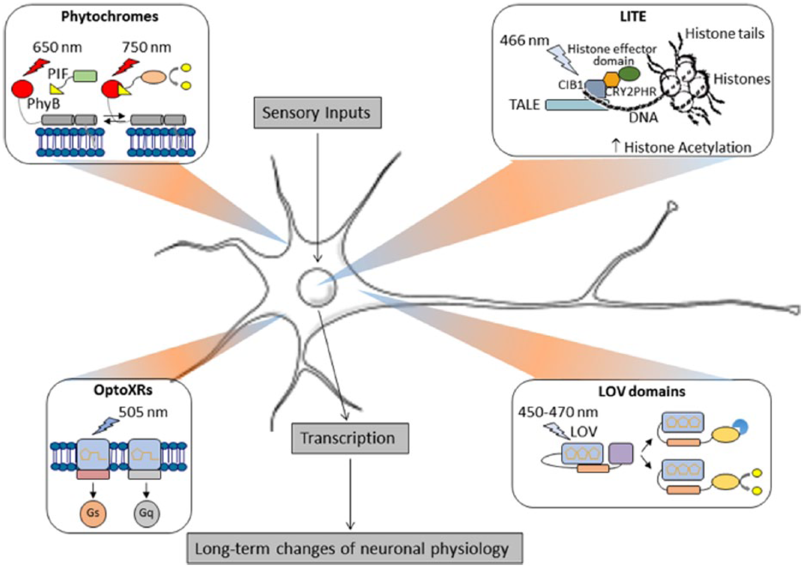
Subcellular targeting of opto-tools. Various light-sensitive systems localize to different intracellular compartments and as such are able to target a specific domain of signalling molecules. For instance, the Phytochromes and the OptoXR system are both membrane associated and have been used to modulate, among other phenomena, peripheral actin dynamics and G-protein signalling. LOV domains are soluble proteins that have been coupled with a number of cytoplasmic effectors. In the LITE system, the transcription activator–like effector (TALE) modules were exploited to target the CRY2-CIB system to the nucleus, thus rendering gene transcription light sensitive. Much like sensory inputs, all the optogenetic tools effectively trigger long-term modifications of neuronal physiology (for specific references, see main text).
Expressing optogenetic actuators in a specific location requires initially the delivery of genetic materials to the nucleus of the correct cell types. Viral transduction is the most common strategy employed in optogenetic studies, intrinsically limiting expression to the injection site. 168 Controlled injection procedures and incubation periods allow anterograde transfection 169 for lentiviruses and AAV or retrograde transport for herpes simplex or rabies viruses, 170 yielding a complete mapping of neural circuits. High infection efficacy in human cells and the absence of associated pathological effects make AAVs the preferred viral vectors for biologists and clinical therapists. 171 AAVs have been used successfully in the delivery of genes56,88 as well as genome editing DNA-binding domains such as zinc finger endonucleases, 172 RNA-guided Cas9 enzymes, 14 or transcription activator–like effector nucleases. 173 Unfortunately, some transcription promoter sequences are quite large, whereas the amount of genetic material packaged in viruses is limited, with AAVs’ cargo limited to 4800 base pairs. To overcome this limitation, scientists make use of the wide variety of CRE recombinase driver lines available to the scientific community in combination with CRE-dependent optogenetic vectors56,88,168 because the CRE promoter sequences are much smaller than selective promoter sequences. Additional tools such as FLEX 174 or lox sites, which are also very short sequences, can be introduced into the viral genome, flanking the gene of interest, to minimize expression leakage into unwanted cell types.168,174
Nanotechnological development has enabled significant advances in synthetic genetic vector systems such as liposomes, 175 nanotubes, 176 and polymer scaffolds. 177 Although these systems allow the packaging of far greater amounts of nucleic acid than viruses, their naturally evolved counterparts remain, for now, the most efficient systems for gene delivery.178,179 A more promising avenue may be the further optimization of virus functionality, such as external receptor targeting 180 or even optogenetic modulation of nuclear translocation, as demonstrated through the insertion of light-sensitive PHY proteins within the capside. 181
Challenges of complex stimulation in vivo
Scattering problem
The scattering of incident light through live tissue is a fundamental problem for non-invasive photo-stimulation of CNS targets. To reach cortical neurons, light has to penetrate through skin, bone, dura, pia, and blood vessels. Deep brain structures are found several millimeters (mice) to tens of centimeters (humans) deeper. The depth of light penetration is proportional to the wavelength, with infrared radiation achieving depths of 2.5 mm, near-infrared (NIR) of 1.4 mm and red of 0.9 mm in explanted human brain tissue. 182 More powerful illumination with NIR has been demonstrated to penetrate through bone and skin samples 183 as deep as 40 mm within the brain. 184 (As an additional tool, the reader is directed to the Deisseroth lab’s tissue penetration calculator: http://web.stanford.edu/group/dlab/cgi-bin/graph/chart.php). Furthermore, the photons’ energy content is inversely proportional to the wavelength, with UV radiation capable of inducing cell death185,186 and DNA damage 187 on its way to the targeted photo-actuators. As such, red and far-red–sensitive proteins such as PHYs 188 or the red-shifted cruxhalorhodopsin Jaws 59 have the potential to be activated non-invasively. Alternatively, blue photo-actuators can be activated distally with 2-photon excitation (2PE). The 2PE technique relies on the convergence of 2 low-energy infrared photons onto a target molecule into a higher energy electronic state 189 and has been shown to work, with some limitations, also on optogenetic proteins. 190 However, this requires a moving objective and a femtosecond laser (both bulky and expensive), so most in vivo studies to date have been performed with blue-sensitive ChR2 and have had to rely on implanted cannulae bearing light guides connected to an external laser (Figure 4A). Considerable resources have now been dedicated to the development of implantable wireless light-emitting diode (LED) optrodes,191–193 but these are still subject to physical (heat dissipation, power requirements) and biomedical (tissue damage, immune response, surgical implantation, discomfort) constraints.

Different approaches to in vivo optical stimulation. Depiction of a rodent brain implanted with a chronic fibre and tethered to stimulation hardware. A coronal section of the brain is used to illustrate in subsequent sub-figures the illumination strategy. (A) Single core fibre optic implant. Here, the light is delivered to an implantable fibre connected extrinsically in the manner of a cannula. (B) Multi-point optical stimulation using an implantable fibre with windows etched along its length, allowing it to deliver light of different spectral compositions (λ) at multiple depths depending on the incident angle (θ) of the light as it penetrates the coupler. 194 (C) 2-dimensional optical stimulation using a spatial light modulator (SLM) which is generally a transmissive liquid crystal display (LCD) or a digital micro-mirror device (DMD). The video signal is focused through a microscope objective onto a fibre optic bundle where each of the fibres transmits a single pixel to an implanted micro-endoscope. 195 The depictions in this figure are neither to scale nor exhaustive.
2-dimensional/3-dimensional light stimulation
The studies described above rely on wide-field excitation, which allows the selective targeting of genetically analogous neuronal populations within an illuminated volume. Spatially selective activation can be achieved in 2 dimensions (2D) with the use of a spatial light modulator (SLM)196,197 (Figure 4C) or a multi-LED array. 198 An elegant example of 2D digital holography performed in vivo in tethered behaving mice consists of a fibrescope set-up combining distinct coherent blue (modulated with a liquid crystal SLM for holographic stimulation of ChR2) and green (modulated with a digital micro-mirror for confocal calcium imaging of genetically expressed GCaMP5-G) light paths. 195
The 2D spatial modulation can be useful when targeting a 2D structure such as the retina, but dissecting neuronal circuits in the brain requires the 3-dimensional sculpting of light into complex volumes. This has been achieved by combining digital holography with 2PE (reviewed thoroughly for their use in optogenetic stimulation in Oron et al 199 ) in vitro, initially for Glu uncaging 200 and more recently for optogenetic activation of fast-acting 201 and red-shifted 202 optogenetic probes. Modulation of the stimulating light in the z dimension in vivo is technically challenging as it requires a moveable objective, making its use in freely behaving animals virtually impossible. In vivo multi-photon imaging of genetically encoded calcium indicators has generated ground-breaking studies in the last decade, relying on 2PE microscopes imaging brain tissue through a cranial window of head-fixed animals.203–205 Although their head is fixed, animals can perform in behavioural experiments based on their navigation atop a 2D treadmill in a projected virtual environment.206,207
Deep brain stimulation
Cranial windows do not allow for deep-brain light stimulation, although this could be achieved with the insertion of a micro-objective or a smaller gradient index lens–based objective. 208 Another possible method for achieving complex light stimulation in deeper structures, without requiring complex lenses, is through the use of single-core, micro-fabricated multimode fibres. 209 This approach is based on digital phase conjugation requiring a calculation of the appropriate wavefront bringing light into defined patterns at the fibre outputs. Further applications of such fibres include advanced microscopy 210 as well as the possibility to deliver spectrally distinct radiations at different depths 194 (Figure 4B). However, there may be complications due to fibre bending, making this approach inappropriate for tethered animals.
Future Perspectives
Chemiluminescence
Optogenetics is an extremely powerful tool to control neuronal excitability and signalling; however, illuminating the cells, a relatively easy task in vitro, becomes the limiting step when trying to apply the same probes to living and behaving animals. Although successful attempts have been made to control animal behaviour by conveying the light through implanted cannulae,34,35,37–40 it is difficult to envisage this method to be the technique of choice for future applications in biomedicine.
In recent years, scientists have started to turn back to what had become an almost obsolete tool, i.e., bioluminescence-emitting luciferases. With the aim of in vivo applications, several groups have tried to engineer red-shifted variants of both luciferases and their substrates.211,212 Interestingly, luciferase-emitted bioluminescence can be used to activate optogenetic probes, provided the emission of the luciferases and the excitation spectra of the optogenetic probes substantially overlap, thus potentially bypassing the need of surgical implants to deliver light from an external source and using systemic injections instead. The use of luciferase to activate optogenetic probes differs from that of chemogenetic modulatory systems such as Designer Receptor Exclusively Activated by Designer Drugs (DREADDs). The former can specifically affect protein function at the intracellular and extracellular level, whereas the later has to make use of signalling pathways originating from membrane-bound receptors. 213 Firefly luciferase could activate Natronomonas NpHR when co-expressed in neurons. 214 Taking a step forward, the bioluminescence resonance energy transfer–based nanolantern probe, formed by the fluorescent protein Venus and Renilla luciferase,215,216 was fused to Natronomonas NpHR. The resulting construct, named inhibitory luminopsin or ‘iLMO’, could silence neuronal firing on coelenterazine (CTZ) administration and modify mouse behaviour when expressed into the striatum and globus pallidum. 217 Similarly, a small luciferase from the sea shrimp Gaussia princeps (Gluc) was fused to ChR2. 218 Using enhanced versions of Gluc, more efficient probes have been recently created by fusing the luciferase with either ChR2 (excitatory luminopsins) or proton pumps (iLMOs). These constructs proved to work also in vivo, as they were delivered to the substantia nigra of living mice and could be activated by both direct and systemic delivery of CTZ. 219
Thus, there is an increasing interest towards the use of bioluminescence as an internal light source. This undoubtedly provides several advantages compared with the ‘classic’ optogenetic approach, chiefly the opportunity to avoid surgery, effectively representing a hardware-free opto-system. This approach is, however, still in its infancy and awaits the availability of a wider panel of luciferases, with different emission kinetics and spectra, as well as of chemically modified and/or caged substrates, which could render luciferase activation more specific in time and space. The combinatorial use of these tools could bring optogenetics a step closer to routine and user-friendly biomedical applications.
Optimization of optogenetic probes
A commonly adopted strategy to tune the photoreceptor photocycle, and thus to adapt it to the specific activity of the effector domain, is to engineer mutations that alter the photocycle time scale by modifying either the chemistry of the cofactor or the properties of the downstream structural moiety. Another possibility is to act directly on the effector domain, for example, by designing mutations that can enhance the interaction with its target. As a speculative example, let us discuss one of the chimeras realized by our group, the AsLOV2-PAH1 construct, 65 where, as effector domain we used the minimal REST-interacting sequence of the corepressor mSin3a. By inspection of the atomic structure obtained by nuclear magnetic resonance studies, it is possible to observe that the REST/PAH1-interacting surface is composed mostly of hydrophobic residues, except for a charged Lys amino acid (K155) on PAH1, which is buried among neutral partners. By performing a computational analysis using a dedicated software, 220 we predicted that changing this residue into a hydrophobic one, such as Phe, would increase the binding free energy of the REST/PAH1 complex by 1.5 kcal/mol. Thus, a chimeric construct carrying the K155F-mutated effector domain should compete more effectively with the endogenous PAH1 binding to REST and give rise to an enhanced binding of the photo-excited chimera.
Recent developments in the field of developmental biology have also yielded new optogenetic tools capable of activating differentiation and developmental features. Examples include the photo-induction of ectopic endodermal cells in zebra fish, 221 an ectopic tail-like structure in Xenopus 222 and the triggering of embryogenesis in Drosophila. 223
Conclusions
In this review, we have tried to summarize the most significant aspects of optogenetics applied to neuronal plasticity. When trying to engineer a novel light-sensitive actuator, but also when ‘simply’ using one of the many tools already available, several challenges have to be faced. This includes the choice of the most appropriate light-sensitive system, of the most efficient way to target it to the cell/brain region under study and of the hardware that could most effectively deliver light while minimizing off-target effects. This may seem an insidious and daunting task; however, despite such complexity, several elegant studies have demonstrated the feasibility of these approaches, showing that synaptic plasticity can indeed be interrogated and/or manipulated using light. The scientific community is putting a continuous effort in optimizing available systems and engineering novel probes, which makes optogenetics a constantly evolving field. Such strive is especially needed to devise optogenetic-based biomedical applications, for which ease-of-use, low-cost, and simple equipment are highly desirable. With the amount of technical and practical knowledge currently in our hands, the advent of such user-friendly tools may not be very far ahead.
Footnotes
Peer Review:
Seven peer reviewers contributed to the peer review report. Reviewers’ reports totalled 1461 words, excluding any confidential comments to the academic editor.
Funding:
The author(s) received no financial support for the research, authorship, and/or publication of this article.
Declaration of Conflicting Interests:
The author(s) declared no potential conflicts of interest with respect to the research, authorship, and/or publication of this article.
Author Contributions
CE, FC, LM, FB, and JFMV jointly developed the structure and arguments of the manuscript. All authors reviewed and approved the final version of the script.