
Abstract
Practical relevance:
As with other species, the skin microbiome of cats has been assessed over the past few years utilizing modern technologies. This has resulted in the identification of many more bacterial and fungal organisms compared with what had been recorded historically on the skin in various states of health and disease using culture-based studies. This information is expanding the knowledge of how microbial communities are impacted by various changes in the skin health of cats. More specifically, how these microbial communities change in the face of health and disease, and how various therapeutic interventions affect the cutaneous microbiome, lends a greater understanding of disease pathogenesis and provides a growing area of research for correcting dysbiosis and improving feline skin health.
Evidence base:
Most studies on the feline skin microbiome thus far have been descriptive in nature. These provide a framework for the next level of investigations on how various states of health and disease impact the products produced by the cutaneous microbiome (ie, the cutaneous metabolome), as well as how targeted interventions may promote the restoration of balance.
Aims:
This review aims to summarize what is currently known about the feline cutaneous microbiome and its clinical implications. The role of the skin microbiome in health and disease, the current state of research in this area and the potential for future studies to produce targeted interventions for cats are a particular focus.
Keywords
The skin microbiome - key players and factors
With expanding knowledge and information comes an improved understanding about what actually makes up the cutaneous ‘microbiome’ (see ‘Terminology’ box). Moreover, the skin microbiome is now recognized not only to exist, but to be an integral part of host health and disease.
In human skin microbiome research, there is an understanding of taxa that are key players,2,3 as well as early information on the potential of microbiome-based therapies,4-6 including probiotics and prebiotics, to treat some of the most common skin conditions. Not surprisingly, research on the canine, and especially feline, skin microbiome is behind relative to human skin studies. However, within the past few years, the microbial communities on these companion animal species have started to be characterized. While most researchers have focused on canine skin microbiota,7-9 several studies dedicated to feline skin microbiota have been performed, including some investigating disease-associated dysbiosis.10-12

The skin represents a unique place for microbes to live, as these organisms have to battle not only against the host immune system and competing microbes, but also the external environment and physiological conditions of the skin. Some features of the skin exist to protect the inner tissues and cells from external damage; for example, an acidic pH and antimicrobial peptides. Cutaneous communities are composed of residents that are able to withstand this tough environment and establish themselves permanently, as well as several transient microbial members, which hold more of a temporary position on the skin and are unable to gain a foothold and incorporate into the cutaneous microbiome. The latter organisms often contribute to infections; dermatophyte species are an example of transient microbial members in cats. Additionally, the skin is not a homogeneous environment across the body, and some sites, such as the axilla, may be less exposed to external influences; differences in hair density, sebaceous secretions or folding may also create niches for certain microbes. 13
Beyond these physiological features that are unique to the skin, there is also a fastidious behavior that should be considered: grooming. Many studies of the feline skin microbiome have recognized the grooming behavior of cats as being potentially important in the context of the skin biome and thus have also investigated samples collected from the oral cavity.10,11,14 While it seems logical that grooming may influence cutaneous communities, the mechanisms and consequences of this remain unknown. The oral cavity may act as a collection site for microbiota, with microbes that are less attached to the skin potentially being easily removed and relocated to the oral cavity. The oral cavity may also act as a source of microbes, with organisms obtained from licking, as well as eating, for instance, potentially being transplanted to the skin. Grooming behavior represents an interesting factor to consider and may have important implications in how the skin microbiota in cats is modulated.
Composition of microbiota in feline skin
Several studies have been undertaken with the goal of describing the feline skin microbiota. The first, performed in 1976 by Krogh and Kristensen, 15 used culture-based methods to identify nine different bacteria across seven body sites. Acinetobacter species, Staphylococcus aureus and alpha-hemolytic streptococci were found to be the predominant taxa. 15 Subsequent investigations of the feline skin microbiota primarily focused on specific microbes rather than providing community-wide descriptions; for example, surveys of staphylococci16–20 or Malassezia species 21 based on culture or case reports of lesions.22-24 With next-generation sequencing (NGS) becoming more accessible, this is now the predominant technique used and has augmented existing understanding of the feline skin microbiota. Unlike historical studies, which were limited to identification of organisms based on culture alone (and the ability of organisms to successfully grow on standard culture media), NGS creates a more robust picture of organisms present in a given sample (see ‘Next-generation sequencing’ box).
The first studies to utilize NGS to investigate the microbiota of feline skin were published in 2017 and focused on the fungal 10 and bacterial 11 communities on healthy and allergic skin. These studies identified a population of microbes on feline skin that was incredibly diverse, much more so than previous studies involving culture-based methods had suggested. Indeed, as with dogs and people, the robust identification of bacterial and fungal organisms previously not reported as part of the cutaneous microbiota, as well as their relative abundances, dwarfed what had been thought to be living on the feline skin.
The fungal microbiome, or mycobiome, was found to be primarily composed of the phylum Ascomycota, which includes many soil-borne fungal organisms. In addition there was a relatively smaller abundance of Basidiomycota, which include many yeast organisms, such as Malassezia species. Dermatophytes, however, were not noted to be part of the normal feline mycobiota. 10 In terms of the bacterial community, Proteobacteria, Bacteroidetes, Firmicutes (including Staphylococcus species), Actino-bacteria and Fusobacteria were found to be the predominant phyla. 11 Interestingly, dysbiosis in the bacterial communities could be observed in allergic cats. This finding, as has been shown in people 2 and dogs, 8 aids our understanding of the pathogenesis of allergic skin disease, as well as the impact the disease condition has on the cutaneous microbiota. The dysbiosis observed may help explain why allergic patients suffer from secondary infections more frequently than individuals in a state of health.
Since these two studies, an additional three have been published on the feline skin microbiota. One of these, a 2019 study, evaluated the influence of both environment and breed on the bacterial and fungal communities, 14 hypothesizing that cats kept completely outdoors would have more diverse communities than those kept indoors. Surprisingly, while some differences in the taxonomic composition of communities were observed based on environment, no differences in diversity were observed in the skin of the cats sampled. Interestingly, the oral cavity showed some differences in bacterial community structure, or beta diversity (see below), based on environment, which were suggested to be due to the difference in food sources available to indoor and outdoor cats. In comparing six different cat breeds, significant differences could be observed in both alpha (within-sample) and beta (between-sample) diversity of both bacterial and fungal communities in some body sites. For the bacterial communities, the dorsum was particularly influenced by breed. In terms of the community composition, cat breeds were shown to differ in relative abundance of Malassezia species, as had been observed in an earlier culture-based study. 21 Overall, the 2019 study of breed and environment showed that both factors may influence the skin microbiome. While it is possible that other influences, such as health status, may be more impactful, breed and environment should be considered when identifying animals for a study, or at least when analyzing data, to ensure they do not act as strong confounding factors. The potential impact of breed and environment should also be taken into account when evaluating microbiome findings in a clinical patient. What may be considered ‘dysbiosis’ in one setting (eg, an indoor-only home) may be a state of normalcy for a patient in another setting (eg, a cat with access outdoors).
The second of the three studies, published in 2021, focused on the staphylococcal communities present on healthy and allergic feline skin. 12 Staphylococcus species are recognized as being important players in human and canine atopic dermatitis, with regards to disease manifestation and exacerbation. While the impact is unclear in feline patients with allergic dermatitis, the 2017 NGS study on healthy and allergic feline skin suggested higher relative abundances of staphylococci on affected animals (Figure 1). 11 The predominant species on feline skin is also unclear, given the range of different species that have been identified across studies.16,19,20,25

Staphylococcus species appear to be more abundant on allergic feline skin (shown here) compared with the skin of a healthy cat
In an effort to identify all the staphylococcal species present, as well as those that may be more allergy-associated, the 2021 study utilized NGS methods that allowed for species-level identification. 12 Regardless of health status, feline skin was shown to harbor diverse communities of staphylococci, with 11 different species identified including Staphylococcus capitis, Staphylococcus epidermidis and Staphylococcus felis. While both healthy and allergic cats showed similar staphylococcal species, some species were found to be more abundant on healthy compared with allergic cats - findings warranting further investigation. More specifically, the majority of staphylococcal sequences from healthy feline samples were classified as S epidermidis, while S capitis was the predominant species found in allergic feline samples. The precise significance of this finding is, as yet, unknown; however, there may be a protective mechanism at play, or this may represent a response to a disease trigger, causing the relative abundances of specific Staphylococcus species to be skewed.
The third study, also published in 2021, looked to further characterize the cutaneous fungal microbiota of Persian cats with severe dermatophytosis, and compare findings with Persians and other longhaired cats with and without a history of ringworm. 26 NGS identified Microsporum canis in all affected Persians, as well as in their home environment. There was a distinct difference in the fungal communities on the skin of affected and non-affected cats, influenced by the large amount of M canis present in the former. Interestingly, however, Persians affected with dermatophytosis did not show decreased fungal diversity in their cutaneous microbiota compared with healthy controls. The presence of fungal organisms remained robust in affected Persians, the difference being the inclusion of dermatophyte fungal species (which were not seen as part of the ‘normal’ mycobiota of healthy controls). In this sense, fungal diversity did not appear to influence the development of clinical disease. This contrasts with what is seen with allergic skin diseases, in which the lack of microbial diversity appears to be a factor in disease development.
These recent studies utilizing NGS - the findings of which are summarized in the box ‘Bacterial and fungal microbiota: comparative descriptions’ - have laid the groundwork for more applied or comparative studies to build upon. Some of the most interesting findings that may be the basis for future research include the significant differences in the relative abundance of Malassezia species between breeds, and the tremendous range of staphylococcal species found on healthy and allergic feline skin.
Role of microbiota in skin health
Traditionally, ‘microbes’ have been discussed in the veterinary literature solely in the context of the state of disease or infection. Microbes are most often thought of as pathogens or, at best, simply commensals that do not contribute to or harm the host. As has been shown, however, microbes can assume a number of different roles, including maintaining a state of health or homeostasis for an individual - the balance of the cutaneous microbiome. The same microbe can even play different roles depending on the health status of the host, 29 with Staphylococcus species providing a good example of this: S epidermidis may be protective in a state of health, yet detrimental during an allergy flare-up in people. In short, microbes can, of course, cause infection and act as allergens, but they can also be important in host protection.
Most historical (and recent) veterinary literature has focused on the description, diagnosis and treatment of infectious microbes in the feline patient. Overarchingly, the bulk of research has been on the management of feline dermatophytosis (Figure 2) 30 - perhaps not unexpectedly given it is one of the most common infectious skin diseases in cats worldwide 30 and tends to affect the most adoptable population of animals (ie, kittens). Several studies have documented a lack of pertinent dermatophytes in the cutaneous mycobiota of cats;10,31,32 rather, these organisms are found when infection is present or in carrier states of certain breeds (most commonly Persians). 26

Persian cat with severe dermatophytosis caused by Microsporum canis
There is, in contrast, a lack of extensive studies on bacterial pyoderma in cats, especially when compared with dogs. This infectious condition seems to be underrecognized, in part due to the differing appearance of feline pyoderma vs its canine counterpart. Whereas dogs with superficial bacterial folli-culitis (the most common form of pyoderma) tend to show classic lesions of papules, pustules, crusts and epidermal collarettes, the clinical appearance of pyoderma in cats is highly variable. In common with other feline dermatoses, bacterial pyoderma may manifest with any of the feline cutaneous reaction patterns, including head, neck or pinnal pruritus (Figure 3), miliary dermatitis, self-induced alopecia, and eosinophilic skin lesions (granuloma, plaque, indolent ulcer; Figure 4). 33
Additionally, feline corneocytes have reduced adherence of staphylococcal organisms compared with the corneocytes of dogs and humans. 34 While low numbers of staphylococci may typically be found on the skin of healthy dogs and people, in cats, given the decreased ‘stickiness’ of bacteria to their skin, small numbers tend to have a larger impact on disease state and should be taken into consideration when recommending treatment interventions. For a cat with representative skin lesions, therapy is typically indicated to treat the infection even if only a small number of bacteria are observed on a cytology sample. The same number of organisms on cytology may not necessitate aggressive infection control in a dog, however.
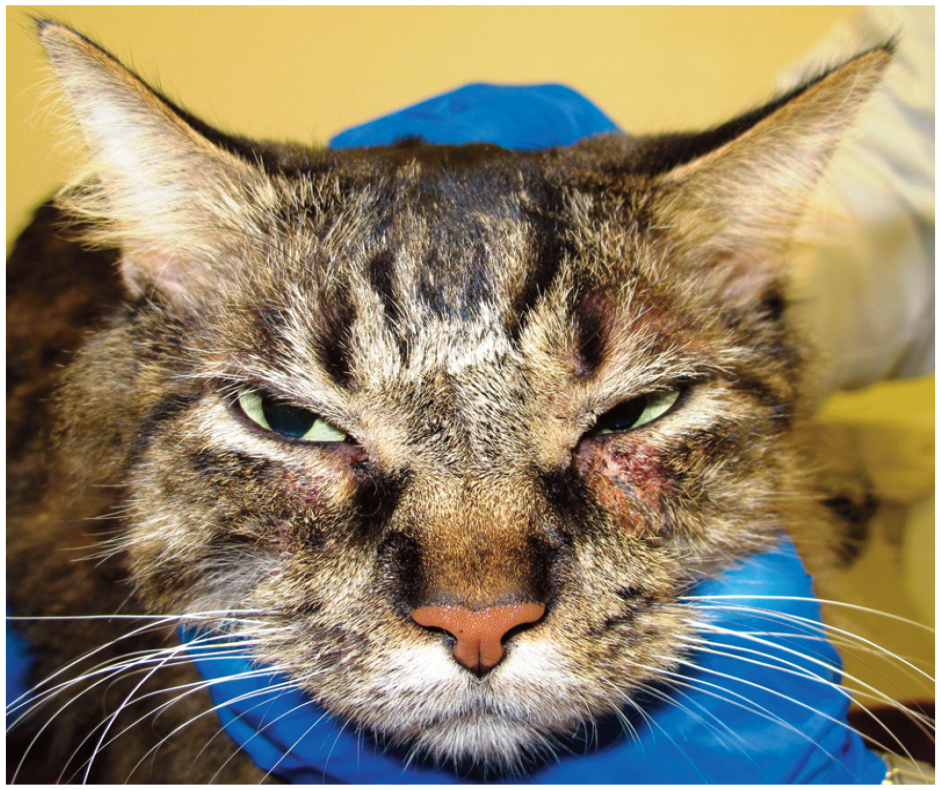
Cervicofacial pruritic dermatitis and secondary bacterial skin infection in a cat

Allergic cat with self-induced alopecia and eosinophilic plaque lesions. A large population of bacterial organisms was identified on cytology samples
Similarly to feline bacterial pyoderma, there is a distinct lack of studies on Malassezia dermatitis in cats. Older literature focused on the finding of severe illness, most commonly neo-plastic conditions, when Malassezia dermatitis was identified in cats.35,36 More recent literature shows close parallels with what is seen in canine patients with Malassezia dermatitis - that cats with allergies, cornification defects and endocrinopathies have some degree of predisposition to yeast overgrowth.37,38 This tends to be seen more commonly in certain cat breeds (eg, Sphynx, Rex).21,39
Either microbes themselves or their products can act as allergens. Of the several fungi that are known to be allergens, 40 the genus Malassezia has received particular attention due to its abundance on human skin 28 and relevance in atopic dermatitis, where it is recognized as an important pathogen and an inducer of IgE production and sensitization.40,41 Examples of microbe-derived products that can act as allergens include superantigens produced by S aureus, which are able to activate the immune system.42,43 Exfoliative toxins produced by some bacteria can also stimulate the immune system and damage the skin barrier, allowing the bacteria and other microbes to enter the skin and interact with the already hyperreactive immune system.44-48
As mentioned, microorganisms can also play a key part in host defense. Many studies have investigated the roles of various staphy-lococcal species due to their importance in skin health.29,49 As well as imparting protective benefits (eg, S epidermidis during a state of skin health), these microbes have been shown to enhance the ability of the innate cutaneous immune system. A few coagulase-negative species have been found to protect the host from more pathogenic strains by producing S aureus-targeting antimicrobial peptides,6,50 and even inducing host-derived antimicrobial peptide production.51,52 Beyond antimicrobial production, bacteria may also improve epidermal barrier function, 53 potentially aiding prevention of infection.
Moving forward on the skin microbiome
Next steps for research
Much of the current microbiome work in cats has, as mentioned, been descriptive in nature, categorizing differences seen in various states of health and disease. In terms of next steps, there is a need to investigate how various interventions impact the cutaneous micro-biome as well as the products that the organisms generate (ie, the cutaneous metabolome). Some of this work is already happening as it pertains to the feline gastrointestinal and oral microbiomes;54,55 however, thus far, changes in the cutaneous microbiota in relation to applied therapy have only started to be evaluated in dogs.56,57
Additionally, the cutaneous metabolome itself needs to be further investigated in the feline patient. Various disease states not only impact the microbial communities on the skin, they also impact the metabolites and products generated by those organisms. What all this means for the state of health or disease has yet to be determined.

Potential applications in clinical practice
As understanding of the feline cutaneous metabolome improves, it is hoped this will allow practitioners to provide more targeted interventions to return a cat to a state of health when microbial dysbiosis occurs (see ‘Microbial-based therapies’ box). This might be in the form of the nutrition needed to promote the healthy microbiome (eg, prebiotics, probiotics) or therapies to target the dysregu-lated population (eg, antibiotics, competitive microbe-derived products). Species-specific considerations will need to be taken into account for cats, however, as many owners have difficulty administering oral medications and many cats are averse to topically administered products. What may be beneficial for regulation of the canine cutaneous microbiome will likely need to be evaluated separately in the feline patient, not only for efficacy but also for feasibility.
Key Points
✜ Comparatively less is known about the feline cutaneous microbiome than that of other species. This is unfortunately in line with feline dermatology as a whole, particularly in relation to what is known/investigated in dogs.
✜ Much work needs to be carried out, not only regarding how the cutaneous microbiome and metabolome is affected in various disease states of the cat, but also how interventions may be able to reverse the dysbiotic state.
✜ NGS techniques are beneficial for research on the cutaneous microbiome and have been integral in framing a more complete picture of the feline skin in states of health and disease. While this technology is becoming more widely accessible with the availability of commercial tests, their use in clinical practice is quite limited at this time. Until there is more information on how specific therapeutics may impact the cutaneous microbiome, NGS is best utilized in a research setting.
Footnotes
Conflict of interest
The authors declared no potential conflicts of interest with respect to the research, authorship, and/or publication of this article.
Funding
The authors received no financial support for the research, authorship, and/or publication of this article.
Ethical approval
This work did not involve the use of animals and therefore ethical approval was not specifically required for publication in JFMS.
Informed consent
This work did not involve the use of animals (including cadavers) and therefore informed consent was not required. No animals or people are identifiable within this publication, and therefore additional informed consent for publication was not required.