
Abstract
Objectives
The aim of this study was to evaluate, using echocardiography, the effects of oral administration of a single dose of gabapentin on the physiologic variables (heart rate [HR], respiratory rate [RR] and systolic blood pressure [SBP]) and systolic and diastolic cardiac function of healthy cats.
Methods
This was a prospective, randomized and blinded study with 40 healthy cats aged between 6 months and 2 years. The cats’ health status was assessed on the first appointment (T1) when they underwent a physical examination, complete blood count, biochemical profile, assessment of physiologic variables and echocardiogram. The echocardiogram was used to measure the left ventricle’s (LV) internal diameter during systole and diastole, isovolumic relaxation time, transmitral flow, E-wave deceleration time and HR. The cats were randomly divided into two groups: (1) a treatment group with 20 cats that received a single oral dose of gabapentin (100 mg/cat); and (2) a control group with 20 cats that received a single oral dose of placebo. All variables of the physiologic and echocardiographic variables were re-evaluated 1–3 weeks after T1 (T2), 90 mins after medication or placebo administration.
Results
There was no difference in the physiologic variables evaluated in both groups. The proportion of cats in the treatment group that had their ventricular filling waves fused on T1 but did not have them fused on T2 was significantly higher (45%) compared with cats in the control group (15%; P = 0.0384).
Conclusions and relevance
There was no difference between the groups in regard to SBP, HR, RR and echocardiographic variables. Gabapentin improved evaluation of diastolic function on echocardiogram because it reduced the fusion of ventricular filling waves during the evaluation of the diastolic function of the LV. Gabapentin did not cause adverse effects on the cardiovascular hemodynamics of young healthy cats.
Keywords
Introduction
Oral gabapentin is a well-tolerated anxiolytic used in cats. It is administered before veterinary appointments and has many benefits.1,2 The main purpose of its use is to reduce anxiety in cats when going to a veterinary clinic or hospital as this can be considered a stressful event. In addition, during the medical consultation, some actions such as restraint, quick and sudden movements and loud sounds can be intimidating, causing hostile reactions in cats. 3 Stress increases sympathetic discharge, which releases epinephrine (adrenaline), norepinephrine (noradrenaline) and cortisol.4,5 Consequently, norepinephrine is released via nerve endings, which elevates the heart rate (HR), respiratory rate (RR), systolic blood pressure (SBP) and rectal temperature. 5
Occasionally, it is necessary to administer drugs that reduce a patient’s anxiety, facilitate handling and reduce its level of stress.3,6,7 The anxiolytic of choice should be one that is safe, well tolerated and one that does not significantly affect cardiovascular and hemodynamic function in order to obtain reliable results in complementary tests and, consequently, avoid an incorrect diagnosis. 8
Gabapentin is a structural analog of gamma-aminobutyric acid. It binds to the alpha2delta (α2δ) accessory subunit of the voltage-dependent calcium channel complexes, mainly in the spinal dorsal horn and the prosencephalon, 9 and provides an inhibiting effect that alleviates anxiety. 10 Recent studies with gabapentin in feline species have shown that it is a safe and effective short-term anxiolytic.1,2 However, its effect on cardiovascular function needs to be further investigated, as only one study to date has evaluated the effect of gabapentin on the physiologic and echocardiographic variables in cats. 8
The objective of the present study was to evaluate the effects of oral gabapentin on the physiologic and echocardiographic variables of healthy cats. The physiologic variables included HR, RR and SBP. The echocardiographic variables were obtained to evaluate systolic and diastolic function. Our hypotheses were: (1) gabapentin does not significantly affect any hemodynamic or echocardiographic variables; and (2) gabapentin facilitates the interpretation of diastolic function in healthy cats by helping to separate the ventricular filling waves of the left ventricle (LV) in stressed or anxious cats.
Materials and methods
Animals
Healthy cats aged between 6 months and 2 years of both sexes and different breeds were selected to participate in the present study between June and December 2021. The cats that participated in the research were later referred to the Feline Medicine Service of the Universidade Federal do Rio Grande do Sul’s Veterinary Teaching Hospital for elective sterilization. The present study was approved by the animal ethics committee (CEUA/UFRGS, approval 40478). The owners signed an informed consent form to participate in this study.
Inclusion criteria included healthy cats aged between 6 months and 2 years, of any sex and any breed. The cats were considered healthy through physical examination, including cardiopulmonary auscultation, measurement of physiologic variables such as HR, RR, rectal temperature and non-invasive SBP by the vascular Doppler method, echocardiogram, complete blood count and serum biochemical profile. Cats were excluded if they showed any alterations in screening tests and/or any structural or functional abnormalities on echocardiographic examination and/or if they were receiving any medications other than periodic antiparasitic drugs.
Study design
This study was prospective, randomized and blinded. On the day of the first (T1) and second (T2) appointments, the cats were examined in the presence of their owners at the Feline Medicine Service at Universidade Federal do Rio Grande do Sul’s Veterinary Teaching Hospital. During both appointments, the cats were acclimated for 15 mins before assessment of the physiologic and echocardiographic variables. The cat carrier was always left open to allow the cat to leave it voluntarily. Cats that did not leave the carrier were gently removed and wrapped in a towel. The same researcher assessed all the physiologic and echocardiographic variables.
A simple randomization table was created in Microsoft Excel in order to divide the cats into two groups and assign them to the corresponding treatment. Each group had the same number of male and female cats. A researcher – who did not participate in the clinical and echocardiographic evaluation of the cats – handed to the cat owners, by the end of the screening appointment (T1), a capsule, with the type of capsule given based on the randomization table. The capsule contained either placebo (composed of magnesium stearate, sodium lauryl sulfate, talc and corn starch) or 100 mg of gabapentin fractioned in a veterinary compounding pharmacy from a drug commercially available for humans (Gabaneurin; EMS Sigma Pharma). The pharmacy follows rigorous quality-control procedures and the standard operating procedures of the Brazilian Ministry of Agriculture, Livestock and Food Supply and the Brazilian Health Regulatory Agency.
The owners were asked to administer the capsule to their cats at home around 90 mins prior to T2, which was scheduled between 1 and 3 weeks after T1. At T2, the owners completed a questionnaire, stating the exact time at which they had administered the capsule to their cats, as well as the degree of difficulty in doing so, and then returned it to the research team. All owners who participated in the study and the researchers who collected the data were blind to the treatment.
Assessment of the degree of relaxation/sedation, and respiratory and heart rates
During the initial physical examination, the degree of relaxation/sedation was assessed to confirm an adequate effect of gabapentin. Subjective assessment of patient behavior after gabapentin ingestion, such as adherence to restraint and degree of spontaneous mobility, was compared with baseline parameters at T1. Afterward, the RR was measured by inspecting and counting the respiratory movements at a distance, without restraining the cat. HR was then determined through cardiac auscultation with a stethoscope. Restraint was kept to a minimum and the cat received a massage on the head, around the ears and under the chin. At this time, a pulmonary auscultation was also performed. For each variable, three consistent measurements were performed, after excluding the first outliers, thus obtaining an average value.
Non-invasive assessment of SBP
SBP was measured by Doppler ultrasonography following a previously reported protocol. 11 Two people performed this procedure; however, the same experienced veterinarian measured the SBP in every cat. The cuff was measured according to 30–40% of the circumference of the cat’s forelimb on the left thoracic limb. The cat was gently restrained using a towel in a way that did not excessively restrict its movements, following a previously reported protocol. 12 Headphones were used to prevent the sound of the device from frightening the cat. The first assessment of SBP was discarded. Then, five additional assessments were performed that had consistent values, indicating that the variability between each was <20%. The mean value was then calculated and recorded. 11
Echocardiography
The echocardiographic examination was performed after the assessment of the physiologic variables and the SBP. Both were performed at the Feline Medicine Service at the Universidade Federal do Rio Grande do Sul’s Veterinary Teaching Hospital. The same evaluator performed the examination using the same ultrasound device in all cases (Siemens ACUSON P500 Ultrasound System; Siemens Healthineers). A sector transducer at a frequency of 4–8 MHz was used. The cat’s thorax had been previously shaved, to improve visualization of the echocardiographic images. The cat was first positioned in right lateral recumbency and then in left lateral recumbency on a table that was specifically designed for the echocardiographic examination. The table had an opening in the center to allow the correct positioning of the transducer on the cat’s thorax.
The LV variables were analyzed. In the right parasternal window, along the transverse axis, the systolic and diastolic diameters of the LV were measured to obtain indicators of systolic function, such as the shortening fraction (SSF) and the ejection fraction (SEF) of the LV. From the apical four-chamber view, in the left parasternal window, along the longitudinal axis, the sample volume of the pulsed Doppler was positioned between the tips of the mitral valve leaflets to measure the transmitral flow in diastole and to evaluate the diastolic function. The early transmitral flow peak (E-wave), late transmitral flow peak (A-wave) and E-wave deceleration time (EDT) were measured. The ratio between the E-wave value and the A-wave value (E:A) was recorded. In cats whose E- and A-waves fused, the maximum velocity of the fused wave and its pressure gradient were obtained. From the five-chamber view, with the sample volume inside the LV, between the septal leaflet of the mitral valve and the LV outflow, the transmitral and aortic flows were registered to obtain the isovolumic relaxation time (IVRT).
After obtaining the ventricular filling waves through transmitral flow, the echocardiographic device calculated the distance between one E-wave and the next to estimate the HR (HR-ECHO). The HR values measured by the echocardiogram were also recorded and analyzed.
All the aforementioned variables were measured three times in order to calculate the mean values. The images were stored digitally and analyzed remotely.
Statistical analysis
A descriptive analysis was performed by calculating: (1) the mean ± SD of the quantitative variables; and (2) the proportions of the qualitative variables. To compare both groups, the difference (delta) between the quantitative variables in T1 and T2 was calculated and then a t-test was used to compare the means of the independent samples. The Shapiro–Wilk test was used to assess the normality of the distributions. The assumption of homoscedasticity of the data was also verified. A binary qualitative variable was created to label the ventricular filling waves as either ‘fused’ or ‘unfused’ in T1 and T2. Based on the new qualitative variables, a test to compare the proportions was performed. The z-test for two independent proportions, which is equivalent to the χ2 independence test, was carried out. As a decision criterion, the significance level was 5%. Statistical analysis was performed using IBM SPSS Statistics 18 (IBM).
Results
Forty-two cats were initially selected to participate in the study; however, two went on to be excluded. The reasons for the exclusion were the presence of the phenotype of hypertrophic cardiomyopathy and clinical suspicion of pyometra. This resulted in a total of 40 cats that met the inclusion criteria. Of these 40 cats, 20 were female and 20 were male. The cats’ mean age was 10 months (range 6–24). The cats did not belong to any specific breed. Mean weight was 3.22 kg (range 2.065–5.32). Patients received the capsule at home approximately 90 mins before the veterinary appointment (Table 1). Mean dose of gabapentin was 31.35 mg/kg (range 18.79–47.61). No adverse effects were observed in the cats given gabapentin.
Time elapsed between the ingestion of the capsule and the beginning of the veterinary appointment in the treatment group (gabapentin) and in the control group (placebo)
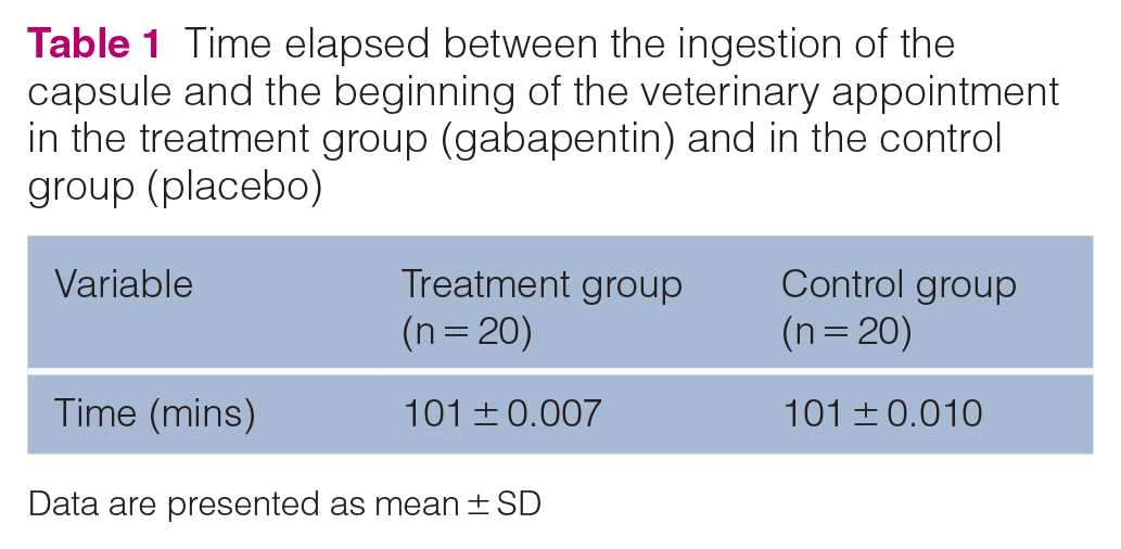
Data are presented as mean ± SD
The data were analyzed and both groups were compared on T2. No statistically significant differences were found between groups regarding RR, HR and SBP (Figure 1). No statistically significant differences were found between the groups regarding echocardiographic variables (ie, EDT, IVRT, SSF and SEF; Table 2). There was no statistical difference between HR measured with a stethoscope and HR measured on echocardiogram (Table 3).

Mean respiratory rate (RR), heart rate (HR), HR measured by the echocardiogram (HR-ECO) and systolic blood pressure (SBP) in the treatment group (gabapentin, 20 cats) and the control group (placebo, 20 cats) at T1 (before treatment) and T2 (after treatment)
Descriptive statistics for the echocardiographic variables in the treatment (gabapentin) and control (placebo) groups at time 1 (T1; before treatment) and time 2 (T2; after treatment)

Data are presented as mean ± SD (range)
EDT = E-wave deceleration time; IVRT = isovolumic relaxation time; LV_SSF = left ventricular fractional shortening; LV_SEF = left ventricular ejection fraction
Descriptive statistics of heart rate (HR) measured with the stethoscope and the echocardiogram in treatment (gabapentin) and control (placebo) groups at time 1 (T1; before treatment) and time 2 (T2; after treatment)

Data are presented as mean ± SD
HR = heart rate;
HR-ECHO = heart rate measured on echocardiography; bpm = beats/min
The proportion of cats in the treatment group that had ventricular filling waves fused at T1 but did not have them fused at T2 was statistically significantly higher (45%) compared with cats in the control group (15%; P = 0.0384).
Discussion
This study did not find significant differences in physiologic and echocardiographic variables after the oral administration of gabapentin in healthy cats. The cat owners administered gabapentin to their cats at home around 90 mins before the appointment at the veterinary clinic. The chosen dose of gabapentin was based on previous studies that elected a dose of 100 mg of gabapentin per cat, with a dose range of 13–29.4 mg/kg 1 and 9.2–47.6 mg/kg,2 and they had positive results in tranquilization. A single measurement was obtained post-treatment, at a time that may or may not correspond to the peak effect, but this interval was chosen based on previous studies that reported that the time at which gabapentin reaches its mean peak serum concentration is 60–100 mins after its administration.9,13
No significant difference was found in RR, HR, HR-ECHO or general SBP considering T1 and T2. This suggests that cats did not experience significant hemodynamic consequences on the echocardiographic examination after receiving oral gabapentin. A recent study reported a mean decrease of 15.2 beats/min (bpm) in HR after a dose of 100 mg of gabapentin. 1 Overall, RR and SBP decreased in cats in the treatment and control groups at T2. This might be related to a decrease in sympathetic tone, 8 since cats were already familiar with the clinic’s environment and may have been less anxious at T2.
According to previous studies, other sedatives cause considerable hemodynamic alterations that hinder correct interpretation of the physiologic and echocardiographic variables.14–18 One study suggested that a combination of butorphanol and ketamine resulted in a significant increase in HR. Moreover, a combination of butorphanol and acepromazine leads to a significant decrease in SBP. 14 Acepromazine – a phenothiazine derivative – can cause paradoxical excitement and hypotension. 15 Butorphanol could be used before the echocardiogram since its agonist–antagonist effects cause sedation, analgesia and low cardiorespiratory depression. 16 However, if butorphanol is administered alone, it only provides light sedation, 17 which would be ineffective in overly nervous and scared cats. A recent study evaluated the effects of oral trazodone before the assessment of physiologic and echocardiographic variables, 18 and did not reveal alterations in echocardiographic rates, although SBP decreased significantly. The present study demonstrated the benefits of administering gabapentin as an anxiolytic before the assessment of physiologic and echocardiographic variables.
Since most cats become stressed under echocardiographic examination, tachycardia is a common occurrence, which leads to a partial or complete fusion of early and late ventricular filling waves and an HR >170 bpm. In this study, an HR >180 bpm, measured by the echocardiogram, resulted in partial or complete fusion of the ventricular filling waves in most cats. The sum of the flow waves leads to interpretation errors and thus must be avoided in the assessment. 19 Therefore, an anxiolytic would be beneficial for cats that are agitated or scared, because the calmer the cat, the lower the probability of fusing of ventricular filling waves (E and A).14,20
In this study, the proportion of cats in the treatment group that had their ventricular filling waves fused in T1 but not in T2 was significantly higher (45%) compared with cats in the control group (15%). Using gentle handling and minimal restraint on cats can help to alleviate stress and prevent E- and A-waves from fusing. The anxiolytic effect can also be beneficial for agitated cats. 19 Both factors were considered in this study, which might have contributed to the statistical difference between both groups. In contrast, the decrease in HR was evident in both groups in T2, with no difference between them, as attested by both the stethoscope examination and the echocardiogram. Nevertheless, the treatment group showed a higher tendency of not fusing the ventricular filling waves. A possible cause for this is that the HR obtained in the echocardiographic examination was 10.3 bpm lower than the HR obtained with the stethoscope. The difference was not statistically significant, but the decrease in HR must be considered because it can influence the fusion of the ventricular filling waves. According to Schober and Chetboul, 19 the complete or partial fusion of the ventricular filling waves can occur with an HR >170 bpm. In the present study, the fusion of the ventricular filling waves was observed with an HR >180 bpm in most cats in the treatment group. A recent study demonstrated a slight decrease in HR after a 100 mg dose of gabapentin. 1 A decrease in HR, even if clinically irrelevant, might prevent the ventricular filling waves from fusing, which was evident in cats in the treatment group at T2.
Gabapentin did not affect systolic function. A recent study demonstrated a decline in the systolic function of healthy cats that received sedative doses of gabapentin. However, all the variables that were measured remained within the existing reference intervals. 8 Similarly, the present study found a slight decline in the systolic function of the LV in the treatment group. In this study, there was no difference between the two groups in relation to the EDT. Some researchers have argued that an increase in HR does not affect EDT in cats or humans.21,22 However, other researchers have reported an increase in HR followed by a decrease in EDT in cats and humans.23,24 In the present study, it was not possible to prove the increase in EDT with the reduction in HR. The results of this study are inconclusive regarding whether EDT really increases under the effect of gabapentin. A study with a larger sample of animals is necessary to validate the hypothesis.
A previous study reported that the higher the HR, the lower the IVRT. 19 This is because the sympathetic stimulus – which results in tachycardia – might decrease the diastolic filling time and thus increase the velocity of the early diastolic elastic recoil of the LV. 21 Based on this study alone, it is not possible to confirm whether the increase in IVRT in T2 was caused by gabapentin, though it might be related to a decrease in HR, which was seen in cats in both groups. One study has claimed that HR does not influence IVRT, but their study only had a small sample, diminishing its statistical power. 24 IVRT varies with age: it is lower in young cats and higher in older cats. 19 In this study, it was unlikely that age affected the results since all cats were <2 years of age.
This study has some limitations. First, a single measurement of variables was performed after treatment, which may not have corresponded to the peak of action of gabapentin. Another point is that we analyzed a small number of healthy cats, and thus it cannot be assumed that gabapentin will not have an effect on the physiologic and echocardiographic variables of older cats or cats with heart disease. In addition, owing to the small sample of the study, the statistical power was reduced. Another limitation is that the results of this study are based on a specific range of gabapentin doses (18.79–47.61 mg/kg), so results can vary with different or repeated doses. Additional studies are needed to evaluate whether gabapentin has a hemodynamic influence in cats that receive this medication daily.
The results of this study suggest that a single oral dose of gabapentin improves the evaluation of diastolic function of the LV because it prevents ventricular filling waves from fusing in the Doppler echocardiogram if it is administered 90 mins before the examination. Furthermore, the results suggest that the use of gabapentin under the conditions described in this article does not significantly change physiologic and echocardiographic variables and is well tolerated by cats. However, additional studies with a larger number of cats are needed in order to confirm our findings.
Conclusions
Gabapentin did not change the physiologic and echocardiographic variables analyzed in this study. Therefore, it can be concluded that gabapentin did not significantly affect the cardiovascular hemodynamics of healthy young cats. Gabapentin improved the evaluation of the diastolic function on echocardiogram because it reduced the fusion of the ventricular filling waves during the evaluation of the diastolic function of the LV. For this reason, gabapentin can be considered as an anxiolytic for healthy young cats before a veterinary visit and echocardiogram.
Footnotes
Conflict of interest
The authors declared no potential conflicts of interest with respect to the research, authorship, and/or publication of this article.
Funding
This study was supported by grants from the National Council for Scientific and Technological Development (CNPq; grant number: 131701/2018-5).
Ethical approval
The work described in this manuscript involved the use of non-experimental (owned or unowned) animals. Established internationally recognized high standards (‘best practice’) of veterinary clinical care for the individual patient were always followed and/or this work involved the use of cadavers. Ethical approval from a committee was therefore not specifically required for publication in JFMS. Although not required, where ethical approval was still obtained, it is stated in the manuscript.
Informed consent
Informed consent (verbal or written) was obtained from the owner or legal custodian of all animal(s) described in this work (experimental or non-experimental animals, including cadavers) for all procedure(s) undertaken (prospective or retrospective studies). No animals or people are identifiable within this publication, and therefore additional informed consent for publication was not required.