
Abstract
Objectives
The aim of this study was to assess the histopathological findings in the uteri and ovaries from clinically healthy queens presented for elective spaying.
Methods
Ovaries and distal uterine horns or complete uteri from 106 female cats were evaluated for pathological alterations.
Results
Pathological alterations of the uterus and/or ovaries were evident in 29 cats; of these, corpora lutea were present on the ovaries of 15 cats. Ovarian cysts were found in 15 cats and were classified as cysts of the Wolffian or Muellerian ducts (n = 4), follicular cysts (n = 4), luteal cysts (n = 1), cystic rete ovarii (n = 2), combinations of different cysts (n = 2) and non-classifiable cysts (n = 2). In 21/106 cats, cystic endometrial hyperplasia (CEH) was present. The incidence of CEH increased with the age of the cat. Six cats had purulent endometritis with or without distension of the uterine lumen. Hyperplastic lesions of the endometrium were detected in two cats. In one cat, a uterine horn malformation with duplication of one uterine horn lumen was diagnosed.
Conclusions and relevance
Whereas the majority of ovarian cysts and slight-to-moderate CEH are unlikely to interfere with an animal’s wellbeing, endometritis must be considered a serious health problem that requires veterinary attention.
Introduction
The reasons to spay female animals differs among species and also between countries. In the USA and Europe, the proportion of privately owned cats reported to be neutered ranges from 27% to 93%. 1 In Austria, legislation requires all female cats with free outdoor access and not explicitly intended for breeding to be spayed. However, this has not so far been verified by the relevant authorities. Therefore, the decision of whether to have their animals spayed is still largely that of the owner. In female cats, commonly also referred to as queens, the main reasons for spaying are to prevent unwanted litters and to inhibit oestrous behaviour, which is often considered annoying by owners. 2
It is widely accepted that the repeated stimulatory effects of progesterone and oestrogens are potential risk factors for the development of proliferative conditions in the uterus and ovaries of dogs.1,3–10 Oestrous cycle-related pathologies of the genital tract have received much less attention in cats than in dogs.11–15 With spontaneous ovulation in queens occurring more frequently than often assumed,16,17 cyclic changes in progesterone and oestrogen concentrations may also predispose female cats to pathologies of the reproductive organs such as cystic endometrial hyperplasia (CEH), purulent endometritis and ovarian cysts.
Histopathological findings in the uteri and ovaries collected from clinically healthy animals at elective ovariohysterectomy have been published for dogs;5,10 however, to the best of our knowledge, such studies in cats are still missing. A review of the recent literature shows that feline reproduction is still a field needing in-depth research. This is true with regard to both physiological pathways and pathologies of the genital tract. 18 Therefore, the objective of this study was to analyse and describe uterine and ovarian pathologies in otherwise healthy female cats that underwent elective spaying.
Material and methods
Animals
Ovaries and distal uterine horns or complete uteri were collected from 106 female cats presented for routine ovariectomy at Vetmeduni Vienna. The animals were aged between 4 and 111 months (21.6 ± 19.8 months). Their body weight ranged from 1.9 to 4.3 kg (3.1 ± 0.5 kg). Physiological data from 89 cats either without pathological alterations of the uterus and ovaries or with only CEH have been reported previously 16 and therefore will not be repeated here.
All cats were adopted from the same animal shelter by their owners no longer than 1 year before surgery. Spaying was offered to the cat owners by Vetmeduni Vienna in cooperation with the animal shelter. In total, cats were clinically examined at least four times before spaying. None of them showed clinical signs of disease and no cat was pregnant on transabdominal ultrasonography of the uterus. According to the owners, queens had not been in contact with gonad-intact males, and no pregnancies were detected in this study.
Because the reason for ovariectomy was not related to our study, no animal experimentation licence was required. Prior consent of the owners for scientific use of the material removed at the time of surgery was always obtained.
Surgical procedures, pathology and histopathology
Anaesthesia and surgical procedures were performed as described. 16 Routine ovariectomy with partial removal of the uterine horns was performed via a small ventral midline incision. In cases of obvious pathological abnormalities (thickening of the uterine wall and/or filling of the uterus; n = 13 cats), the incision was extended and the uterus was removed completely.
Surgically removed ovaries and uterine horn segments or uteri were examined macroscopically and any abnormalities were noted. Directly thereafter, they were transferred into 4% neutral buffered formalin and stored for 48 h at room temperature. Ovaries were cut longitudinally and uterine horn segments transversally 1 cm caudal to the horn tip. After dehydration in ethanol, tissues were embedded in paraffin wax. Sections (3 μm thick) were cut from the tissue blocks with a microtome, subsequently mounted on coated slides, dried overnight at 37 °C and stained with haematoxylin and eosin for histological evaluation. For further examination, slides were assessed with the Aperia CS2 image capture device (Leica Biosystems). Sections were cut to size using an Adobe Photoshop raster graphics editor (Adobe Systems). Measurements were conducted with the Icy open community platform for bioimage informatics (bioimage-analysis.org). All histological evaluations were performed by the same senior veterinary pathologist (MR).
CEH was defined as proliferation and cystic distension of the endometrial glands. The diameter of the endometrial glands was measured (mean of two lines orthogonal to each other). The severity of CEH was defined as mild with a minimum of four dilated glands per microscopic visual field (× 200 magnification) with a diameter >100 μm. Moderate CEH was defined as at least four dilated glands >400 μm in diameter and a classification of severe CEH was defined as at least four dilated endometrial glands with a diameter >800 μm.
Ovarian cysts were defined as fluid-filled structures with a diameter >3.5 mm in, on or adjacent to the ovaries. In cats, the most common cysts in the ovary are periovarian cysts of the Wolffian and Muellerian duct, follicular or luteinised cysts and cystic rete ovarii. To distinguish among cysts, their location, type of lining cells (granulosa, theca, epithelial or ciliated cells) and the shape of these cells (cuboidal, palisade-shaped, flattened) were evaluated. Finally, stromal cells and tissues were described (smooth muscle cells, connective tissue, blood vessels).16,19,20
Results
Pathological changes of the uterus and/or the ovaries were detected in 29/106 female cats (27.4%). In cats aged 3–5 years (n = 22), 50.0% were affected and in those aged >5 years (n = 6), 83.3% were affected. Of the 29 female cats with pathological alterations of the genital tract, 15 had one or several corpora lutea on their ovaries.
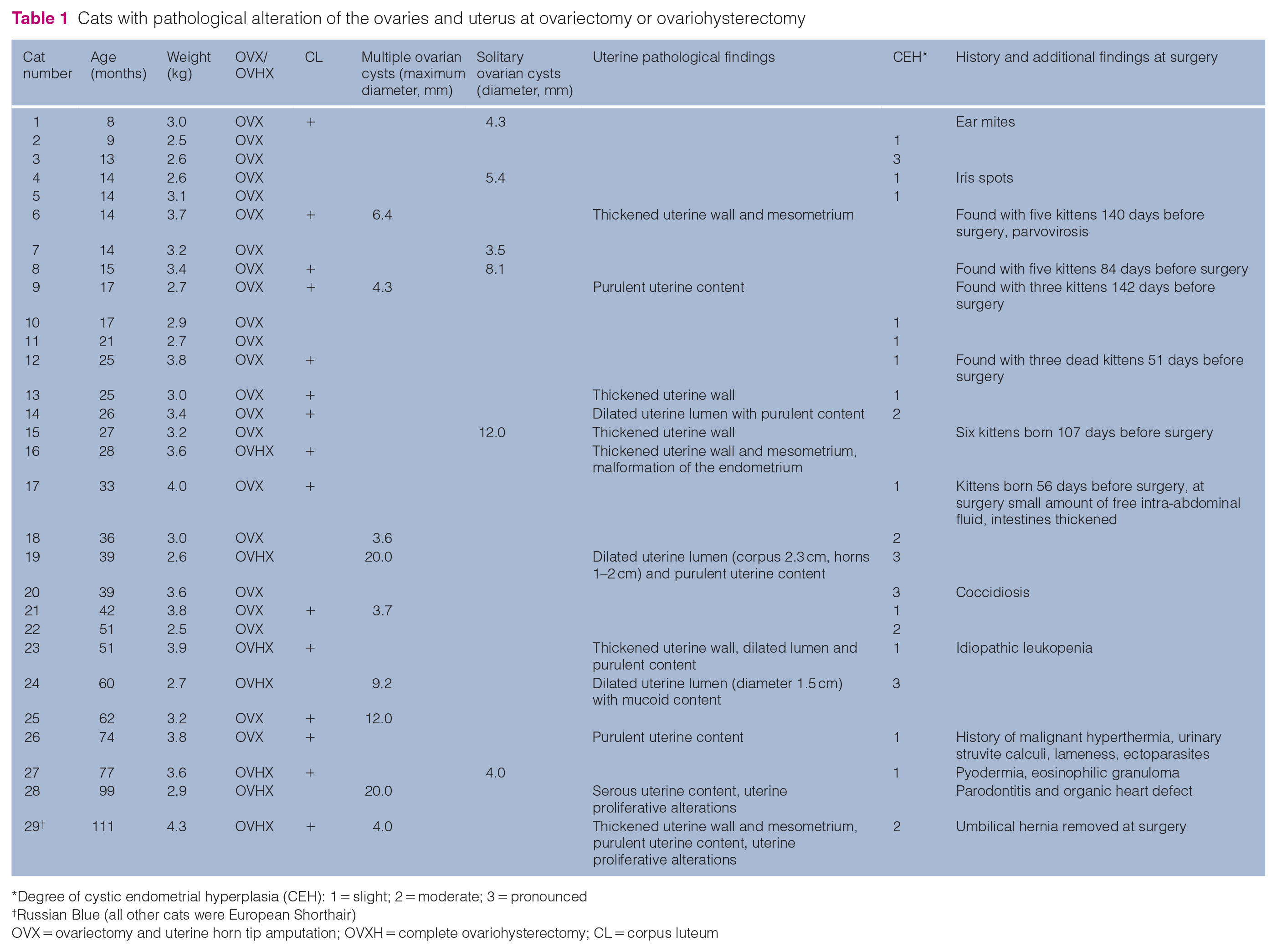
Ovarian cysts with a diameter of >3.5 mm were found in 15 cats (14.2% of the total number studied). Of the cysts, 40% (n = 6) had a diameter between 8 and 25 mm. In six cats, only one single cyst was present, whereas in nine cases, multiple cysts on both ovaries were present (Table 1, Figure 1). Histological examination of the cystic structures resulted in classification as either cysts of the Wolffian or Muellerian duct (n = 4), follicular cysts (n = 4), luteinised cyst (n = 1), cystic rete ovarii (n = 2), combination of different cysts (n = 2) and non-classifiable cysts (n = 2; Table 2).
Cats with pathological alteration of the ovaries and uterus at ovariectomy or ovariohysterectomy
Degree of cystic endometrial hyperplasia (CEH): 1 = slight; 2 = moderate; 3 = pronounced
Russian Blue (all other cats were European Shorthair)
OVX = ovariectomy and uterine horn tip amputation; OVXH = complete ovariohysterectomy; CL = corpus luteum
Examples of ovaries with cystic alterations: (a) rete ovarii cyst in cat 7; (b) cyst of the Wolffian or Muellerian duct in cat 15; (c) luteinised cyst in cat 1; and (d) non-classifiable cysts in cat 28 (haematoxylin and eosin staining, bar = 3 mm)
Occurrence and histopathological description of ovarian cysts

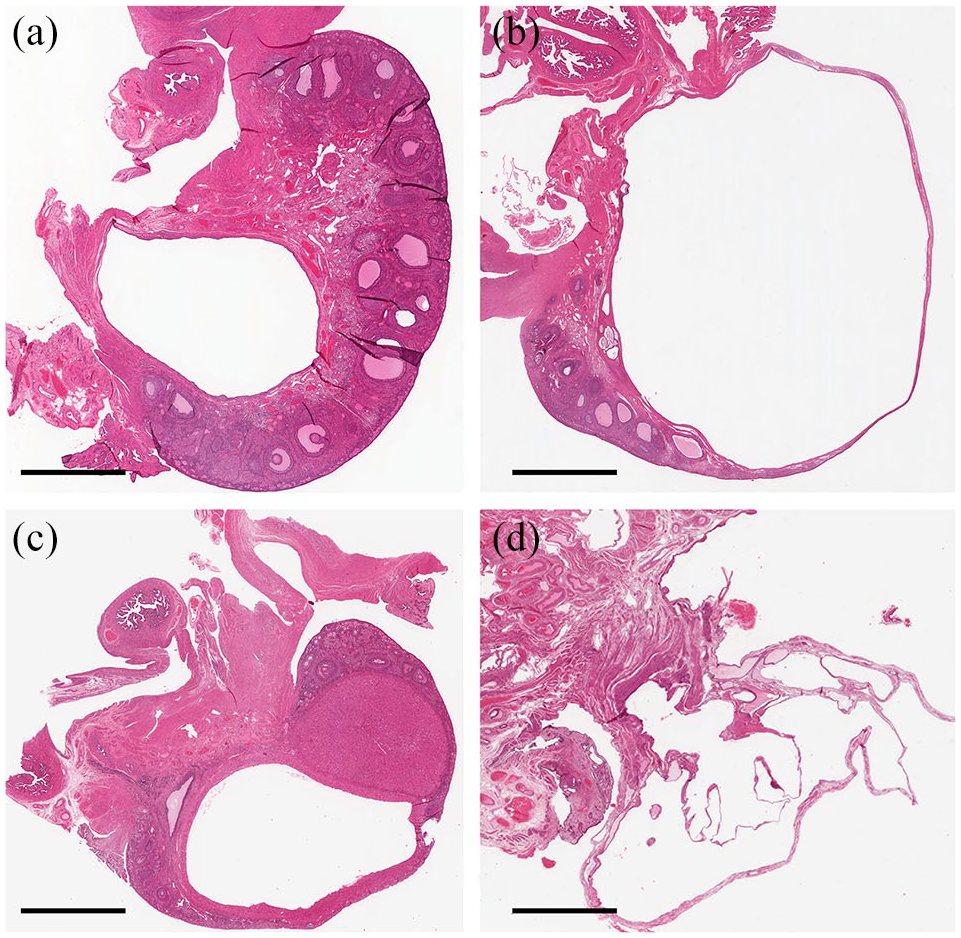
Based on histopathological findings, 19.8% of the cats (n = 21) had CEH. In 57.1% (n = 12) of these cats, CEH was classified as mild, in 19.1% (n = 4) as moderate and in 23.8% (n = 5) as severe. The incidence of CEH increased with the age of the animals (Figure 2).
Examples of uterine horn segments with: (a)moderate, cat 22; and (b) severe cystic endometrial hyperplasia, cat 24 (haematoxylin and eosin staining, bar = 1500 μm)
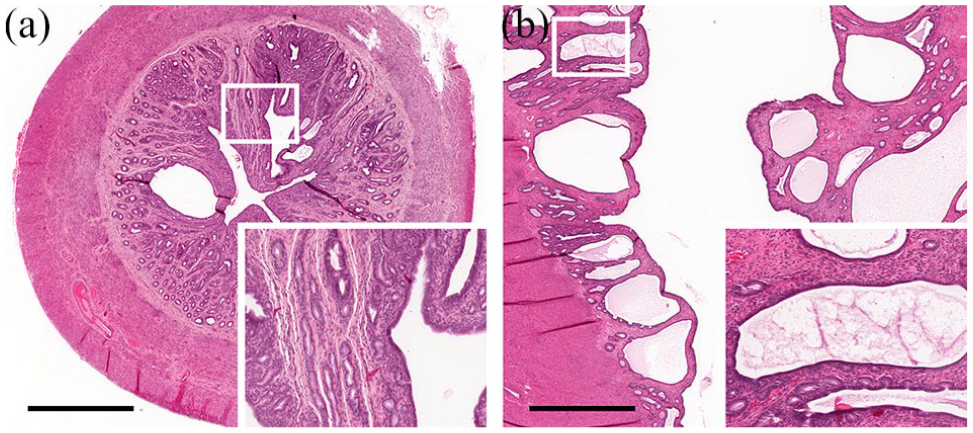
Six cats suffered from varying degrees of endometritis with or without distension of the uterine lumen. In all these cases, histopathology revealed an immigration of macrophages, granulocytes and lymphocytes into the endometrium (Figure 3). A mucoid intrauterine content and inflammatory cells in the uterine lumen were evident in cat 24 (Figure 3d), and a serous uterine content without inflammatory cells was present in cat 29.
Examples of uterine horn segments with different degrees of endometritis: (a) cat 14; (b) cat 23; (c) cat 19; and (d) mucometra, cat 24 (haematoxylin and eosin staining, bar = 3 mm)
Uterine proliferative alterations were detected in two cats. In both cases, complete ovariohysterectomy was performed because the uterine wall was thickened. With an age of approximately 8 and 9 years, respectively, these were the oldest cats in our study. In cat 28, histological examination revealed multiple cystic glandular spaces of different sizes lined by proliferating epithelium in both uterine horns. Some cysts contained proteinaceous fluid and cell debris. In one of these dilated glands, a papillary proliferation was covered by a single layer of epithelial cells. Cells showed a distinct anisocytosis and anisokaryosis, karyopyknosis and a slightly increased mitosis rate. Single giant nuclei and some multinucleate giant cells were present. In cat 29, multicentric papillary tumour proliferations were present in the form of papillary growths in numerous dilated glands, as well as in the central lumen. Anisokaryosis with varying chromatin content, often with several nucleoli, was visible. Cells showed anisocytosis and single-cell necrosis. Furthermore, we found multiple multinucleate giant cells and karyopyknosis (Figure 4).

Hyperplastic uterine lesions in (a) cat 28 and (b) cat 29 (haematoxylin and eosin staining, bar = 3 mm)
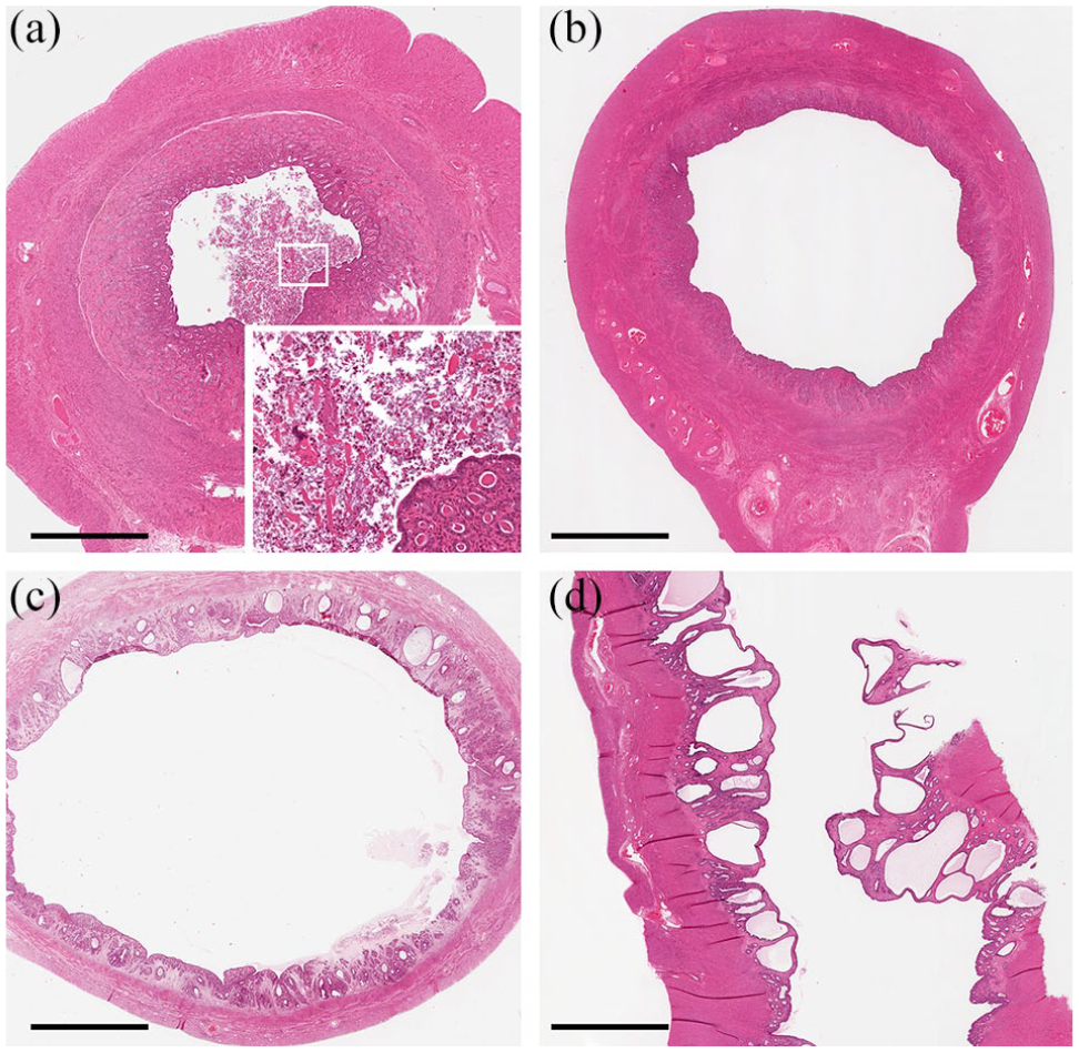
In one 28-month-old cat (cat 16), a malformation of the endometrium was diagnosed. The lumen of the left uterine horn was bi-partitioned by a thin endometrial wall covered on both sides with regular luminal epithelium. The smaller part of the lumen and the lumen of the other horn was filled with proteinaceous fluid (Figure 5).
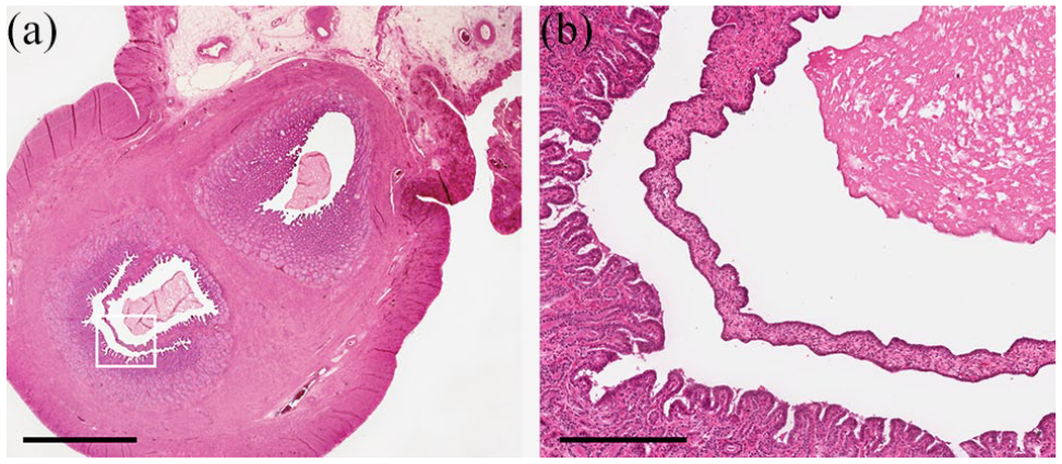
Malformation of the left uterine horn in cat 16: (a) overview of the malformation at the level of the uterine bifurcation; and (b) magnification of the bi-partitioned lumen covered on both sides with regular luminal epithelium (haematoxylin and eosin staining; [a] bar = 3 mm, [b] bar = 300 μm)
Discussion
In this study, 50% of female cats older than 3 years of age and 83% of cats over 5 years of age showed more or less pronounced pathological alterations of the uterus or ovaries. All cats had passed through the same animal shelter. Medical records from at least 3 months before spaying and detailed information on housing conditions of the cats with their current owners was available. Because none of these cats showed clinical signs of general or gynaecological disorders, neither in a preliminary examination nor at the pre-surgery examination, there was no information on whether pathological alterations of the uterus and ovaries would have resulted in future health problems.
Although all cats were housed inside under the close supervision of their owners and were at no time in contact with gonad-intact males, 15/29 cats with pathological alterations of the uterus and/or ovaries had at least one corpus luteum. As spontaneous ovulations occur regularly in female cats,16,17,21 pathological alterations potentially associated with the feline oestrous cycle should be further investigated. However, most domestic cats are spayed at an early age. 22 Therefore, the long-term sequelae of exposure to gonadal steroid hormones such as CEH, endometritis, pyometra and ovarian cysts are difficult to examine in this species.
Endometritis was evident in almost 6% of our cases and is a finding that may cause health problems. This can be expected if the condition develops into pyometra, that is, purulent endometritis with filling of the uterus. A retrospective study from Sweden suggested that the incidence of pyometra defined as purulent uterine content associated with signs of general illness in female cats is much higher (depending on the breed, between 0.9% and 14.1%, with a mean case fatality rate of 5.7%) than often assumed and increases with age. 23 CEH was even more frequent in our study (as already described recently17) and was detected in approximately 20% of cats. In cats, as in dogs, 24 prevalence increases with age. This may indicate a cumulative effect of repeated gonadal steroid hormone stimulation in subsequent oestrous cycles.4,11,12,13,15 To what extent CEH finally develops into pyometra in the female cat and whether this is associated with spontaneous ovulation remains unanswered.
Ovarian cysts were found in several cats in this study and were traced back to different ovarian and non-ovarian structures. Only in two cases with polycystic ovaries and multiple cysts of >20 mm diameter, the tissue from which they originated could not be determined. When expanded cysts alter their morphology under pressure, their origin may become unclear and, in addition, the cells lining the cysts lose their original shape with expansion of the cysts. 25 The present findings agree with reports on different ovarian cyst types in dogs.19,26 We suggest that ovarian cysts in cats are mostly incidental findings without clinical relevance. They can, however, replace the surrounding physiological ovarian tissue, which may result in a secondary functional loss.27,28
Hyperplastic uterine lesions were evident in the two oldest cats of our study. Such proliferative lesions may indicate prolonged endometrial repair post-delivery. Because of the animals’ age, previous pregnancies in these cats cannot be excluded. Such lesions are occasionally confused as being neoplastic changes of the endometrium in female cats. 29 Uterine tumours are rare in cats and account for approximately 0.3% of all feline neoplasms, with endometrial adenocarcinoma and uterine leiomyoma being the most common tumours of the feline genital tract.29,30 Although uterine neoplasia cannot completely be ruled out, hyperplastic lesions potentially associated with hormonal stimulation are more likely. Uterine adenocarcinomas are frequently diagnosed in rabbits. The rabbit is an induced ovulator and therefore laboratory and pet rabbits housed individually are under near-constant oestrogen stimulation. 31 In contrast, spontaneous ovulations with a subsequent luteal phase in cats are more frequent than previously assumed. 17 Furthermore, female cats are often spayed. 1 Together, this explains the much lower prevalence of uterine neoplasia in cats vs rabbits.
The doubled uterine horn lumen in one cat is an accidental finding. A similar unilateral double malformation of one uterine horn and oviduct in a cat has been reported previously, but in that cat all layers of the uterine horn wall were duplicated, 32 while in our cat, the two parallel parts of the uterine horn were separated by endometrium only. Urogenital anomalies are twice as common in cats than in dogs, 33 and have been assumed to occur during the fourth and fifth week of embryonic development, which is a phase especially sensitive to disruption of reproductive organ development in cats. 32
Conclusions
Pathohistological alterations of the uterus and ovaries were present in approximately 25% of female cats presented for elective spaying. Most of these alterations (eg, the majority of ovarian cysts and slight-to-moderate CEH) are unlikely to interfere with the animals’ wellbeing. However, uterine disease such as purulent endometritis should be considered as a potential cause of serious health problems.
Footnotes
Conflict of interest
The authors declared no potential conflicts of interest with respect to the research, authorship, and/or publication of this article.
Funding
The authors received no financial support for the research, authorship, and/or publication of this article.
Ethical approval
This work involved the use of non-experimental animals only (including owned or unowned animals and data from prospective or retrospective studies). Established internationally recognised high standards (‘best practice’) of individual veterinary clinical patient care were followed. Ethical approval from a committee was therefore not necessarily required.
Informed consent
Informed consent (either verbal or written) was obtained from the owner or legal custodian of all animal(s) described in this work (either experimental or non-experimental animals) for the procedure(s) undertaken (either prospective or retrospective studies). No animals or humans are identifiable within this publication, and therefore additional informed consent for publication was not required.