
Abstract
Objectives
The aim of this study was to describe the treatment and outcome of acetabular and other pelvic fractures in cats with patellar fracture and dental anomaly syndrome (PADS) and to provide advice on how to manage these cases in practice.
Methods
Data were collated on cats with PADS that were reported to have sustained pelvic fractures or had fractures or fissures of the pelvis identified on submitted radiographs. The details of the fractures were recorded, in addition to any treatment and outcome information.
Results
Of the 215 cases reported with PADS, 58 cats (27%) were found to have pelvic fractures, none of which were known to have resulted from significant trauma. There were 101 fractures in total and of these 15 were treated with surgery, including 10 acetabular fractures, two ilial, two pubic and one ischial fracture. Screw loosening and loss of fracture reduction was seen in four of the surgically treated fractures (two pubic fractures, one ilial and one acetabular fracture). Fourteen cats were euthanased as a direct result of a fracture occurring. While most pelvic fractures healed uneventfully, some cats remained intermittently lame, but it was not always possible to determine the cause of the lameness from the information available and because all cats had concurrent patellar fractures.
Conclusions and relevance
Many of the pelvic fractures healed with conservative management. Fractures involving articular surfaces such as acetabular fractures may benefit from surgical stabilisation as surgery may offer the benefits of articular fracture repair with improved joint congruency and a faster return to normal activity.
Introduction
Patellar fracture and dental anomaly syndrome (PADS), formerly known as knees and teeth syndrome, is recognised as a pathological disorder in cats involving insufficiency fractures of the patella and dental abnormalities. 1 Previous studies report that many of these cats develop non-traumatic fractures to other bones, preceding or subsequent to the patellar fractures. Pelvic fractures are the most frequent additional insufficiency fractures in cats suspected to be suffering from PADS.1–3
An insufficiency fracture occurs when physiological muscular forces are applied to abnormal bone. 4 Surgical treatment for patellar insufficiency fractures has been reported, but it is challenging and often results in failure of the repair, non-union or further fragmentation of the patella.1,2 Although surgical treatment for insufficiency patellar fractures has a poor prognosis, there is evidence that surgical management for insufficiency fractures to other bones in cats affected by PADS is successful,2,3 and these fractures can heal normally, in a similar manner to a surgically treated traumatic fracture; however, there is insufficient published literature to support this assertion.
This report aims to document the treatment and outcome following preceding and subsequent pelvic fractures in cats with PADS.
Materials and methods
Details on cats with PADS have been collected since 2005, following a letter requesting cases. 5 Recently, a combined database was established based on information of cases acquired at the University of Bristol (UK) and Exclusively Cats Veterinary Hospital, Michigan (USA). Following the initial letter, publications in the veterinary literature, and a page on Facebook (https://www.facebook.com/felinepatellafracturestudy), owners and veterinarians with cats they believed to be affected by this syndrome have continued to make contact. Cats were included in the databases if they had the following clinical features: transverse patellar fracture(s) with or without dental anomalies, including one or all persistent deciduous teeth, unerupted deciduous or adult teeth; and one or more atraumatic fracture(s) to other bones. The PADS database was searched for all cats with pelvic fractures up until June 2020. Owners and veterinarians were contacted by email or telephone to obtain follow-up information. A request was made to complete a questionnaire (see supplementary material) to obtain long-term follow-up. The questionnaire requested information on the cat’s current condition and fractures to other bones; radiographs and histories were requested for details on the treatment and outcome.
For this article, radiographs were reviewed for evaluation of treatment and information was recorded from the data submitted regarding postoperative progress and clinical and current status.
The signalment of the affected cat, date or age the fracture occurred, details of unilateral or bilateral patellar fracture, and whether dental anomalies were present, were recorded for each cat. The treatment method and outcome, including complications, clinical outcome and final outcome, of the cats were documented; when the cats were reported to have died the cause of death was noted, if known.
The University of Bristol Ethical Committee approved the use of the acquired information.
Results
Information was obtained and registered for 215 cats with suspected PADS. Of these, 39.1% (n = 84) had sustained preceding or subsequent fractures to other bones, and in 58 of these cats, fractures involved the pelvis. In these 58 cats, bilateral (41 cats) or unilateral (17 cats) patellar fractures were present and 31/58 cats had dental anomalies, mainly persistent deciduous teeth. In these 58 cats there were 101 pelvic fractures of which 47.5% (n = 48) were acetabular, 30.7% (n = 31) ischial, 7.9% (n = 8) ilial, 6.9% (n = 7) pubic and 6.9% (n = 7) were unspecified pelvic fractures. Twenty-eight (48%) of the 58 cats with pelvic fractures had only one part of the pelvis affected (unilateral acetabular, ischial, ilial or pubic); 23 cats had two fractures, three cats had three fractures, two had four fractures and two cats had five fractures. The details of the numbers of pelvic fractures, patellar fractures and whether there were dental anomalies reported in these 58 cats are summarised in Table 1.
Summary of the numbers of pelvic and patellar fractures and dental anomalies in cats with patellar fracture and dental anomaly syndrome
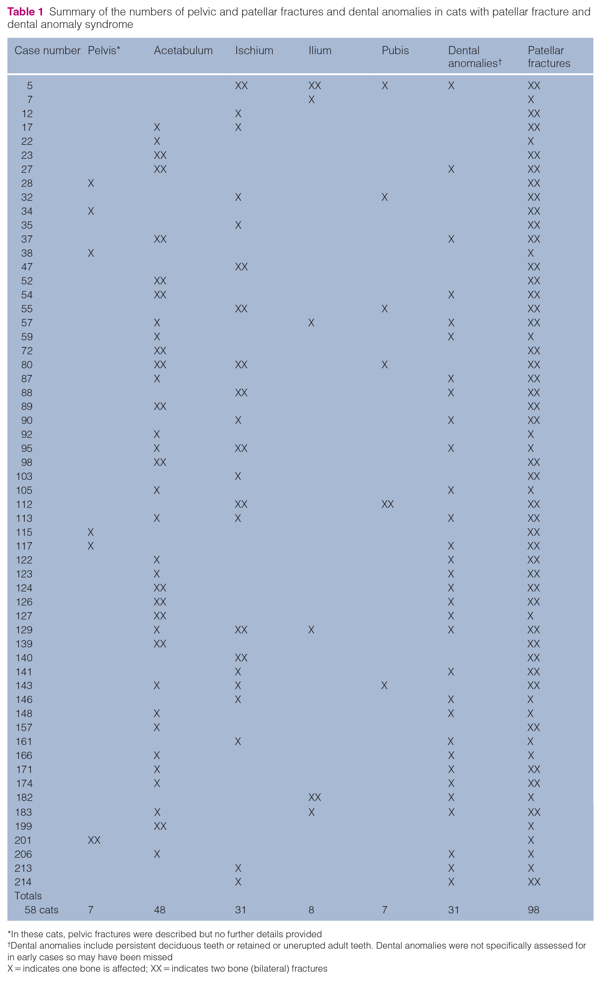
In these cats, pelvic fractures were described but no further details provided
Dental anomalies include persistent deciduous teeth or retained or unerupted adult teeth. Dental anomalies were not specifically assessed for in early cases so may have been missed
X = indicates one bone is affected; XX = indicates two bone (bilateral) fractures
Thirty-four cats had 48 acetabular fractures; of these, 20 were unilateral and 14 were bilateral fractures. Age at which the acetabular fracture occurred was reported for 27 fractures (range 11 months to 7 years 2 months; mean 3 years to 9 months). There was information on the original fracture configuration for 27 fractures (before healing) and exact location; these fractures were all simple fractures with transverse configuration located cranially, centrally or caudally, and with minimal-to-moderate displacement.
Ten of the acetabular fractures were managed surgically (Figures 1 and 2) by stabilisation with a plate and screws and the fracture was not primarily treated, but femoral head and neck ostectomy was performed in three cats. One postoperative complication was reported with screw loosening and loss of fracture reduction in case 80. Fifteen acetabular fractures were reported to have been treated conservatively (cage rest and analgesia; Figure 3), and in 20 cats the treatment method was not known, not reported or not specifically prescribed as the fractures may have been undiagnosed until they were identified incidentally or the cat was euthanased before treatment. There were 26 healed acetabular fractures and two fractures in one cat that developed non-unions. Six cats with acetabular fractures were ultimately euthanased as a direct result of fracturing a bone (either the acetabulum or different bones). A summary of the details of treatment and outcome for acetabular fractures is listed in Table 2.

Ventrodorsal projection of the pelvis of case 17. A simple acetabular fracture was stabilised with a four-hole 2.0 mm acetabular plate and screws. Healing is complete, with mild degenerative joint disease evident. The surgical approach involved a greater trochanter osteotomy that has been stabilised with pins and a tension band wire. There is a concurrent left ischial fracture, which has not healed. This cat had bilateral partial patellectomy as treatment for the patellar fractures

Ventrodorsal projection of the pelvis of case 23. Bilateral plate and screw fixation using seven-hole 2.0 mm dynamic compression plates for bilateral acetabular fractures. There is some reaction around the greater trochanters bilaterally, associated with gluteal tenotomies that were performed as part of the surgical approach to the acetabular fractures. The cat has a simple transverse right patellar fracture and the left patellar fracture is evident along with periarticular ossification, mineralisation and enthesophytosis

(a) Lateral and (b–d) ventrodorsal projections of the pelvis of case 27. (a,b) At 11 months of age the cat developed a spontaneous cranial right acetabular fracture, which was treated conservatively. (c) At 14 months of age the right acetabular fracture has healed and there is callus around a recent left acetabular fracture. (d) Further callus development and healing of the left acetabular fracture is causing mild pelvic canal narrowing. There is coxofemoral incongruency with mild hip subluxation and very mild periarticular osteophytosis. The right acetabular fracture has healed and remodelled well
Details of treatment and outcome in the cats with patellar fracture and dental anomaly syndrome and acetabular fractures

Euthanased owing to fractures; therefore, outcomes of fracture not reported
LTFU = lost to follow-up; NR = no report; PTS = put to sleep; FHO = femoral head ostectomy; NT = no treatment; NK = not known; NA = not available
Details on treatment and outcome of ischial fractures are described in Table 3. Twenty-two cats were reported to have 31 ischial fractures of which 13 were unilateral and nine bilateral. Fracture configuration was similar between cats, being simple transverse or oblique fractures lines through the ischial body. Details on treatment methods were reported for 15 fractures. One cat with bilateral fractures had stabilisation of the left ischial fracture with bone plate and screws; the right side was treated conservatively. Conservative therapy was the most common treatment method for 14 fractures; the remainder of the fractures were not treated as they were incidental findings or no treatment method was reported. Regarding the outcome of the ischial fractures, in the majority of the cats (20 cats) the fractures were reported as healed (Figure 4). Five cats were euthanased later in life because they went on to fracture other bones.
Details of treatment and outcome in the cats with patellar fracture and dental anomaly syndrome and ischial fractures

NR = no report; R = right; L = left; LTFU = lost to follow-up; CT = computed tomography; PTS = put to sleep; GI = gastrointestinal; NK = not known; NT = no treatment

Ventrodorsal projection of the pelvis of case 95. Bilateral healed ischial fractures with smooth callus formation. There is callus associated with the cranial aspect of the right acetabulum suggestive, but not conclusive, of a prior fracture in this region
Six cats were reported to have eight ilial fractures; information on the configuration of the fracture was available for seven fractures (five cats), with a transverse or short oblique fracture line through the ilial body being the most frequent. Only two cats (one ilial fracture each) had information on treatment methods, one had stabilisation with plate and screws, which failed and the outcome was a non-union, and the other fracture was stabilised with pins and figure-of-eight wire. The outcome for these fractures was reported as healed in one cat and complications and a non-union in one other cat. The clinical outcome was not reported in three cats; two cats were reported to be stiff or constantly lame. One cat was euthanased owing to multiple fractures. Details on the treatment and outcome of cats with ilial fractures are summarised in Table 4.
Details of treatment and outcome in the cats with patellar fracture and dental anomaly syndrome and ilial fractures

Euthanased owing to fractures; therefore, outcome of fracture not reported
NR = no report; R = right; LTFU = lost to follow-up; L = left; NA = not available; NT = no treatment; PTS = put to sleep
Information on treatment for pubic fractures was recorded for six cats with seven fractures. Three of these fractures had conservative treatment and two fractures in one cat were managed surgically by stabilisation with a plate and screws. This cat suffered postoperative complications, with infection and screw loosening, and loss of fracture reduction. Outcome information was available for five cats, all fractures healed but two cats were euthanased later owing to other fractures, one died from unrelated causes, three were stiff and lame, and two were walking normally. Table 5 describes the information on the treatment and outcome in cats with pubic fractures.
Details of treatment and outcome in the cats with patellar fracture and dental anomaly syndrome and pubic fractures

NR = no report; R = right; L = left; LTFU = lost to follow-up; GI = gastrointestinal; PTS = put to sleep
Discussion
Fractures of the pelvis are the commonest fracture seen in cats with PADS in addition to the patellar fracture(s). The majority of the non-articular pelvic fractures healed with conservative management. Ilial and pubic fractures were the least common fracture type, with only four fractures treated surgically, and conclusions could not be drawn from such a small number of cases. Surgical stabilisation was most commonly employed for the acetabular fractures with good outcomes, when follow-up was available. Conservative management for acetabular fractures was also successful in terms of bone healing, albeit the bones healed with malunions and mild joint incongruity.
Comparative aspects on concurrent diseases
Pelvic insufficiency fractures are common in geriatric humans with osteoporotic bones and other predisposing factors such as rheumatoid arthritis, metabolic bone disease, neurological disorders, prior irradiation, corticosteroid therapy and bisphosphonate therapy have also been described.4,6 Cats affected by PADS do not appear to have osteoporotic bones or history of other predisposing factors; on the contrary, some of them had increased bone density, 3 which has also been reported as a predisposing factor for insufficiency fractures in humans. 7 Results of a variety of blood tests on PADS cats, including analysis of calcium and vitamin D levels, have not shown consistent or significant abnormalities. Histopathological analysis is presently being performed on bone from cas with PADS that have died, and the results of this analysis will be published in a future article.
Aetiology
Pelvic fractures in cats occur frequently as a result of major trauma such as road traffic accident or falls from a height, and comprise 20–32% of all fractures seen in cats.8,9 The fractures in the cats in the present study were usually sustained under circumstances of normal activity and were often missed owing to mild insidious onset lameness or because the lameness was attributed to the patellar fractures, hence, frequently identified late as an incidental finding. Some insufficiency fractures in humans may be asymptomatic and discovered accidentally, 10 and are the result of a low energy mechanism. 11
In humans, multiple stress fractures are rare.12,13 Approximately 50% of the cats with PADS in this study with pelvic fractures only had single pelvic fractures. It is highly unusual in cats with traumatic pelvic fractures to only have one fracture line as multiple fractures usually occur. 8
Incidence
The acetabulum was the most commonly affected part of the pelvis affected in the cats with PADS, followed by the ischium, ilium and pubis. In traumatic feline pelvic fractures, the pelvic floor is the most commonly affected with 72% pubic fractures and 51% suffering ischial fractures, ilial fractures accounted for 51% and the least commonly affected bone was the acetabulum (26%). 8 So the fractures seen in cats with PADS do not concur with the types of fractures usually seen after trauma.
General treatment of insufficiency fractures
Treatment for insufficiency fractures will depend on the location, fracture extent and displacement, functional status of the patient and concomitant diseases. Historically, feline pelvic fractures have been treated conservatively;14,15 however, in recent years, several studies have shown the importance of surgical management of selected pelvis fractures.9,16–18 Indications for surgery have included pelvic canal narrowing, disruption of the weightbearing axis (acetabular, ilial body or sacroiliac luxations), nerve impingement, intractable pain, inability to ambulate within a few days of injury and bilateral/concomitant orthopaedic injuries. 18 In humans, traumatic pelvic fractures are usually treated surgically and it has been demonstrated that outcome of conservative therapy for either traumatic or insufficiency fractures is poor, with loss of social and physical independence and autonomy. 19 In cats, complications associated with pelvic fractures have been associated with narrowing of the pelvic canal as a risk factor for obstipation/constipation. 20 In humans, reported complications with surgical treatment are associated with osteopenia, with poor bone stock compromising internal fixation and screw purchase, and increasing the risk of non-union. 21 In the pelvic fractures in the cats with PADS treated surgically, and reported on in this paper, there were just a few cases of screw loosening, which did not appear to affect outcome. The majority of the pelvic fractures, where information was available, were reported to have healed, with only five reported as non-unions. In many cats follow-up radiographs were not taken so these figures may not be an accurate representation.
Acetabular fractures
Acetabular fractures in cats comprise 17.5–26% of pelvic fractures,15,17 and are usually unilateral and frequently contain two fragments. 8 Meeson and Geddes 9 published a survey of 43 cats with traumatic pelvic fractures of which the acetabulum was the least commonly affected bone (n = 12/43) and all of the acetabular fractures were unilateral. In that publication, seven fractures occurred along the caudal acetabular rim, or were comminuted and were managed conservatively; of these, two had concurrent femoral head and neck excision. Only five acetabular fractures were managed surgically, and the outcome in the cats with these acetabular fractures was not specifically reported. 9 Haine et al 22 reported 16 cats with 17 acetabular fractures, all associated with concurrent orthopaedic injuries and all managed surgically. Of the 16 cats, three were judged from client history to regain full function and 13 had acceptable function after treatment. 22 Of the acetabular fractures in the cats with PADS approximately a quarter of the cats were managed directly with surgery to reduce and stabilise the fracture, or indirectly by femoral head ostectomy. Unfortunately, because of concurrent patellar fractures, it was difficult to ascertain whether surgical management made a difference in outcome in the cats in the present study, but evidence from other studies is supportive of the benefits of surgical stabilisation of articular fractures with a quicker return to function and improved joint congruency. 22
Outcome
Some of the cats were reported to be lame, but the cause of lameness could not be determined, whether from the pelvic fracture or the patellar fracture. Meeson and Geddes 9 reported that of 43 cats with treated pelvic fractures, 36 showed no sign of lameness and seven had some degree of permanent lameness. 9 Approximately 20% of the cats with PADS reported on in this study were euthanased as a direct result of either the pelvic fractures or fractures in other bones.
Limitations of the study
In a retrospective study such as the present, gathering data is complex and complete information for many of the cats was not available. Surgical procedures were performed by different surgeons and follow-up times were different for each case. However, this is the first paper that describes the treatment of the most common coincidental fractures in cats with PADS with enough data to give a prognosis on the likelihood of healing following treatment.
Conclusions
Many of the pelvic fractures showed radiographic evidence of bone healing regardless of the treatment method; hence, our conclusion is that many pelvic fractures in cats with PADS, with the exception of the acetabulum (and the ilium), do not require stabilisation surgery. Acetabular fractures may heal with conservative management, but surgical stabilisation should be considered as it may give a quicker return to function and improve outcome. There were insufficient ilial fractures to be able to draw definitive conclusions on the optimal treatment method, but the authors would suggest that, similar to traumatic ilial fractures, the option of surgical stabilisation of these fractures in cats with PADS should be given careful consideration.
Footnotes
Acknowledgements
The authors would like to thank Mark Longley for assisting in the collection of data on the cats and all the veterinarians and owners who both treated and provided details, including histories, images and radiographs, concerning these cases and without whom this study would not be possible.
Supplementary material
Conflict of interest
The authors declared no potential conflicts of interest with respect to the research, authorship, and/or publication of this article.
Funding
The authors received no financial support for the research, authorship, and/or publication of this article.
Ethical approval
This work involved the use of non-experimental animals only (including owned or unowned animals and data from prospective or retrospective studies). Established internationally recognised high standards (‘best practice’) of individual veterinary clinical patient care were followed. Ethical approval was therefore not specifically required for publication in JFMS.
Informed consent
Informed consent (either verbal or written) was obtained from the owner or legal custodian of all animal(s) described in this work (either experimental or non-experimental animals) for the procedure(s) undertaken (either prospective or retrospective studies). No animals or humans are identifiable within this publication, and therefore additional informed consent for publication was not required.