
Abstract
Objectives
The aim of this study is to describe the treatment and outcome of humeral condylar fractures and humeral intracondylar fissures in cats with patellar fracture and dental anomaly syndrome (PADS) and to provide advice on how to manage these cases in practice.
Methods
Data were collated on cats with PADS that were reported to have sustained humeral fractures or had fractures or fissures of the humerus identified on radiographs. The details of the fractures were recorded in addition to any treatment and outcome information.
Results
Of the 207 cases reported with PADS, 18 cats (8.7%) were found to have humeral condylar fractures, none of which was known to have resulted from significant trauma. Where treatment occurred, it involved the placement of transcondylar positional or lag screws. In some cases additional implants, including supracondylar bone plates and screws or Kirschner wires (K-wires), were used. Follow-up data revealed that only two cats were euthanased owing to the presence of the humeral fractures, with at least eight achieving some degree of recovery of function.
Conclusions and relevance
These humeral fractures all have the characteristics of stress insufficiency fractures, being simple isolated fractures that are short oblique, with increased radio-density at the fracture line and occurring following minimal or no trauma. Humeral intracondylar fissures were identified in two cats and it is possible that some of the other fractures may have occurred secondary to pre-existing fissures. To our knowledge, no prior reports exist of fissures in cats that do not meet the criteria for PADS. Surgical repair primarily consisted of the placement of transcondylar lag or positional screws with, in some cases, adjunct implants such as bone plates and screws or K-wires. Though there were insufficient data to determine the prognosis for these fractures in the long term, unlike patellar fractures, many of these fractures will heal if treated appropriately.
Introduction
A pathological disorder, ‘patellar fracture and dental anomaly syndrome’ (PADS), formerly known as ‘knees and teeth syndrome’, has been identified in cats. 1 The syndrome involves stress fractures of the patella and dental abnormalities, including persistent deciduous teeth and unerupted permanent teeth. These dental anomalies can lead to the development of osteomyelitis or abscesses. 2 Additionally, many of these cats develop non-traumatic fractures to another bone(s), either preceding or subsequent to the patellar fracture(s).1,3–5 Of these other fractures, the pelvis, tibia and humeral condyle are the sites most frequently affected. To our knowledge, at the time of writing, no reports exist of atraumatic fractures of the humeral condyle in cats that do not meet the criteria for PADS.
The aetiology of PADS is currently unknown and it is hoped that by increasing the awareness of the syndrome and collecting more data we may advance our understanding of the condition. The objective of this study was to describe the recommended treatment options for cats with PADS that sustain humeral condylar fractures in order to inform the veterinary community and provide owners with more accurate information concerning the prognosis for these fractures.
Materials and methods
The cats in this study were sourced from records at the University of Bristol (UK) and Exclusively Cats Veterinary Hospital, Michigan (USA) after a request was made for cases that met the criteria for PADS in the Veterinary Record, 6 the Veterinary Information Network and social media pages (https://www.facebook.com/felinepatellafracturestudy). These criteria included transverse patellar fracture(s) with or without dental abnormalities such as persistent deciduous or unerupted adult teeth and atraumatic fracture(s) of other bones.
Long-term follow-up data were obtained by requesting owners and veterinarians to complete a questionnaire (see supplementary material) relating to the cats’ progress since diagnosis or treatment. Data were sourced from radiographs, questionnaires and patient histories of cats with humeral fractures in addition to PADS. Where available, the outcome of the cats was noted, including the cause of death.
The use of this information was approved by the University of Bristol Ethical Committee.
Results
Information was obtained for 207 cats with suspected PADS. Of these cats, 18 (8.7%) had sustained humeral condylar fractures, with 24 humeral fractures in total (12 cats with unilateral and six with bilateral fractures) (Table 1). The nature of the fracture was recorded in 13 of the cats, with 12 involving the lateral condyle, three the medial condyle and one a Y-fracture. The fractures were generally simple, isolated and short oblique, with varying degrees of displacement.
Details of humeral condylar fractures in cats with patellar fracture and dental anomaly syndrome

MN = male neutered; DSH = domestic shorthair; NR = not reported; ME = male entire; LTFU = lost to follow-up; FN = female neutered; DLH = domestic longhair; PTS = put to sleep; R = right; L = left; HIF = humeral intracondylar fissure; y = years; mo = months;
Humeral intracondylar fissures (HIFs) were diagnosed in two cats (Figure 1). Chronic humeral fractures were observed in two cats.
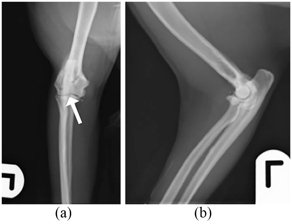
(a) Craniocaudal and (b) mediolateral radiographic views of a suspected humeral intracondylar fissure with a radiolucent line extending from the articular surface halfway up the condyle in the left humerus of case 17 (arrow). There is moderate and well-established osteoarthritis with osteophytosis affecting the radial head and ulna notch and mineralisation in the region of the medial joint capsule
The age at which the first humeral fracture occurred was recorded in 9/18 cats (mean 4.2 years; range 24–101 months). Body weight was recorded in 8/18 cats (mean 4.9 kg; range 3.8–7.0 kg). Sex was recorded in 16 animals (with nine males [one entire, eight neutered] and seven females [all neutered]). Breed was recorded in 16 cats (11 domestic shorthairs, two domestic longhairs, two Russian Blues and one Siamese cross).
The cause of the humeral fracture was documented in 8/18 cats, with none of these being reported as having occurred as a result of external direct trauma.
Treatment
Of the 24 humeral fractures, six were recorded as having undergone surgical treatment: three were repaired with transcondylar lag or positional screws (Figures 2–7), one with a transcondylar lag screw in addition to a supracondylar dynamic compression plate and screws (Figures 3 and 4), one with a transcondylar lag screw and anti-rotational Kirschner wires (K-wires) (Figure 5), and one with a transcondylar lag screw and metaphyseal screw (Figures 6 and 7). Two fractures were managed conservatively with analgesia and anti-inflammatory medication.
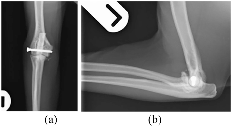
Immediate postoperative (a) craniocaudal and (b) mediolateral radiographic views demonstrating transcondylar 2.4 mm/22 mm positional screw implantation to stabilise the left humeral intracondylar fissure in case 17

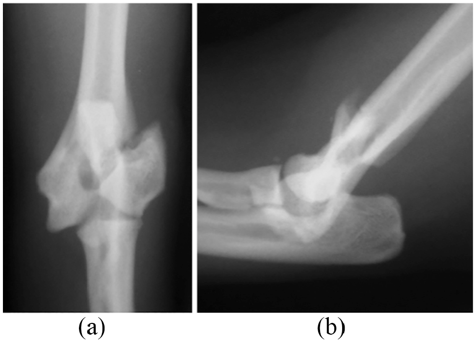
(a) Craniocaudal and (b) mediolateral radiographic views of a fracture of the lateral aspect of the right humeral condyle of case 17. There is moderate, well-established osteoarthritis with osteophytosis affecting the radial head and ulna notch and mineralisation in the region of the medial joint capsule

Immediate postoperative (a) craniocaudal and (b) mediolateral radiographic views demonstrating a combination of an supracondylar contoured five-hole 2.0 mm dynamic compression plate and screws with a transcondylar 2.4 mm/22 mm lag screw to stabilise the fracture of the lateral aspect of the humeral condyle in case 17. There is severe and well-established osteoarthritis with osteophytosis affecting the radial head and ulna notch and mineralisation in the region of the medial joint capsule

Immediate postoperative (a) craniocaudal and (b) mediolateral radiographic views demonstrating a combination of a transcondylar lag screw and two anti-rotational Kirschner wires to stabilise the fracture of the lateral aspect of the humeral condyle in case 95
(a) Craniocaudal and (b) mediolateral radiographic views of a displaced fracture of the lateral aspect of the humeral condyle in case 37

Immediate (a) craniocaudal and (b) mediolateral radiographic views demonstrating a combination of a transcondylar lag screw and a metaphyseal lag screw placed to stabilise the fracture of the lateral aspect of the humeral condyle in case 37
Outcomes
Outcomes were recorded for 11/18 cats; two were euthanased owing to the humeral fracture(s) and one owing to the presence of other fractures. For the remaining eight cats, three recovered to be fully weightbearing on the affected limb(s) and for the other five cats, their causes of death were not attributable to having sustained the fractures (although it is not clear to what degree they recovered).
Discussion
Fractures of the humeral condyle are rare in cats, with most feline humeral fractures being diaphyseal. 7 Feline humeral condylar fractures that have been previously reported have involved high-energy trauma primarily due to road traffic accidents, 8 producing comminuted fractures as opposed to the oblique condylar fractures found in cats with PADS. However, none of the cats in this study were reported to have developed these fractures as a result of external direct trauma. Furthermore, the fact that many of these cases involve indoor-only cats renders a high-energy traumatic cause of the fractures unlikely. Therefore, these cases are more likely to represent stress insufficiency fractures. These occur following minimal or no trauma, when normal or physiological stresses are placed on abnormal bones,9,10 producing simple isolated fractures, transverse or short oblique, and often with increased radio-opacity at the fracture line, as demonstrated on radiographs (Figures 3a and 6a). This contrasts with stress fatigue fractures that occur when repetitive forces act upon normal bone.11,12
One of the major risk factors for condylar fractures in dogs is the presence of HIFs, which are areas of weakness in the bone. 13 Recently, the aetiopathogenesis of HIFs has been disputed. The generally accepted theory was that HIFs develop secondarily to incomplete ossification of the humeral condyle.14,15 The humeral condyle develops from two ossification centres: the medial centre comprising the medial aspect and trochlea, and the lateral involving the capitulum and lateral aspect of the condyle. In the cat, these two ossification centres should fuse by 14 weeks of age. 16 However, it has been discovered that HIFs can develop after ossification has successfully occurred, as observed with CT 17 and MRI 18 studies carried out pre- and post-fissure formation. Accordingly, some argue for an alternative hypothesis involving a previously ossified condyle undergoing remodelling and stress fatigue, leading to fissure formation. HIFs have only been reported in cats with PADS and their presence would support the theory of stress fractures.
The majority of the fractures in this study involved the lateral aspect of the condyle, as is generally the case with canine humeral condylar fractures. In dogs, these fractures are over-represented in certain breeds, such as English Springer Spaniels and French Bulldogs.19,20 The two groups most commonly affected are skeletally immature dogs around 4 months of age and older, skeletally mature patients. 20 In skeletally immature dogs, the fracture is a Salter–Harris Type IV classification, involving the growth plate, metaphysis and epiphysis. These are usually associated with minor trauma, with a possible history of low-grade lameness prior to the event. 21 In older dogs, HIFs are believed to render this part of the bone more vulnerable to fracture, thus explaining why even minor forces acting on the limb are capable of producing fractures.22,23
To our knowledge, HIFs have not previously been described in cats not afflicted by PADS, yet HIFs were observed in two of the cases in this study and suspected in others. In this study, the data indicate that these fractures occurred in skeletally mature cats of at least 2 years of age; however, it is possible that fissures could have been present before the other fractures were sustained.
The pathophysiology of the fracture in dogs is that a force applied to the foot (via a fall or jump) results in impact of the radial head across the humeral condyle, producing an indirect shear fracture. The lateral aspect of the humeral condyle is thought to fracture more commonly than the medial aspect owing to its articulation with the radius resulting in these forces acting laterally. Additionally, the smaller lateral supracondylar ridge is weaker than the medial ridge. 24
Several anatomical factors explain why cats sustain fewer condylar fractures than dogs. 21 Cats have a supracondyloid foramen at the level of the distal humerus as opposed to the perforate supratrochlear foramen in dogs; additionally, feline supracondylar ridges are relatively straight and wide in comparison to their canine counterparts (Figure 8).19,24 These differences render the feline humerus less vulnerable to fracture than its canine counterpart.

Cranial aspect of canine (left) and feline (right) humeri of the right limb. Note the presence of (left) the perforate supratrochlear foramen in the canine humerus and (right) the supracondyloid foramen in the feline humerus
As fractures of the humeral condyle are rare in cats, it is possible that they may be overlooked with any accompanying elbow pain treated as arthritis if imaging is not performed. In 4/207 cases with PADS, sudden-onset elbow pain was observed; however, no radiographs were taken and it is conceivable that these cats sustained undiagnosed condylar fractures. Indeed, of the cats in the present study, one (case 68) was treated for elbow arthritis for several years and chronic displaced condylar fractures (Figure 9) were only discovered on radiographs after the cat sustained a tibial fracture.

(a,b) Craniocaudal and (c,d) mediolateral radiographic views of a chronic healed displaced fracture of the lateral aspect of the humeral condyle in case 68
In a cat with suspected PADS, a history of forelimb lameness localising to the elbow and associated with minimal trauma should raise suspicions of a humeral condylar stress insufficiency fracture. The lameness may be variable in severity, particularly if the fracture is incomplete or if an HIF is present. Crepitus and swelling may not be readily observable in the early stages, although elbow pain is likely to be elicited on manipulation. Although diagnosis of these cases was carried out radiographically, CT is the most sensitive diagnostic method and considered the gold standard imaging modality for these fractures, particularly in cases where HIFs are suspected, given the challenge of identifying these on plain radiographs. Should radiographs be taken, orthogonal views are essential. MRI or arthroscopy could also be considered.
Where treatment occurred, this generally involved the placement of transcondylar positional or lag screws to achieve interfragmentary compression in the manner used conventionally to treat condylar fractures in dogs. 7 Repair may be carried out with open reduction and internal fixation, or by closed reduction with fluoroscopic guidance and a minimally invasive technique. 25 Additional implants such as supracondylar bone plates and screws, anti-rotational K-wires, Steinmann pins or additional supracondylar screws may be used to limit rotation and increase the stability of the fracture fragments. In cats, care should be taken during surgical repair to avoid the brachial artery and median nerve that pass through the supracondyloid foramen and ensure appropriate placement of implants to prevent further damage. 14
Prophylactic surgical repair is indicated in dogs where an HIF is suspected given the high rates of condylar fracture associated with these fissures. 8 In feline cases, it seems prudent to follow this recommendation as there was a suspicion in some of these cases that fractures also occurred secondary to HIFs. Transcondylar screws in addition to a bone plate applied to the lateral supracondylar crest may be advisable in such cases. 7 In dogs, it is suggested that these fissures do not heal even after surgical repair. 26 As such, the aim of the fissure repair is rather to resolve lameness and lower the risk of progression to a complete condylar fracture.
Should conservative management of fractures be attempted, this is likely to result in chronic, possibly severe, lameness that will require long-term analgesia and anti-inflammatory medication. Conservative management of HIFs in dogs, and conceivably cats, is associated with a high risk of progression to complete fracture of the humeral condyle. 8
Of the 11 cats in this study where outcome data are present, in only two cases was the humeral fracture listed as the reason for euthanasia. It is not possible to conclude that all the remaining cases recovered to achieve full function based on the information available. However, a good level of recovery was recorded in three of the cases and for the other six cats the humeral fractures were not impairing the animals’ quality of life enough to be a primary motivation for euthanasia.
One of the cats (case 98) was found to have a chronic healed humeral condylar fracture as an incidental finding. This implies that even without surgical intervention, some humeral condylar fractures may be capable of repair. Similarly, in another cat (case 68), adequate weightbearing was achieved with medical management alone, despite chronic bilateral humeral condylar fractures (Figure 9). This contrasts with patellar fractures in cats with PADS where in one study, 3 even with surgical treatment, 33/34 cats demonstrated non-unions in radiographs taken up to 2 years postoperatively. Unlike these patellar fractures, humeral condylar fractures appear to be capable of healing. Following appropriate treatment, the cats should regain use of the affected limb(s) thus offering a less guarded prognosis. Nevertheless, given that 14/18 of these cats sustained fractures in other bones aside from the humerus and patella, it is important to warn owners that regardless of the prognosis for the humeral fractures, the risk of other frac-tures occurring in cats with a diagnosis of PADS is relatively high. 5
The limitations of this study were that radiographs, treatment information and follow-up data were not available for all animals. Nevertheless, despite these limitations, we feel that this study provides useful information relating to the existence and treatment of other fractures occurring in cats with PADS that are likely to be overlooked and misdiagnosed owing to a current lack of awareness of the condition. It is hoped that this information will be useful for discussion on treatment options and the prognosis of affected animals, while alerting veterinarians to the possibility that cats can be affected by this syndrome.
Conclusions
These humeral condylar fractures have all the characteristics of stress insufficiency fractures, being simple isolated fractures, short oblique, with increased radio-opacity at the fracture line and occurring following minimal or no trauma. HIFs were present in two cats and it is possible that other cases developed complete fractures subsequent to a fissure. Surgical repair primarily consists of transcondylar lag or positional screw placement with adjunct implants such as bone plates and screws or K-wires. Although the prognosis for these fractures cannot be stated with certainty, unlike patellar fractures in cats with PADS, these fractures have the potential to heal if treated appropriately.
Supplemental Material
Supplementary Material
Knees and Teeth Syndrome (KaTS) follow-up survey
Footnotes
Acknowledgements
The authors would like to thank Mark Longley for his assistance in the collection of data on the cats, and all the owners and veterinarians who both treated and provided details concerning these cases.
Supplementary material
Conflict of interest
The authors declared no potential conflicts of interest with respect to the research, authorship, and/or publication of this article.
Funding
The authors received no financial support for the research, authorship, and/or publication of this article.
Ethical approval
This work involved the use of non-experimental animals only (owned or unowned), and followed established internationally recognised high standards (‘best practice’) of individual veterinary clinical patient care. Ethical approval from a committee was therefore not necessarily required.
Informed consent
Informed consent (either verbal or written) was obtained from the owner or legal custodian of all animal(s) described in this work for the procedure(s) undertaken. No animals or humans are identifiable within this publication, and therefore additional informed consent for publication was not required.
References
Supplementary Material
Please find the following supplemental material available below.
For Open Access articles published under a Creative Commons License, all supplemental material carries the same license as the article it is associated with.
For non-Open Access articles published, all supplemental material carries a non-exclusive license, and permission requests for re-use of supplemental material or any part of supplemental material shall be sent directly to the copyright owner as specified in the copyright notice associated with the article.