
Abstract
Objectives
The aim of this study was to document the incidence of preceding and subsequent fractures to the patellar fractures in cats with patellar fractures and dental anomaly syndrome.
Methods
Records of cats with patellar fracture and dental anomaly syndrome were retrieved from the combined databases at the University of Bristol, UK, and Exclusively Cats Veterinary Hospital, USA. A request was made to complete a questionnaire to obtain long-term follow-up of these cats with respect to their current status and fractures to other bones; radiographs and histories were requested and were reviewed for treatment of ongoing fractures and outcome.
Results
Of the 191 cases reported with this syndrome, 92 cats (48.2%) had dental anomalies and 78 (40.8%) had fractures to other bones; 21 cats sustained the fractures preceding the patellar fractures and 57 subsequently. In total, there were 175 fractures: acetabulum (25%), tibia (22%), ischium (15.4%), humeral condyle (13.7%), calcaneus (5.1%), ilium (5.1%), pubis (3.4%) and other bones (10.2%). The majority of these fractures were characteristic of insufficiency (stress) fractures with a very similar configuration in each bone.
Conclusions and relevance
A high proportion of cats with patellar fracture and dental anomaly syndrome will have preceding or subsequent fractures to their patellar fractures. In this study, >10% of cats suffered characteristic fractures preceding the patellar fractures. The presence of these fractures should alert the veterinarian to the possibility that the cat is affected by patellar fracture and dental anomaly syndrome.
Keywords
Introduction
A pathological disorder in cats involving insufficiency fractures of the patellar and dental abnormalities, including persistence of deciduous teeth and unerupted permanent teeth, has been identified. 1 The colloquial term ‘knees and teeth syndrome’ (KaTS) has been used to describe this condition, but we are now terming it ‘patellar fracture and dental anomaly syndrome’ (PADS). Previous studies report a mean age of 28 months (range 4 months to 8 years) at the time of sustaining the first patellar fracture and a mean gap of 3 months before sustaining the contralateral patellar fracture in >50% of these cats. 2 Persistent deciduous teeth and/or unerupted permanent teeth have been reported in cats with patellar fractures; these dental anomalies can lead to skull and jaw problems, including maxillary or mandibular abscesses and osteomyelitis.1–3 In addition, some of these cats develop non-traumatic fractures to other bone(s), preceding or subsequent to the patellar fractures.1,4
The objective of this report is to document the incidence of preceding and subsequent fractures to the patellar fractures in cats with this syndrome and to describe these fractures, in order to increase awareness of this condition in the veterinary community and to be able to give owners more accurate information about the prognosis for the disease. Treatment methods and outcome for these fractures will be described in a separate publication.
Work is ongoing to establish the cause for this condition and it is only by increasing awareness and obtaining data and samples from more affected cats that we can hope to make progress in this area.
Materials and methods
Cases were recruited from the combined databases held at the University of Bristol, UK, and Exclusively Cats Veterinary Hospital, Waterford, MI, USA. These cases had been collected since 2005 from owners and veterinarians who contacted us following an initial request for cases 5 and from information on the Veterinary Information Network. Following the initial letter, publications in the veterinary literature and on a Facebook page (https://www.facebook.com/felinepatellafracturestudy) continued, and owners and veterinarians with cats they believed to be affected by this syndrome continued to contact us with case details. Cats were included in the databases if they had the following clinical features: transverse patellar fracture(s) with or without dental anomalies, including one or all persistent deciduous teeth, unerupted deciduous or adult teeth, and one or more atraumatic fracture(s) to other bones.
For this part of the study efforts were made to contact all owners by email or telephone, to obtain follow-up information. A request was made to complete a questionnaire (see supplementary material) to obtain long-term follow-up. The questionnaire asked for information on the cat’s current condition and fractures to other bones; radiographs and histories were requested, in addition to details on the treatment and outcome.
The radiographs and questionnaires were reviewed for all cats with fractures to other bones in addition to the patellar fractures and teeth anomalies. The signalment of the affected cat, bone(s) affected, date or age the fracture occurred, details of the fracture and whether dental anomalies were present, were recorded for each cat. The final outcome of the cats was documented; when the cats were reported to have died the cause of death and age of death were noted.
The University of Bristol Ethical Committee approved the use of the acquired information.
Results
Information was obtained and registered for 191 cats with suspected patellar fracture and dental anomaly syndrome. Of these, 48.4% (n = 92) had dental anomalies and 40.8% (n = 78) had sustained preceding or subsequent fractures to other bones. Of these 78 cats, 27% (n = 21) had unilateral patellar fracture and 73% (n = 57) had bilateral patellar fractures. Altogether, 51.2% (n = 40) of the cats were reported as having dental anomalies, including retained or persistent deciduous teeth and impacted permanent teeth. Twenty-one of the 78 cats (26.9%) had fractures to other bones (acetabulum, tibia, ischium, humeral condyle and calcaneus) prior to the occurrence of the patellar fracture(s).
Details of the cats with affected bones and teeth are summarised in Table 1. The 78 cats had 175 preceding or subsequent fractures to other bones (range of 1–6 fractures per cat), excluding the patellar fractures. Of these fractures to other bones, 53 cats had 90 pelvic fractures, of which 47.7% (n = 43) were acetabular, 30% (n = 27) ischial, 10% (n = 9) ilial, 6.7% (n = 6) pubic and 5.6% (n = 5) were unspecified pelvic fractures. Twenty-eight cats had 39 tibial fractures; 18 cats had 24 fractures of the humeral condyle; seven cats had nine calcaneal fractures; three cats had femoral fractures; two cats had three olecranon fractures; one cat had two tarsal fractures; one cat had a sternal fracture; one cat had a jaw fracture; one cat had an accessory carpal bone fracture; and one cat had an atlas fracture. The fractures occurred following no or minimal trauma or an unobserved cause for the onset of the lameness. Forty-three of 78 cats (55%) sustained a preceding or subsequent fracture to more than one bone (excluding the patella), at different time points. Three cats had six fractures, five cats had five fractures, nine cats had four fractures, nine cats had three fractures and 17 cats had two fractures.
Details of cats with patellar fracture and dental anomaly syndrome
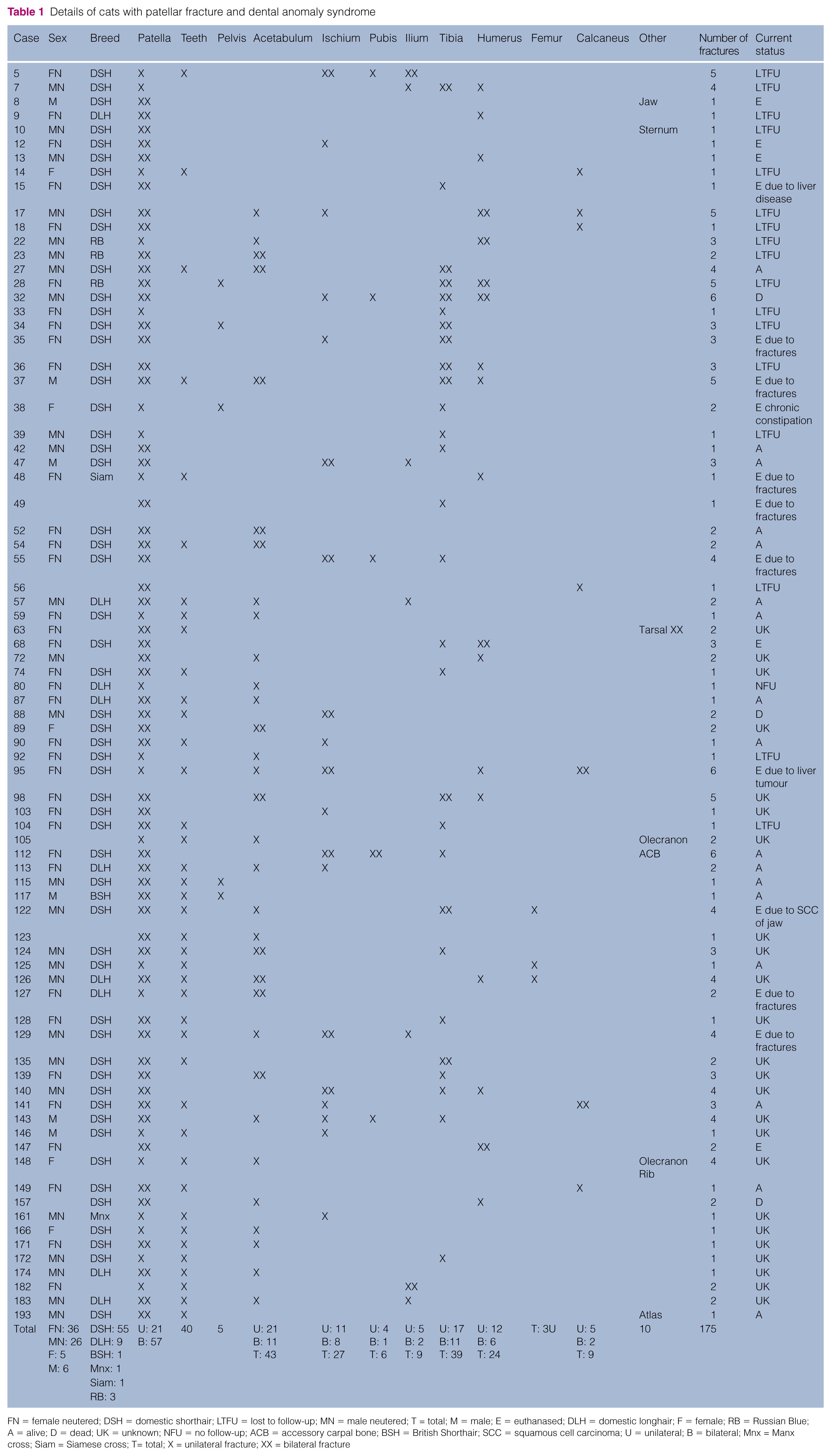
FN = female neutered; DSH = domestic shorthair; LTFU = lost to follow-up; MN = male neutered; T = total; M = male; E = euthanased; DLH = domestic longhair; F = female; RB = Russian Blue; A = alive; D = dead; UK = unknown; NFU = no follow-up; ACB = accessory carpal bone; BSH = British Shorthair; SCC = squamous cell carcinoma; U = unilateral; B = bilateral; Mnx = Manx cross; Siam = Siamese cross; T= total; X = unilateral fracture; XX = bilateral fracture
The age at which the cats sustained the additional fracture(s) was recorded in 44/78 cats; mean age was 5.5 years (range 5–156 months). Sex was recorded for 73/78 cases; there were 32 males (26 neutered and six entire) and 41 females (36 neutered and five entire). Breed was recorded for 70/78 cats; there were 55 domestic shorthairs, nine domestic longhairs, three Russian Blues, one British Shorthair, one Siamese cross and one Manx cross.
The majority of the fractures had a similar configuration, with a single fracture line and a transverse pattern. Radiographic changes of bone density were consistent in the preceding and subsequent fractures, as well as for the patellar fractures; sclerosis was exhibited in all affected bones around the fractures lines. The most common location and typical fracture pattern for the different affected bones are shown in Figure 1. Of the 78 cats, nine had altered skeletal bone density (cases 63, 88, 90, 104, 105, 146, 148, 166 and 172), which varied from generalised sclerosis to focal marked density increase within the same bone (Figure 2).
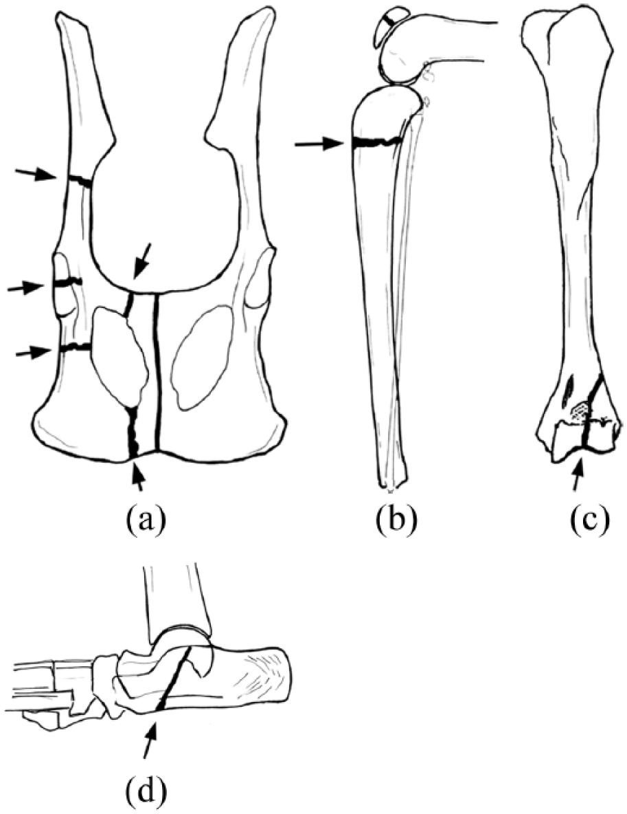
Diagrams of the (a) pelvis, (b) tibia, (c) humerus and (d) calcaneus showing the typical fracture lines (arrows) seen in cats with suspected patellar fracture and dental anomaly syndrome

(a) Radiograph showing altered bone density in an 8-month-old cat (case 146) with patellar fracture. (b) Radiograph of the same cat (case 146) 4 years and 4 months later when it sustained an ischial fracture. (c) Radiograph showing altered bone density in a 6-month-old cat with an olecranon fracture (case 105)
Forty-three acetabular fractures were reported in 32 cats (Table 2); of these, 21 had unilateral and 11 had bilateral fractures. The fractures were cranial, central or caudal with transverse configuration and mild-to-moderate, or no, displacement (Figure 3) in 13 cats. Nine cats had chronic healed acetabular fractures, identified when radiographs were taken for other reasons. Five cats had acetabular fractures diagnosed prior to the occurrence of the patellar fractures. Age at which the acetabular fracture occurred was reported for 17/32 cats and it ranged from 11 months to 8 years, with a mean of 3.7 years.
Details of cats with preceding and subsequent acetabular fractures
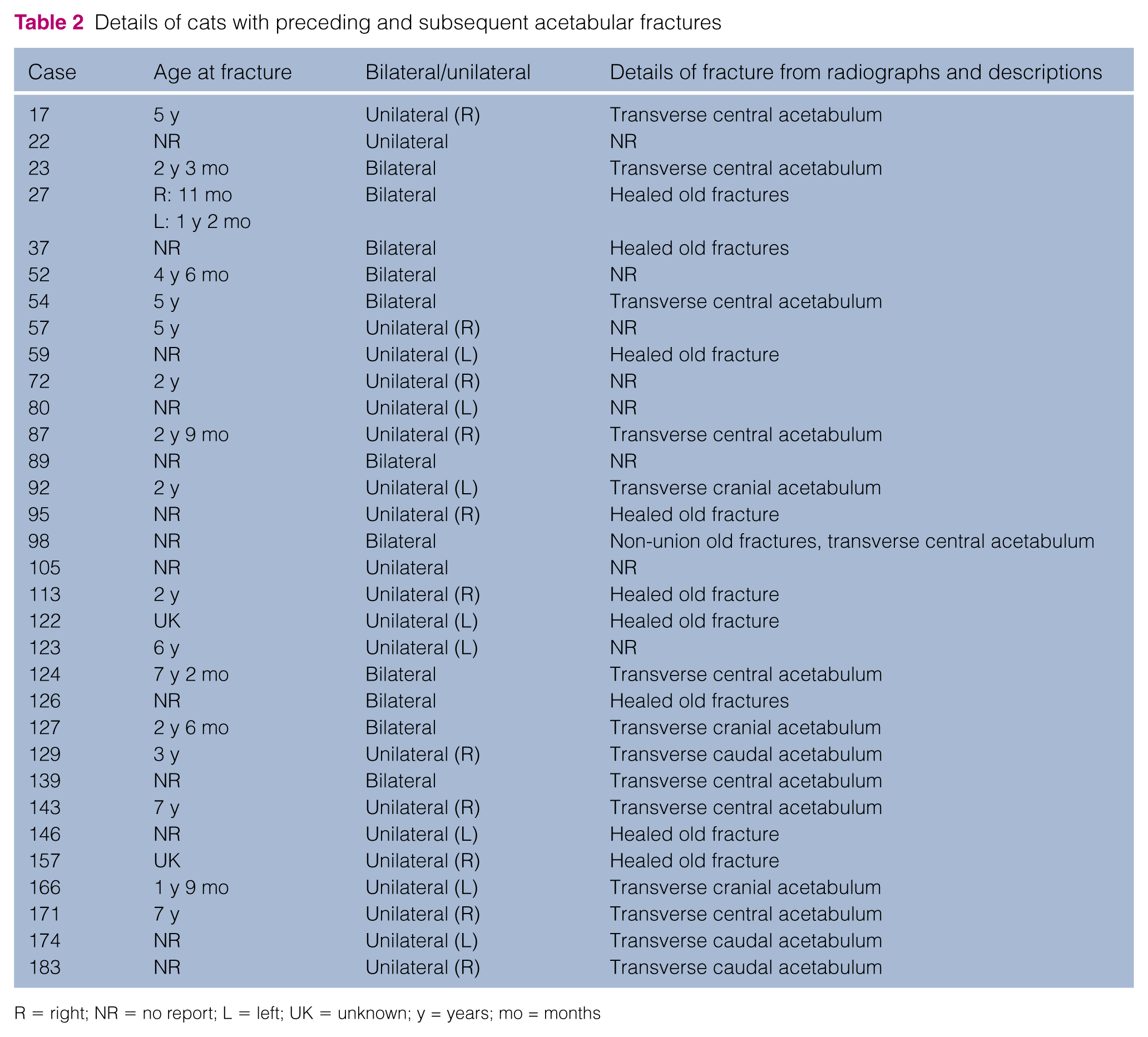
R = right; NR = no report; L = left; UK = unknown; y = years; mo = months

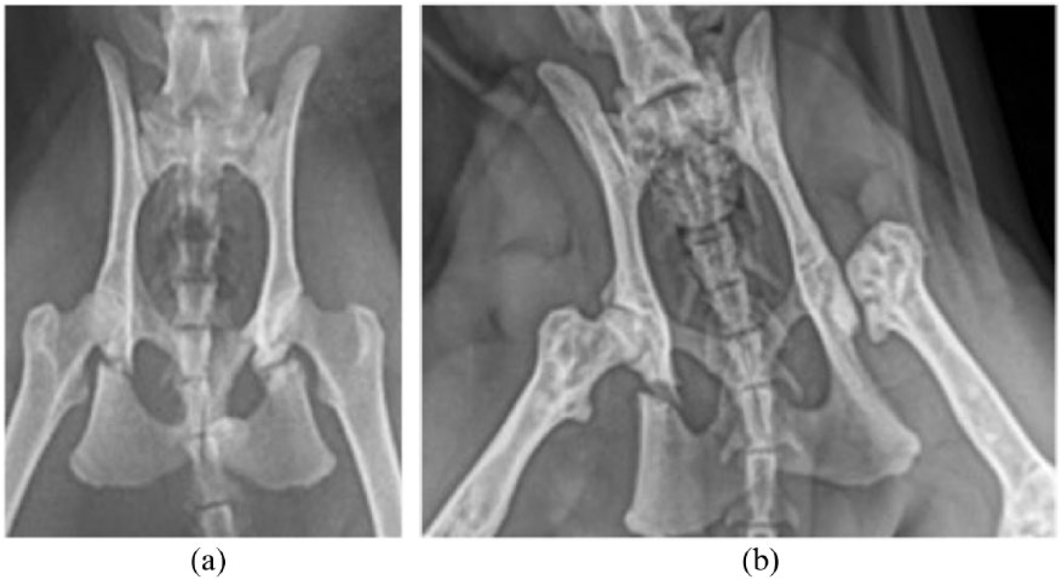
(a) Lateral view and (b) ventrodorsal view of an isolated mildly displaced central transverse acetabular fracture (white arrows) in a cat aged 7 years and 2 months (case 124). Note the sclerosis at the fracture site, the minimal displacement and the lack of other pelvic fractures, which would be likely to occur if this was a traumatic fracture
Nineteen cats had 27 ischial fractures (Table 3), of which eight were bilateral and 11 unilateral. The majority occurred through the ischial body and were transverse or short oblique fractures (Figure 4) and the mean age at presentation was 5.1 years.
Details of cats with preceding and subsequent ischial fractures
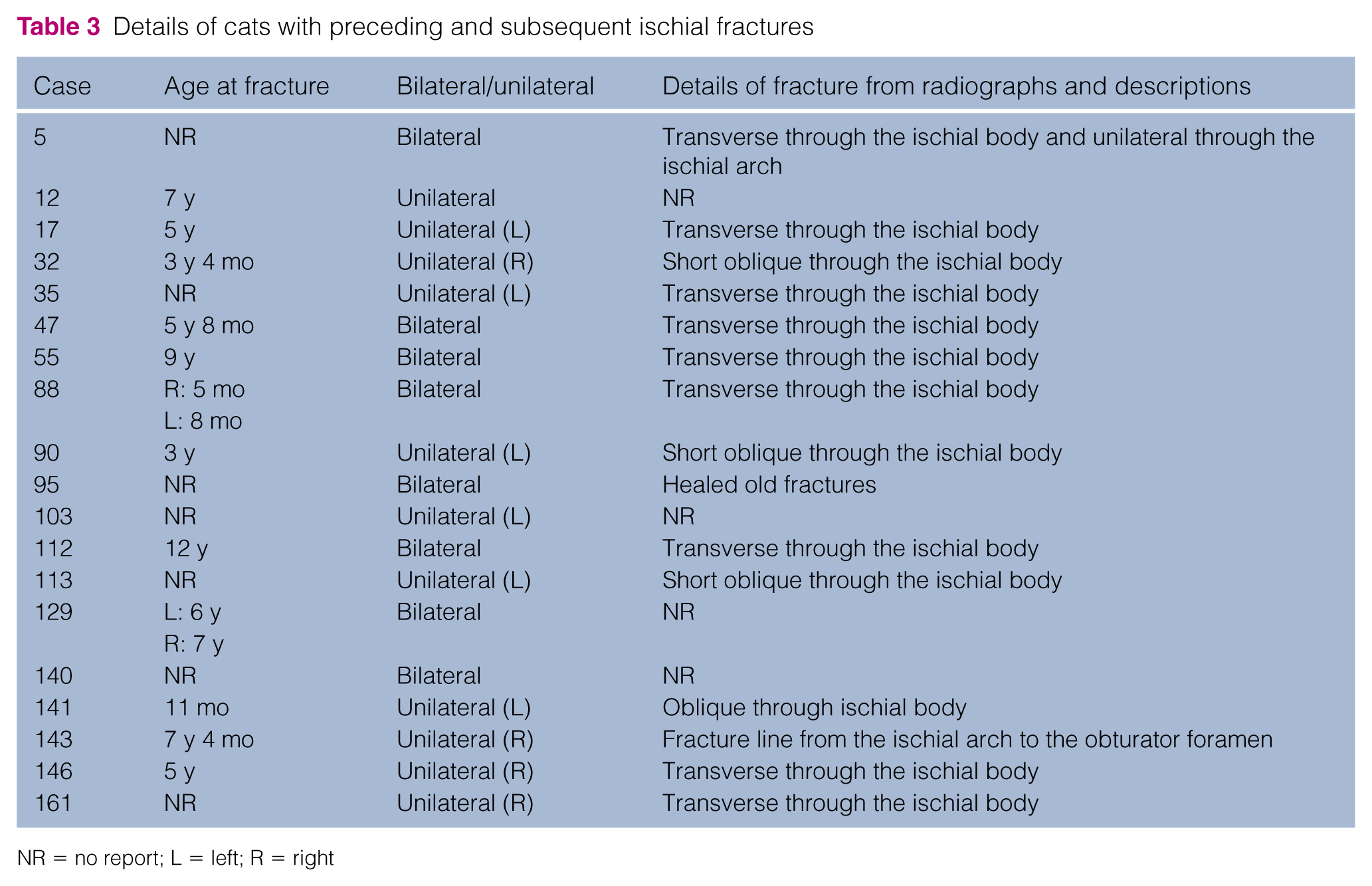
NR = no report; L = left; R = right
(a) Radiograph showing bilateral transverse ischial body fractures in a cat aged 5 years and 8 months (case 47). (b) Radiograph showing a right unilateral short oblique fracture in a 3-year-old cat (case 90). The cat had a previous left femoral head and neck excision for a femoral capital fracture
Ilial and pubic fractures were the least common of the pelvic fractures. Two cats sustained bilateral and five cats unilateral ilial transverse fractures; all of them occurred through the ilial body (Figure 5). Four cats sustained unilateral pubic fractures, these were transverse and occurred through the caudal branches of the pubis; one cat had bilateral transverse fractures of the left and right cranial branches.

(a) Radiograph showing right unilateral transverse ilial body fracture (case 183). There is minimal fracture displacement and sclerosis at the fracture line. (b) Radiograph showing bilateral transverse ilial body fractures, bilateral ischial fractures and pubic fractures (case 5)
Details of cats with tibial fractures are summarised in Table 4. Twenty-eight cats had 39 fractures (11 of the cats had bilateral and 17 had unilateral fractures); of these, 22 were recorded as transverse proximal diaphyseal or proximal metaphyseal fractures (Figure 6). The age at presentation of the tibial fractures ranged from 2.5–13 years (mean 8.1 years).
Details of cats with preceding and subsequent tibial fractures
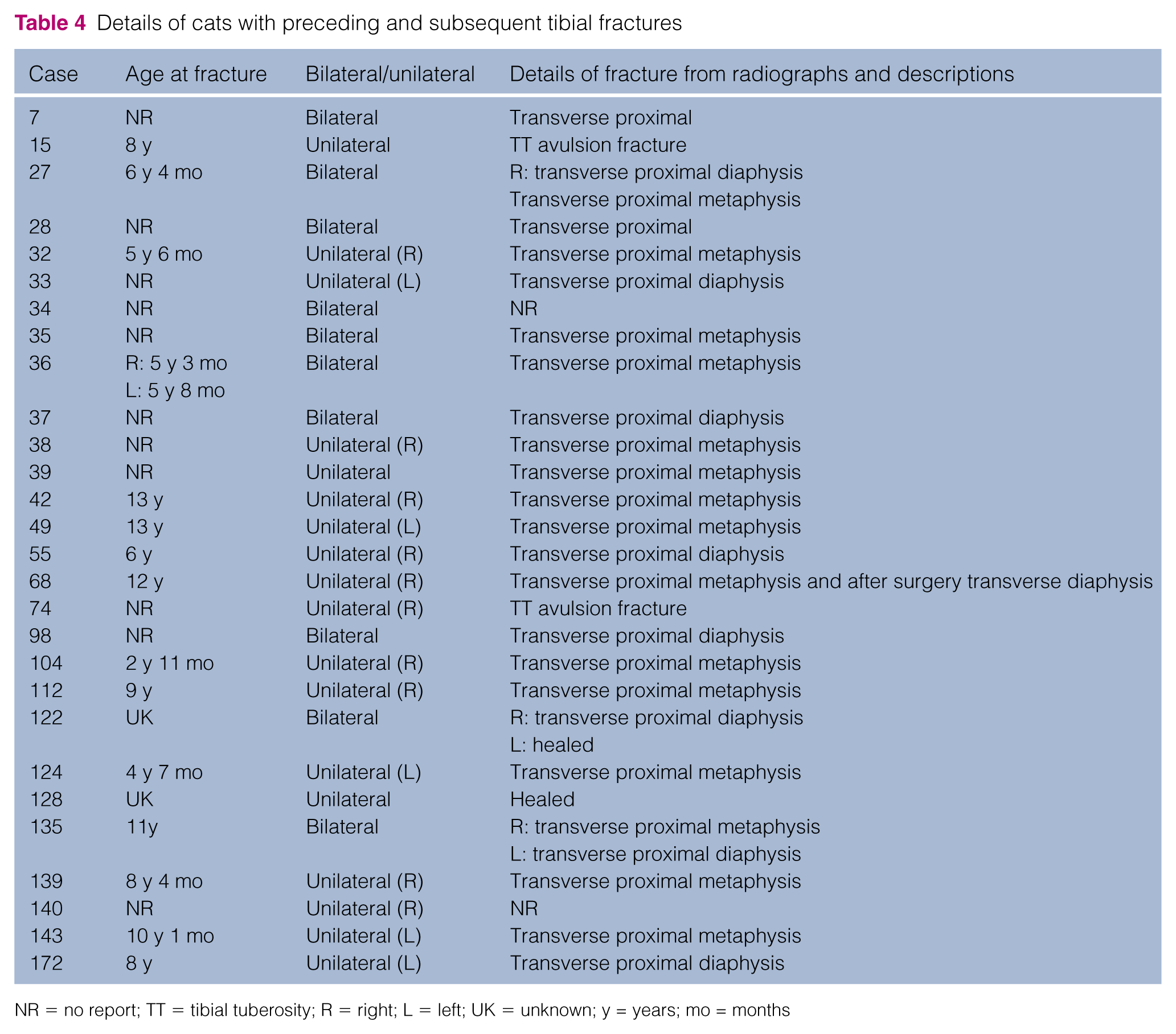
NR = no report; TT = tibial tuberosity; R = right; L = left; UK = unknown; y = years; mo = months
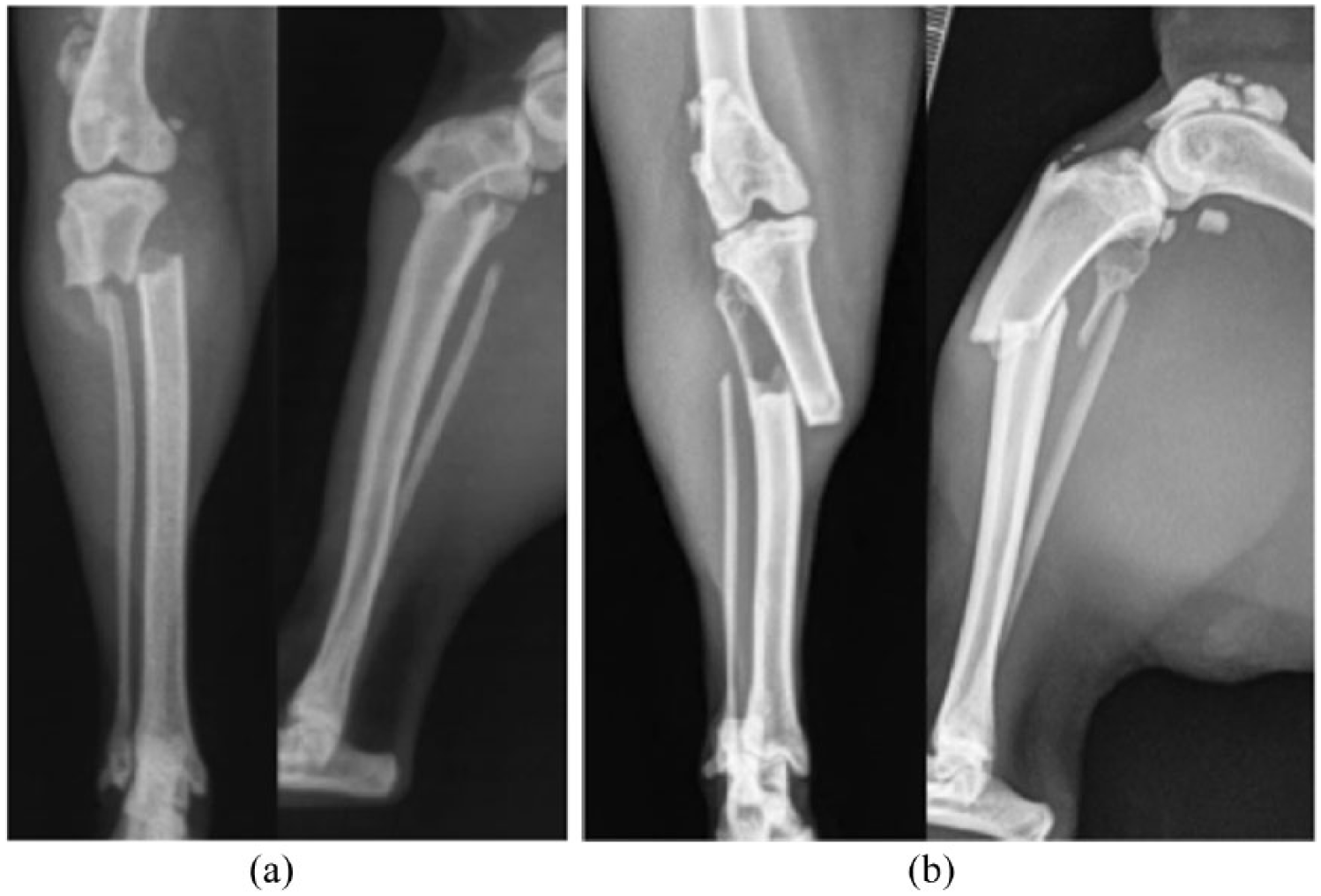
(a) Mediolateral and craniocaudal radiographic views of a transverse proximal metaphyseal tibial fracture (case 143). (b) Mediolateral and craniocaudal radiographic views of a transverse proximal diaphyseal tibial fracture (case 98). There is sclerosis of the bone at the fracture sites, particularly affecting the cranial cortices
Eighteen cats sustained 24 humeral fractures (six cats with bilateral and 12 with unilateral fractures; Table 5). Two of the cats fractured the medial aspect of the humeral condyle and 13 the lateral aspect (Figure 7a), one cat had a Y fracture and two cats had radiographic evidence of bilateral chronic healed fractures (Figure 7b). Two cats had incomplete fractures or humeral intracondylar fissures (cases 17 and 147). The mean age at which the humeral fracture occurred was 5.1 years (range 2–8.5 years).
Details of cats with preceding and subsequent humeral fractures

NR = no report; R = right; L = left; HIF = humeral intercondylar fissure; y = years; mo = months
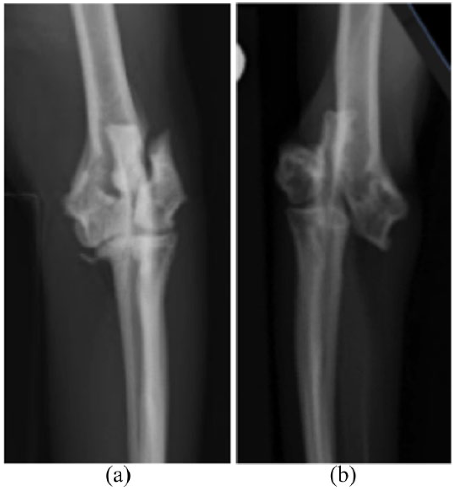
(a) Craniocaudal radiographic view of an acute fracture of the lateral aspect of the humeral condyle (case 17). (b) Craniocaudal radiographic view of a chronic healed fracture of the lateral aspect of the humeral condyle (case 68)
Seven cats suffered nine calcaneal fractures (Table 6). All the fractures had a similar configuration, with a short oblique fracture line present at the base of the calcaneus (Figure 8). Age was reported for 5/9 fractures; the mean age for the occurrence of the fractures was 4.2 years (range 3–8.7 years).
Details of cats with preceding and subsequent calcaneal fractures
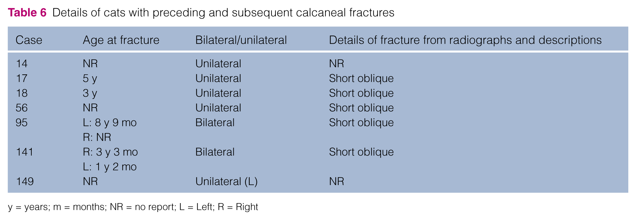
y = years; m = months; NR = no report; L = Left; R = Right

Mediolateral radiographic view of a calcaneal fracture in (a) an 8-year-old cat (case 95) and (b) a cat aged 1 year and 2 months (case 141). The fractures in the two cats have similar configurations, with a distoproximal caudocranial oblique fracture line and sclerosis and remodelling of the fracture gap
Discussion
Information was available for 191 cats with suspected patellar fracture and dental anomaly syndrome. Of these, 10.9% sustained insufficiency fractures to other bones preceding the patellar fractures. Of the 191 cats, 40.8% developed spontaneous fractures to other bones; the pelvis, tibia, humerus and calcaneus were the most frequently affected. The high predisposition of these cats to the development of further fractures without a traumatic event, the similar configuration of the fractures and their consistent location for each bone adds further evidence to the hypothesis that there is a primary bone disorder in these cats leading to a high number of insufficiency fractures.
Stress fractures are caused by prolonged, repetitive muscular activity on bones that are not able to accommodate the increased pull of the involved muscles. Aetiologically, two types of stress fractures exist: fatigue fractures and insufficiency fractures. 6
Fatigue fractures occur when abnormal stresses are applied to normal bone; this is a common condition reported in human athletes and military recruits, and can involve several bones, with the patella being affected fairly infrequently.7,8 Fatigue fractures are fairly common in athletic dogs such as Greyhounds, but minimal literature exists on fatigue fractures in other small animals.2,4,9–11
Insufficiency fractures are the result of normal loading upon abnormally weakened bone. Predisposing and risk factors include metabolic bone disease, osteoporosis, osteogenesis imperfecta, rheumatoid arthritis, osteopetrosis, neurological disorders, prior radiation, total hip replacement, corticosteroid therapy, high-dose fluoride therapy and bisphosphonate therapy, among others.6,12–14 In humans, insufficiency fractures occur most commonly in the bones of the pelvic girdle, including the sacrum, followed by the femur and the vertebral bodies; infrequently, the tibia, fibula, calcaneus and metatarsal bones are also affected. Additionally, the incidence of these fractures is higher in elderly people, mainly owing to osteoporosis or osteomalacia. 14 The cats in this study sustained the majority of fractures in the pelvic bones followed by the tibia, the humerus and the calcaneus. The age at which these cats sustained their first insufficiency fracture was variable, the youngest cat being 5 months and the oldest being 13 years of age. The theory that these cats sustain insufficiency fractures has been strongly suggested in other reports1,2,4 but has not yet been validated nor its aetiology elucidated.
To the best of our knowledge, insufficiency fractures have not been specifically reported in companion animals (excluding studies of cats with patellar fracture and dental anomaly syndrome); however, as detailed above, insufficiency fractures are a subtype of stress fracture. Most reports of stress fractures in the literature are fatigue fractures; these have been shown to be constant in various bones and species. In companion animals fatigue fractures have been reported in horses and Greyhounds. The fracture lines through the affected bones are very similar in position and direction and, as in the cats in our study, the bones and the fracture lines also present as sclerotic, which suggests that remodelling occurs in response to chronic stress and is likely to represent a process of changes preceding the fatigue fracture.15–17 Other reports also support this evidence as fatigue fractures of the tibia in horses were evaluated and it was found that all of them occurred through the same site and with a very similar pattern.18,19 Similar results were obtained when evaluating fatigue fractures of the proximal equine phalanx: 81.5% of the fractures were identical in configuration and site. 20 In our study, most of the insufficiency fractures in each anatomical area had similar fracture morphology. The fractures resembled avulsion type fractures triggered by loading stress forces and tension rather than trauma.
Persistence of deciduous teeth and/or unerupted permanent dentition is rarely reported in cats in the veterinary literature. 21 The original focus of our study in 2005 was on cats with patellar fractures and their poor rate of healing. 5 Subsequently, veterinarians were not specifically asked about the presence of dental anomalies in the original questionnaire. It was only when several cats in the first case series were noted to have persistent deciduous teeth, and appreciating how unusual this condition was in the cat, were we alerted to the fact that this may be a significant finding. 2 Simultaneously to collecting cats with patellar fractures in the UK, information on cats with patellar fractures and persistent deciduous teeth was being collected in the USA. The dental anomalies and the impact of these anomalies have since become a focus of our studies. 3 Reported teeth anomalies in affected cats include persistent deciduous teeth, and unerupted or impacted permanent teeth. The presence of unerupted or impacted teeth may not be immediately obvious on oral examination; imaging such as a CT scan or dental radiography is required to identify these teeth. It is therefore possible that the number of cats with this syndrome affected by dental pathology is higher than reported herein.
In this study, the acetabulum was the most commonly fractured pelvic bone followed by the ischium, ilium and pubis. Pelvic fractures in cats are commonly the result of a major trauma such as road traffic accidents or fall from a height22,23 and comprise 7–25% of fractures in cats.24,25 The box-like structure of the pelvis predisposes it to having multiple fractures, 24 with the most frequent sites of fracture being the pelvic floor (90%) and ilial body (48.5%). In humans, pelvic fractures with no evident trauma are related to athletic activities and are very uncommon; they comprise 1.6–7.1% of all stress fractures, with the pubis being the most frequently affected bone. 26
In the cats in our study, the nature of the pelvic fractures in the absence of reported trauma were suggestive of insufficiency fractures. The pelvic fractures were usually isolated injuries sustained following minimal trauma and many of them were picked up as incidental findings when investigating other problems in the cats, such as lameness perceived to be from the patellar fractures. Some of the pelvic fractures had a chronic appearance on radiography, with the absence of sharp fracture lines and the presence of early callus formation and bone remodelling. Additionally, the fracture fragments were often only minimally or mildly displaced, and if displaced it was in the direction of the tension forces from the attached muscles.
Stress fractures of the acetabulum are rare in dogs and cats; there is one report on acetabular fatigue fractures in racing Greyhounds. 17 The radiographic appearance of the fractures in these Greyhounds was identical in all dogs, with little or no fragment displacement and a fracture line evident in the caudal third of the acetabulum. 17 Similarly, the fractures in the cats were mostly mildly displaced or non-displaced with radiographic evidence of a fracture line through the central acetabulum. Acetabular fatigue fractures are also uncommon in other species; however, there are reports of such fractures in athletes and the military population following repetitive forces on the bones. Acetabular fractures in human beings are more commonly insufficiency supra-acetabular fractures, reported mainly in elderly people with an underlying bone disease, and women are more predisposed. 27 In contrast, the feline acetabular insufficiency fractures reported herein occurred at 11 months to 8.5 years of age, and no significant sex predisposition was detected (41% males and 52.5% females).
Cats can sustain ischial fractures following trauma and these fractures are usually accompanied by other pelvic fractures and/or other injuries.28,29 In humans, avulsion fractures affecting the ischial tuberosity usually occur in young athletes.30,31 The ischial fractures in the cats in this study occurred mainly through the ischial body and table (Figure 4).
The tibial fractures in our cases affected the proximal bone with a transverse configuration through the metaphysis or diaphysis. Only two cats (cases 15 and 74) sustained avulsion fractures of the tibial tuberosity, which is also reported to be a stress fracture in young Greyhounds. 32 Concurrent tibial fractures in cats with unilateral or bilateral patellar fractures have been previously reported and more detail is given in Langley-Hobbs et al. 4 In that publication the mean age was 8.4 years, similar to 8.1 years in the present study.
The humeral fractures identified in our cases all involved the humeral condyle, with the majority affecting the lateral aspect. Fractures of the humeral condyle are uncommon in cats; to our knowledge, no reports of simple fractures in this area exist. In comparison, such fractures are very common in dogs.33,34 These fractures are seen in immature dogs after drops, jumps or falls, and in older dogs after minimal trauma when they are thought to be associated with an intracondylar fissure or incomplete ossification of the humeral condyle. 35 As in the cats in our study, in dogs fracture of the lateral aspect of the humeral condyle is more common than fracture of both the medial and lateral aspect (a Y or T fracture) or fracture of the medial aspect alone. 36 Interestingly, some of the cats presented here had chronic displaced fractures. Potentially, as fracture in this area is not common in the cat, radiographs may not be taken and the fractures are therefore not identified, with the elbow pain treated as arthritis. In addition to the humeral fractures documented in the case records or questionnaires, there were four cats where elbow arthritis or pain, sometimes of sudden onset, was reported. It is possible that these cats may also have suffered condylar fractures but were undiagnosed, as imaging was not performed. One affected cat had been treated for elbow pain for several years and the fractures were only diagnosed (Figure 6) when images were taken of these joints while the cat was sedated for imaging for a tibial fracture. Two cats were also noted to have a humeral intracondylar fissures. This condition has not been reported in cats, but it is common in dogs, particularly Spaniel breeds, and progression to complete fracture can occur.34,37
All calcaneal fractures in the cats in our study had similar configuration: simple with a short oblique fracture line extending from the distocaudal bone at the level of the distal third in a proximocranial direction (Figure 8). There was mild sclerosis and thickening of the caudal cortex in all cases. Details of one cat with bilateral calcaneal fractures, persistent deciduous teeth and no patellar fractures have been previously reported, and two of this cat’s siblings had retained deciduous teeth and patellar fractures. 9 Two cats with bilateral calcaneal stress fractures and other suspected stress fractures affecting the ribs have been reported, with no conclusions made as to whether they were insufficiency or fatigue stress fractures. 10 In these animals there were no reports of teeth or patellar abnormalities, but the fracture configuration was very similar to the cases reported here. Calcaneal fractures are uncommon in cats and a traumatic aetiology is usually reported. 38 Greyhounds have been reported to suffer fatigue fractures of their calcaneus, 39 but the configuration is different to that seen in the cats in our study, probably because in Greyhounds the pathogenesis is related to racing around a track on a flat surface. In the cats the calcaneal mechanism is an area subject to significant tensile forces during jumping, which may make it susceptible to avulsion fracture. 10
Initial thoughts were that cats with patellar fractures were suffering from brittle bone disease, or osteogenesis imperfecta. 2 However, we have now seen several affected cats with generalised increased bone density (Figure 2) and this fact, combined with typical features of the patella being sclerotic at the time of fracture, means we are now suspicious that the underlying bone disease in these cats is a form of osteopetrosis. There are several different presentations of cats with patellar fracture and dental anomaly syndrome. Some cats present only with fracture of their patellae; others have patellar fractures and dental anomalies; and others have patellar fractures, dental anomalies and fractures to one or multiple other bones. Only a small proportion of cats have markedly increased skeletal density (osteopetrosis), dental anomalies, patellar fractures and fractures to other bones. Work is ongoing in this area to perform histopathology of samples from affected cats. Research into osteoclast function and dysfunction is being investigated.
When reviewing the information supplied by the owners and veterinarians, we found reports of two cats with dental anomalies and bilateral atraumatic insufficiency fractures (calcaneus and acetabulum). Two other cats were reported to have only dental anomalies. None of these cats are included in our database owing to the absence of a patellar fracture(s). However, they will be closely followed, as >10% of the cats in the present study had preceding fractures to the patellar fracture and the abnormalities of the teeth may be detected at an earlier age.
The limitations with this study comprise the varying data obtained for the cats, given the fact that radiographs and long-term follow up was not available for all cases. This is, however, the largest study to date of cats presenting with spontaneous fractures and dental abnormalities. Despite the limitation on available follow-up, we feel that the information on preceding and subsequent fractures will be useful for discussion on prognosis of affected animals and alerting veterinarians to the possibility that cats are affected by this syndrome.
Conclusions
Our study provides further evidence that transverse patellar fractures in cats are likely to be insufficiency fractures, a subtype of stress fracture. Many affected cats will have persistent deciduous teeth and other dental anomalies. By obtaining follow-up on these cats we have acquired information on additional fractures in nearly half of the affected population. Fractures are seen in the pelvis, tibia, humerus and calcaneus, all with characteristics of insufficiency fractures, being simple and isolated, as well as transverse or short oblique, fractures. Sclerosis at the fracture line was present in all fractures and the fractures occurred following minimal or no trauma. In the majority of cases other fractures occurred following the patellar fracture, but in >10% of cases the other fractures occurred preceding the patellar fracture. If fractures as described in this article are seen in cats, we would recommend the cat is investigated for patellar fracture and dental anomaly syndrome. The owners should be counselled about this syndrome and the possibility that further fractures and dental issues may occur.
Supplemental Material
KaTS_survey – Supplemental material for Incidence and types of preceding and subsequent fractures in cats with patellar fracture and dental anomaly syndrome
Supplemental material, KaTS_survey for Incidence and types of preceding and subsequent fractures in cats with patellar fracture and dental anomaly syndrome by Natalia A Reyes, Mark Longley, Steven Bailey and Sorrel J Langley-Hobbs in Journal of Feline Medicine and Surgery
Footnotes
Acknowledgements
The authors thank all the owners and veterinarians who provided details on their cats and their cases.
Supplementary material
Knees and teeth syndrome (KaTS) follow-up survey.
Conflict of interest
The authors declared no potential conflicts of interest with respect to the research, authorship, and/or publication of this article.
Funding
The authors received no financial support for the research, authorship, and/or publication of this article.
References
Supplementary Material
Please find the following supplemental material available below.
For Open Access articles published under a Creative Commons License, all supplemental material carries the same license as the article it is associated with.
For non-Open Access articles published, all supplemental material carries a non-exclusive license, and permission requests for re-use of supplemental material or any part of supplemental material shall be sent directly to the copyright owner as specified in the copyright notice associated with the article.