
Abstract
Objectives
The aim of the study was to investigate retrospectively the prognostic impact of variables such as sex, neuter status, breed, age, number of lesions, location and size of the tumour, tumour extension beyond the nasal planum, ulceration and lymph node status, among others, in a population of cats treated with high-dose rate brachytherapy.
Methods
This study reviews the outcome of 58 cats with cytologically and/or histologically confirmed squamous cell carcinoma of the nasal planum, treated at the Clinic Alliance (Bordeaux, France) with high-dose rate brachytherapy from 2010–2016. The total radiation dose delivered was 30 Gy, administered in two different schedules: five fractions of 6 Gy for a period of 4 days (Tuesday–Friday) or four fractions of 7.5 Gy for a period of 3 days (Tuesday– Thursday). Data were collected from cats’ clinical records.
Results
Complete response was achieved in 72% (n = 36) of the cats, partial response in 24% (n = 13) and 2% (n = 1) did not respond. Median progression-free survival and overall survival times were 316 and 835 days, respectively.
Conclusions and relevance
Results indicated that sex (P = 0.045), extension of the tumour from the nasal planum to the upper lip (P = 0.015), tumour size (P = 0.015; P = 0.001), the existence of a previous treatment (P = 0.043) and the tumour response to high-dose rate brachytherapy (P = 0.038; P <0.001) are prognostic factors for cats with squamous cell carcinoma of the nasal planum following high-dose rate brachytherapy.
Introduction
Squamous cell carcinoma (SCC) is one of the most common malignant tumours of the skin of cats (about 15%).1–3 The development of cutaneous SCC is strongly related to sunlight exposure: ultraviolet radiation causes skin damage if the fur or pigmentation do not grant an adequate protection to the skin by reflecting the light and preventing it from reaching the skin’s surface.4–8 Therefore, higher incidence rates are associated with cats with light-coloured fur, lack of protective pigment and sparsely haired/hairless areas such as the ears, eyelids, nasal planum and temples, which make these the most affected areas.1,8,9
SCC located on the nasal planum is relatively common in the cat. 9 The main goal of the treatment is to achieve the best local control of the tumour, given its locally invasive character and low metastatic rate. 10 Although surgical excision is considered the first option for cutaneous SCC, in the case of the nasal planum location a complete tumour excision with proper margins could be challenging and could lead to an aesthetically disfiguring outcome, and therefore other therapeutic modalities should also be considered.5,10,11
Radiation therapy (RT) has been used for many years in cats with the purpose of treating nasal planum SCC.7,11–21 The number of studies published so far regarding the use of RT in the treatment of cats with SCC is relatively small. The use of photon beam (x-ray) RT,7,13,15 hypofractionated proton beam RT, 14 hypofractionated electron beam RT,16,20 Strontium-90 plesiotherapy17,18 and boron neutron capture therapy 19 has been reported. However, the results are controversial and wide ranging in terms of clinical impact and prognosis, and none of them report the use of brachytherapy.
Brachytherapy is a method of delivering radiation at a short distance through an after-loading machine (curietherapy system) that offers two treatment possibilities based on the dose rate: pulsed-dose rate and high-dose rate (HDR).22–25
HDR brachytherapy has been used in nasal planum SCC to irradiate superficial lesions in a fast treatment modality (dose rate over 12 Gy/h).22,26 This treatment has little effect on the surrounding normal tissues and low dissipation of radiation to the room, as well as to the technicians; although the room should still be properly shielded. 25 It is a fast therapeutic modality and can be completed in less than a week, with a very short overall treatment time. The veterinary literature on this topic is scarce, with no published studies regarding HDR brachytherapy in feline SCC of the nasal planum.
In the absence of literature data, the main purpose of this retrospective study was to describe the effect of some epidemiological and tumour variables on progression-free survival and overall survival, and ascertain prognostic factors for cats with SCC of the nasal planum following HDR brachytherapy.
Materials and methods
Case selection
Cats with SCC of the nasal planum treated with HDR brachytherapy at Clinique Vétérinaire Alliance, in Bordeaux, France, during a 6 year period from 1 June 2010 to 31 July 2016 were retrospectively included.
Data acquisition
Data collected from clinical records comprised sex, neuter status, breed, age, number of lesions, location and size, extension to the haired skin, extension to the upper lip (without involvement of the oral cavity), ulceration, lymph node involvement, lung metastases, any previous treatment, tumour response to HDR brachytherapy, local recurrence and overall survival. Information about the side effects of the treatment was unavailable for the majority of the clinical cases and therefore not included in this study.
Concerning the number of lesions, two categories were considered: a single lesion or multiple lesions. Regarding the location of the tumour, based on the clinical history data, six locations were considered: nasal cavity when the tumour invaded the nasal cavity; only the nose tip (the edge of the nose); dorsal if it was in the dorsal side of the nasal planum; rostral if it was in the rostral side of the nasal planum; nostril wing when the wing of the nostril was affected; and a combination when the lesion affected two or more of these locations (Figure 1). For tumour size, tumours were categorised into two groups: T ⩽2 cm and T >2 cm (adaptation of the World Health Organization [WHO] staging system). 27 In the case of multiple lesions, the lesion with the largest diameter was the selected for evaluation of tumour size. Diagnosis was made by fine-needle aspiration cytology and/or biopsy.
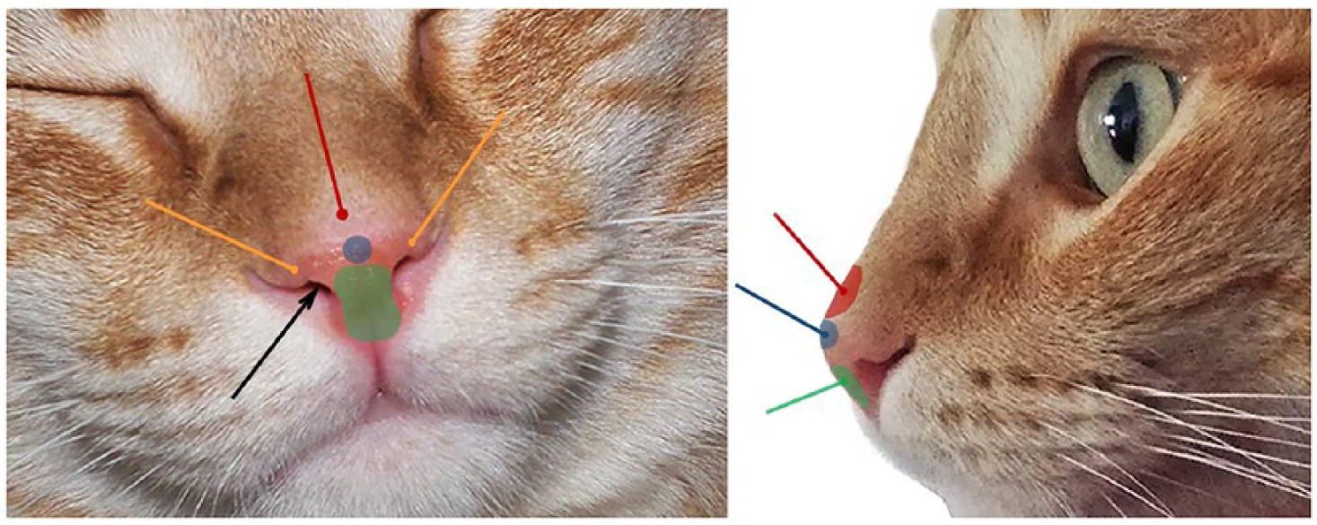
Nasal planum squamous cell carcinoma anatomical locations considered for this study. Black arrow = nasal cavity; blue shadow and arrow = nose tip; red shadow and arrow = dorsal face; green shadow and arrow = rostral face; orange arrow = nostril wing
Regional lymph node involvement was assessed by fine-needle aspiration cytology. Thoracic radiographs or CT scans were performed to assess lung metastasis. Clinical staging evaluation was assessed using the WHO recommendations. 27
Treatment
All cats were submitted to the same treatment protocol. The anaesthetic protocol consisted of premedication with buprenorphine 0.02 mg/kg and diazepam 0.2 mg/kg IM, induction with propofol 4 mg/kg IV and maintenance with isoflurane. There was no need for further analgesia, in addition to premedication with buprenorphine, during the hospitalisation time. The HDR brachytherapy protocol consisted of a total dose of 30 Gy, administered in two different schedules, according to owner compliance: five fractions of 6 Gy distributed over a period of 4 days (Tuesday–Friday, as follows: Tuesday morning and evening, Thursday morning and evening, and Friday morning) or four fractions of 7.5 Gy distributed over a period of 3 days (Tuesday–Thursday, as follows: Tuesday morning and evening, Thursday morning and evening). The planning target volume as defined by the International Commission on Radiation Unit was the same as the clinical target volume (CTV) because internal margins and set-up margins were considered to be non-existent. The CTV was defined by the gross tumour volume extended by 3 mm.
Tumour response to brachytherapy was categorised as complete response (CR), partial response (PR) and progressive disease (PD), as previously described. 17
Progression-free survival (PFS) was defined as the time from the beginning of treatment until first recurrence. Overall survival (OS) was defined as the length of time cats lived from the beginning of treatment until death by the tumour or the end of the study.
Statistical analysis
SPSS version 24.0 (IBM) was used for statistical analysis. ANOVA was used for continuous variables. For categorical variables, we used the χ2 test and Fisher’s exact test when appropriate.
Survival curves were generated by the Kaplan–Meier method and the survival rates were compared using the log-rank test. In this study, no cats were lost to follow-up; therefore, censored observations correspond to animals that died from causes other than the neoplastic disease or that remained alive at the end of the study period.
Continuous variables were expressed as mean ± SD. In all statistical comparisons, P <0.05 was accepted as denoting statistically significant differences.
Results
Fifty-eight cats were included in this study; however, owing to its retrospective nature, it was not always possible to obtain information for all the analysed variables. Therefore, for each one of the characteristics described below, the percentage presented refers to the total number of cats for which it was possible to obtain information.
Regarding sex, 31/58 (53.4%) cats were male and 27 (46.6%) were female. Only one cat was intact and 57/58 (98.3%) were neutered. The majority of cats (n = 56/58) were European Shorthair (96.6%) and two cats were Turkish and Birman crosses. The median age was 11 years (range 3–17 years) and this variable was categorised into two groups for survival analysis: the first was composed of cats aged ⩽11 years (n = 29/57; 50.9%), and the second with cats aged >11 years (n = 28/57; 49.1%).
Based on the number of lesions on the nasal planum, 12/57 (21.1%) cats presented with multiple lesions and 45/57 (78.9%) had a single lesion. Information about the location of the tumour on the nasal planum was available for 56 cats: 1/56 cats (1.8%) had the tumour in the nasal cavity, 2/56 (3.6%) on the nose tip, 3/56 (5.4%) had it on the dorsal face, 4/56 (7.1%) on the rostral face, 19/56 (33.9%) had the wing of one of the nostrils affected and 27/56 (48.2%) had a combination of two or more of the above.
The extension of the tumour from the nasal planum to the haired skin was present in 38/45 (84.4%) cats and absent in 7/45 (15.6%). Extension to the upper lip was observed in 24/52 (46.2%) cats and was absent in 28/52 (53.8%). Tumour ulceration was present in 46/55 (83.6%) cats and absent in 9/55 (16.4%).
Regarding the size of the tumour, in 21/48 cats (43.8%) the tumour was ⩽2 cm, and in 27/48 (56.3%) the tumour was >2 cm. Tumour size was the only variable that was statistically significantly associated (P = 0.015) with the tumour response to brachytherapy, showing that small tumours had a better response to treatment.
According to the records, 6/55 (10.9%) cats received a previous treatment: three were treated with surgical resection, one with electrochemotherapy, one with surgical excision followed by external radiotherapy, electrochemotherapy and chemotherapy, and one with electrocauterisation. The remaining 49 cats (89.1%) did not receive any type of treatment prior to HDR brachytherapy.
Regarding the tumour response to HDR brachytherapy, 36/50 cats (72%) achieved CR, 13/50 (26%) achieved PR and 1/50 (2%) showed PD, corresponding to a response rate and clinical benefit of 98% (n = 49/50). Tumour recurrence was observed in 29/43 (67.4%) cats. Concerning the side effects associated with this therapy, and as mentioned in the ‘Materials and methods’ section, it was not possible to collect enough data from the patients’ files to describe precisely the side effects of brachytherapy in this location. However, although we were not able to record the precise number of cats, nor the severity of the side effects, in some cats acute radiodermatitis was observed that resolved with symptomatic treatment (data not shown).
Follow-up information (PFS and OS) was available for 43/58 cases. All 43 tumours were in a local stage of disease. Disease progression (when it occurred) was only observed as a local recurrence of the disease, and none of the cases developed regional or distant metastasis during follow-up. At the end of the study, 22/43 cats (51.2%) were dead owing to tumour-related causes (in all cases as a consequence of the deterioration of the general condition, depression, inappetence, weakness, anorexia and/or increasing pain); of the remaining 21 cats (48.8%), 13/21 (61.9%) were alive and 8/21 (38.1%) were dead from other causes.
The mean and median times of PFS were 583 days (95% confidence interval [CI] 406.9–759.4 days) and 316 days (95% CI 76.6–555.4 days), respectively. A previous treatment before HDR brachytherapy (P = 0.043) and tumour response to brachytherapy (P = 0.038) revealed a statistical significant association with a shorter PFS (Figure 2).
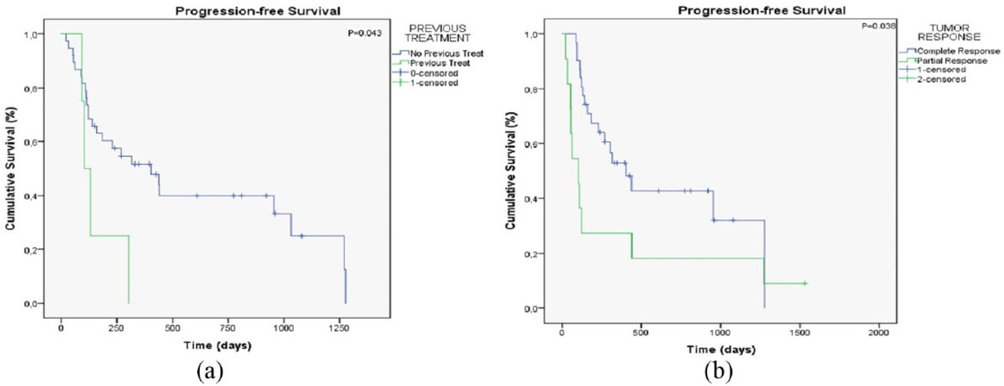
Kaplan–Meier curves comparing the (a) progression-free survival (PFS) of cats treated with high-dose rate (HDR) brachytherapy with and without a previous treatment (P = 0.043) and (b) PFS of complete or partial tumour response to HDR brachytherapy (P = 0.038)
The mean and median times of OS were 963 days (95% CI 707.4–1218.9 days) and 835 days (95% CI 84.8–1585.2 days), respectively. A statistically significant association was found with regard to sex (P = 0.045). Male cats showed a decreased OS vs female cats (354 days vs 1817 days; Figure 3a). Cats that presented an extension of the tumour from the nasal planum to the upper lip showed a significantly shorter (P = 0.015) OS time (267 days vs 1817 days; Figure 3b). A larger tumour size was also found to significantly affect OS (267 days vs 1817 days [P = 0.001]; Figure 3c). Regarding tumour response to HDR brachytherapy, cats that experienced CR had a better prognosis and a longer OS (P <0.001) than those with a PR (1024 days vs 639 days; Figure 3d). Detailed information about the relationship of the variables with PFS and OS is summarised in Table 1.

Kaplan–Meier curves comparing overall survival of (a) male and female cats treated with high-dose rate (HDR) brachytherapy (P = 0.045); (b) cats without and with extension of the tumour from the nasal planum to the upper lip, treated with HDR brachytherapy (P = 0.015); (c) cats treated with HDR brachytherapy with a tumour size ⩽2 cm and >2 cm (P = 0.001); and (d) cats with a complete, partial or progressive tumour response to HDR brachytherapy (P <0.001)
Relationship of variables with progression-free and overall survival at days 500 and 1000

Bold denotes statistical significance
Non-computable (SPSS system information)
CR = complete response; PR = partial response; PD = progressive disease
Discussion
Nasal planum SCC in cats is a relatively frequent tumour characterised by its high local aggressiveness and recurrence rates. 10 Surgical excision is sometimes difficult owing to the nasal location; therefore, HDR brachytherapy is a promising therapeutic alternative.5,10,11 Usually, HDR brachytherapy is used instead of surgery; however, there is a report of its use in the postoperative period for treatment of fibrosarcoma in cats with promising results. 28
As stated before, there are some publications on the use of external RT in the management of feline SCC of the nasal planum.7,13–20 However, studies on HDR brachytherapy are lacking. Therefore, all the comparisons between the present results and the data from the literature refer to other RT modalities.
The studied population of 58 cats was almost half divided by sex, with a slightly larger number of males than females (male:female ratio 1:1), a result similar to those already obtained by others.7,13,16,18 These data also supported the inexistence of a sex predisposition to feline SCC, already known from other publications.1,16,29
European Shorthair cats were over-represented in this study (96.6%) and so it was not possible to observe whether a short-haired breed or any pure breed would be more susceptible, as referenced in the literature. 1 Owing to the variety of characteristics and multiple coat colours possible among the European Shorthair cats, these results also sustain the lack of a breed predisposition to SCC.1,16,29
The mean and median age of the cats in this study was 11 years. This confirms, once again, the tendency of these tumours to affect older cats (10–12 years old).1,5,30,31
In this work, a single lesion occurred in the majority of cats, in contrast to a 1991 publication cited by Rassnick, 4 where it was reported that multiple lesions were observed in approximately 45% of the cats with cutaneous SCC. However, in that study, cutaneous SCCs were in distinct locations and not exclusively on the nasal planum, which prevents solid comparisons with the present study.
To our best knowledge, there are no data available in the literature about the clinical and prognostic impact of tumour extension to the haired skin or to the upper lip. According to the results of this study, cats with an affected upper lip lived significantly shorter than the others without an affected upper lip (median OS 267 days vs 1817 days). This aspect has never been described before and might represent promising new prognostic information to have when evaluating nasal planum SCC in cats. However, the present results should be analysed with caution as it is not possible to compare our results with those from studies already published, as the treatment modalities were different.
Tumour size was the only clinical characteristic that showed a significant association with tumour response to HDR brachytherapy in the present study. These results are in accordance with Cunha et al, 20 who stated that T1 lesions showed better RT treatment responses (CR in 62.5%) than T3 and T4 lesions (CR in 31.3%). Tumour size was also found to be statistically associated with OS, revealing that cats with larger lesions have shorter OS times, which is in agreement with other studies.1,7,13,16,20,32,33 In fact, cats with tumours ⩽2 cm lived significantly longer than cats with larger lesions (median OS of 1817 days vs 267 days). The present results are in line with the already published results by Théon et al, 7 and emphasise that tumour size is a prognostic factor for RT treatment efficacy in these tumours.
Tumour ulceration was present in the majority (83.6%) of the lesions, although without a significant prognostic value. A variety of publications report the usual observation of ulceration in nasal planum SCC without prognostic impact.1–3,15,17,19,29,34–38
The therapeutic benefit of HDR brachytherapy observed in this study was very high. A CR was achieved by the majority of the cats (72%), which was a satisfactory result compared with the results already published for tumour response to external RT.14–18,20 In 26% of the cases the cats experienced PR, which is also in agreement with previous publications, although with a wider range of values.14,17–19 There was one cat (2%) in this study that showed a PD, indicating that, as in RT, a small percentage of cases did not respond to therapy.14,20
Tumour recurrence was observed in 67.4% of the cats. The possible occurrence of new lesions in the nasal planum but not exactly in the same place as the treated tumour is difficult to discern, and even harder if it is a different evaluating veterinarian, which often happens owing to other facilities referring cats for RT. These limitations were also mentioned in the publication of Hammond et al; 18 moreover, there was the possibility of an overestimation of the number of recurrences by the owners, or a progression of a stable lesion and not reporting it to their veterinarian immediately, wrongly extending the PFS times. Recurrence has been reported in studies using external RT treatment protocols,13–16,20 where it ranges from 11.1–70%. This wide range of values reflects the need for prospective, well-controlled studies where the evaluation follow-up procedures should be judicious and systematic, and preferably include CT evaluations to properly assess tumour volume, and therefore reduce the possibility of an incorrect treatment approach.
Interestingly, sex was found to affect OS. This association has been described in human medicine where some studies identified men at higher risk of cutaneous SCC.39,40 Even though there are no other reports of similar data in cats, our results advise special attention to be paid to the disease evolution when male cats are affected.
Tumour response to brachytherapy was found to be statistically associated with PFS and OS. Cats that achieved a CR showed longer local tumour control times and lived longer, which is in agreement with Hammond et al, 18 who used Strontium-90 plesiotherapy and cats with a CR had a longer OS than cats with PR (P <0.001).
One of the limitations of the present study is the lack of comparison of our results with previous literature as the number of HDR brachytherapy studies is small or non-existent.
The protocol used in this study demonstrated ease of application, being performed during a 1 week period, requiring only sedation and monitoring of vital signs for each session.
Our data revealed that HDR brachytherapy can be considered as an additional therapeutic option to treat SCC of nasal planum in cats with a good therapeutic response.
Conclusions
In cats with nasal planum SCC treated with HDR brachytherapy, male sex, tumour extension from the nasal planum to the upper lip, larger tumour size and a partial tumour response to HDR brachytherapy instead of complete remission showed to be of prognostic value, being directly associated with a worse prognosis.
Footnotes
Conflict of interest
The authors declared no potential conflicts of interest with respect to the research, authorship, and/or publication of this article.
Funding
This work was partially supported by research projects UID/AGR/04033/2013 and UID/MULTI/00211/2013, all financed by Portuguese Foundation for Science and Technology.