
Abstract
Intrathecal administration is an important route for drug delivery, and in pharmacology and toxicology studies, cerebrospinal fluid (CSF) collection and analysis is required for evaluating blood–brain barrier penetration and central nervous system exposure. The characteristics of CSF in commonly used nonrodent models are lacking. The purpose of this study is to evaluate and provide some insights into normal cellular and biochemical composition of CSF as well as diffusion potential following intrathecal injection across several nonrodent species. Cerebrospinal fluid samples were collected from the cerebellomedullary cistern of beagle dogs, cynomolgus monkeys, and Göttingen minipigs and analyzed for clinical chemistry and cytological evaluation. Diffusion into the intrathecal space following intrathecal injection was assessed following administration of a contrast agent using fluoroscopy. The predominant cell types identified in CSF samples were lymphocytes and monocytoid cells; however, lymphocytes were represented in a higher percentage in dogs and monkeys as opposed to monocytoid cells in minipigs. Clinical chemistry parameters in CSF revealed higher Cl− concentrations than plasma, but lower K+, Ca2+, phosphorus, glucose, creatinine, and total protein levels consistent across all 3 species. Diffusion rates following intrathecal injection of iodixanol showed some variability with dogs, showing the greatest diffusion distance; however, the longest diffusion time through the intervertebral space, followed by monkeys and minipigs. Minimal diffusion was observed in minipigs, which could have been attributed to anatomical spinal constraints that have been previously identified in this species.
Introduction
Intrathecal delivery of drugs into the cerebrospinal fluid (CSF) is a convenient way to target the central nervous system (CNS). In preclinical drug development, CSF analysis is often required for evaluating blood–brain barrier penetration and CNS exposure of new drug candidates. 1 With neurology being the second most frequent indication pursued by drug development companies, 2 evaluation of CNS drug distribution and pharmacodynamics in large animal species has become an important aspect of preclinical drug testing. 3 Cerebrospinal fluid analysis may also be used to help identify a wide variety of conditions affecting the CNS, including drug-induced neurotoxicity. 4
Cerebrospinal fluid collections are generally carried out as part of pharmacology and toxicology studies conducted in large animal species, including dogs, swine, and nonhuman primates. The CSF is a metabolically active and dynamic substance playing many important functions such as structural, hydrodynamic, metabolic, and immunologic aspects. 5 However, limited data are available concerning CSF characteristics between species, and there is a need for more sensitive and specific biomarkers in the CSF relevant across animal models that could allow for the prediction of neurotoxicity, which could translate from nonclinical to clinical data. Biomarkers in the CSF are particularly valuable as CSF localizes with target neural tissues and can be indicative of biochemical changes in these tissues due to drug-induced neurotoxicity 4 as well as other pathological conditions such as inflammation (infectious or noninfectious) involving the brain, spinal cord, and meninges.
Nonhuman primates are ideal animal models for the translation of drug testing results to humans as they share many similarities with the human anatomy and physiology; however, the use of nonhuman primates can present both ethical and economical obstacles in drug studies. Hence, alternative nonrodent models are also necessary. The minipig has been an extensively used animal model for translational neurological research and is an ideal species for epidural and intrathecal injection studies. 6,7 In addition, dogs are also frequently used as a nonrodent animal model for efficacy and drug safety studies because of their similarities in anatomical morphology and physiology to humans, but also because these animals are very easy to handle compared to minipigs and nonhuman primates in the laboratory.
The purpose of this study is to evaluate and provide some insights into normal cellular and biochemical composition of CSF as well as diffusion potential in beagle dogs, Göttingen minipigs, and cynomolgus monkeys. To characterize interspecies differences, CSF clinical chemistry was evaluated in comparison to blood plasma, CSF cytology was analyzed, and intrathecal diffusion was assessed following injection of a contrast agent into the intervertebral space.
Methods and Materials
Statement on Use and Care of Animals
During the study, care and use of animals were conducted in accordance with principles outlined in the current Guide to the Care and Use of Experimental Animals published by the Canadian Council on Animal Care and the Guide for the Care and Use of Laboratory Animals published by the US National Institutes of Health (NIH publication no. 85-23, revised 2010). Citoxlab’s facility is AAALAC accredited. All procedures were conducted as per Standard Operating Procedures.
Animal Housing
Beagle dogs
Ten female in-house colony female Beagle dogs (Canis familiaris) were selected for this study. The original source of the animals was Marshall Bioresources (North Rose, New York). The animals were between 0.8 and 1.3 years old and weighed between 6.7 and 8.8 kg. The animal room environment was controlled (temperature: 21°C ± 3°C, humidity: 30%-70%, 12-hour light, 12-hour dark, 10-15 air changes per hour), with temperature and relative humidity being monitored continuously. A standard certified commercial dog chow (Certified 25% Lab Dog Diet #8727C, Envigo Teklad, Madison, Wisconsin) was available daily.
Göttingen minipigs
Six males and 3 females in-house colony Göttingen minipigs (Sus scrofa) were selected for this study. The original source of the animals was Marshall Bioresources. The animals were between 6.3 and 6.9 months old. Body weights ranged between 13.2 and 17.7 kg and between 13.7 and 16.6 kg for males and females, respectively. The animal room environment was controlled (temperature: 21°C ± 3°C, humidity 30%-70%, 12-hour light, 12-hour dark, 10-15 air changes per hour), with temperature and relative humidity being monitored continuously. A standard certified commercial chow (Miniswine Diet #7037C, Envigo Teklad) was available twice daily.
Cynomolgus monkeys
Six males and 4 females colony Chinese cynomolgus monkeys (Macaca fascicularis) were selected for this study. The animals were between 5.2 and 6.9 years old. Body weights ranged between 5.2 and 9.6 kg and between 3.6 and 5.2 kg for males and females, respectively. The animal room environment was controlled (temperature: 21°C ± 3°C, humidity: 30%-70%, 12-hour light, 12-hour dark, 10-15 air changes per hour), with temperature and relative humidity being monitored continuously. A standard certified commercial primate chow (Certified Hi-Fiber Primate Diet 7195C, Envigo Teklad) was available twice daily.
Experimental Methods
Cerebrospinal fluid collection and analysis
Cerebrospinal fluid samples were obtained from the cerebellomedullary cistern under anesthesia with isoflurane. Animals were positioned in lateral recumbency and the skin at the site of puncture was aseptically prepared. The head was held at a right angle to the vertebral column and the animal was observed for adequate ventilation during the entire procedure. A triangular landmark composed of the occipital protuberance and the wings of the atlas was identified with the puncture following in the middle of this triangle. The cerebellomedullary cistern was punctured between the occipital bone and the atlas using a 20- or 22-guage spinal needle (BD, Ontario, Canada). The needle was carefully inserted and was considered in the right place once the puncture of the dura meter and atlanto-occipital membrane was felt. Cerebrospinal fluid was allowed to drip into the collection tube to minimize blood contamination. One sample (2 mL) was obtained in plain tube for measurement of electrolytes (Cl−, Na+, K+, Ca2+, and phosphorus), total protein, glucose, urea, and creatinine using the Cobas 6000 Module c501 clinical chemistry analyzer (Roche Diagnostics Ltd, Indianapolis, Indiana). A second sample (0.5 mL), collected into tubes containing K2EDTA as anticoagulant (Sarstedt, Quebec, Canada), was used for cytological evaluation. Cerebrospinal fluid samples were placed in the sample port of a cytochamber containing a tan cytopad and were cytocentrifuged for 5 minutes at 1,200 rpm (Hematology Slide Stainer/CytocentrifugeAerospray 7120, Wescor Inc, Logan, Utah). As the sample was centrifuged, the cells present in CSF were forced into a monolayer within a 7-mm diameter circle area on the slide. A portion of the fluid was absorbed by the cytopad, producing a more concentrated area of cells on the slide. Cytospin smears were stained with the Aerospray hematology slide stainer (Hematology Slide Stainer/CytocentrifugeAerospray 7120, Wescor Inc) using a 2-part aqueous Romanowski stain. Differential cells count was evaluated on a total of 200 cells per slide.
Blood collection and analysis
Blood samples (1.1 mL) were collected into tubes containing clotting activator gel (Sarstedt) for clinical chemistry evaluation. Levels of electrolytes (Cl−, Na+, K+, Ca2+, and phosphorus), total protein, glucose, urea, creatinine, albumin, globuline, total bilirubin, cholesterol, and triglycerides were measured and the activity of alanine aminotransferase, alkaline phosphatise, and aspartate aminotransferase was measured, using the Cobas 6000 Module c501 clinical chemistry analyzer (Roche Diagnostics Ltd).
Intrathecal injection
To assess the diffusion into the intrathecal space following intrathecal injection, iodixanol (Visipaque), a nonionic radiographic contrast agent, was administered to 5 animals of each species. Anesthesia was maintained by isoflurane inhalation and a 20-guage spinal needle was inserted perpendicular to the skin at the level of the L3 to L4 intervertebral space (Figure 1). Needle placement was confirmed by aspiration of CSF, then 0.5 mL of Visipaque (320 mgI/mL, GE Healthcare, Ontario, Canada) was injected slowly. Diffusion of iodixanol into the intrathecal space was visualized by fluoroscopic imaging (SIREMOBIL compact L fluoroscope, Siemens, Malvern, Pennsylvania).
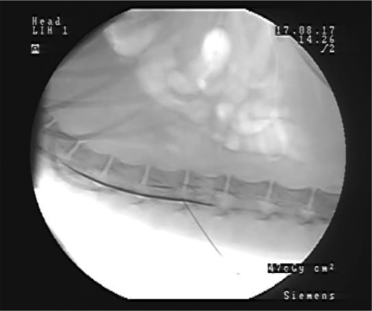
Fluoroscopic imaging of iodixanol (Visipaque) injection in L3 to L4 intrathecal space in a beagle dog.
Statistical analysis
Results are presented as mean ± standard deviation. Student t test was used to compare the means for clinical chemistry parameters between the CSF and the blood to identify statistically significant differences in concentrations.
Results
Cerebrospinal Fluid Differential Cell Count
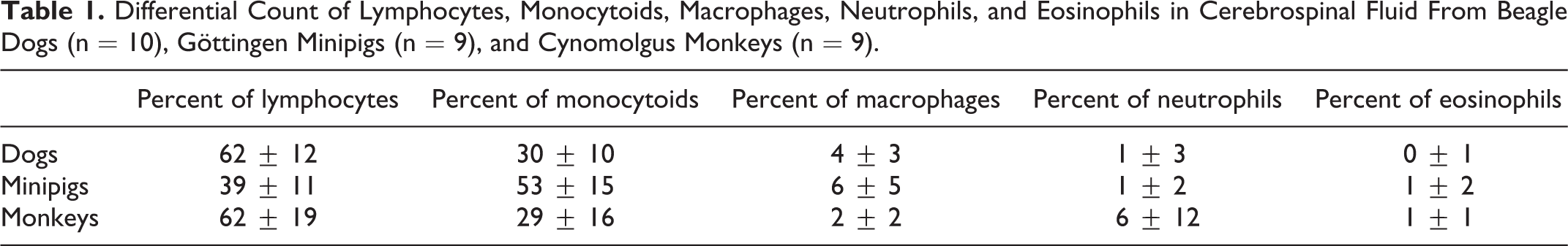
The predominant cell types identified in CSF samples were lymphocytes and monocytoid cells (Table 1). Together, lymphocytes and monocytoid cells accounted for approximately 90% of the total cell count. In dogs and monkeys, respectively, lymphocyte counts were 62% ± 12% and 62% ± 19%, while monocytoid cell counts were 30% ± 10% and 29% ± 16%. In minipigs, the percentage of monocytoid cells was higher than lymphocytes, with differential count of 53% ± 15% and 39% ± 11%, respectively. Macrophages were present in a small proportion in all 3 species (4% ± 3%, 6% ± 5%, and 2% ± 2% for dogs, minipigs, and monkeys, respectively). Lastly, a small percentage of neutrophils were detected in CSF for the 3 species, most likely due to minimal blood contamination during collection. For one male monkey, however, blood contamination was obvious and resulted in a neutrophil relative count of 54%; therefore, the results from this animal were excluded. Eosinophils were also detected in some samples, but at a very small percentage (<3%).
Differential Count of Lymphocytes, Monocytoids, Macrophages, Neutrophils, and Eosinophils in Cerebrospinal Fluid From Beagle Dogs (n = 10), Göttingen Minipigs (n = 9), and Cynomolgus Monkeys (n = 9).
Clinical Chemistry
Analysis of clinical chemistry parameters in CSF revealed higher Cl− concentrations than plasma but lower K+, Ca2+, phosphorus, glucose, creatinine, and total protein levels (Tables 2 and 3). These differences between CSF and plasma were consistent across the 3 species. Clinical chemistry parameters in blood plasma were within normal ranges based on in-house historical databases across all 3 species.
Clinical Chemistry Evaluation in Cerebrospinal Fluid From Beagle Dogs (n = 10), Göttingen Minipigs (n = 9), and Cynomolgus Monkeys (n = 9).
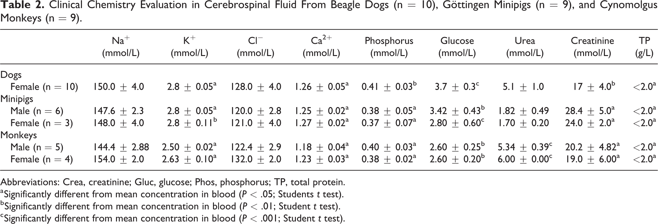
Abbreviations: Crea, creatinine; Gluc, glucose; Phos, phosphorus; TP, total protein.
a Significantly different from mean concentration in blood (P < .05; Students t test).
b Significantly different from mean concentration in blood (P < .01; Student t test).
c Significantly different from mean concentration in blood (P < .001; Student t test).
Clinical Chemistry Evaluation in Blood From Beagle Dogs (n = 10), Göttingen Minipigs (n = 9), and Cynomolgus Monkeys (n = 9).
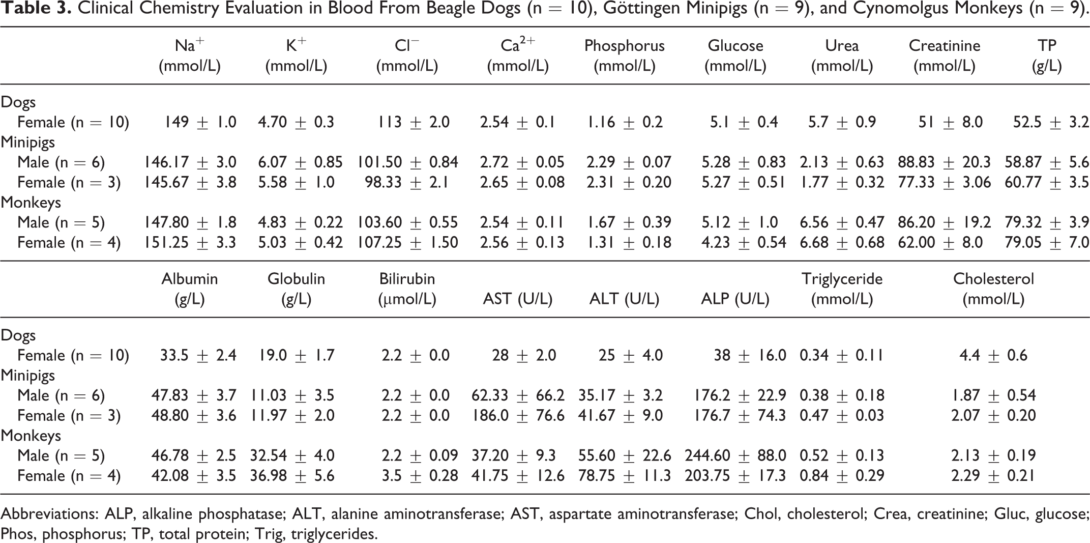
Abbreviations: ALP, alkaline phosphatase; ALT, alanine aminotransferase; AST, aspartate aminotransferase; Chol, cholesterol; Crea, creatinine; Gluc, glucose; Phos, phosphorus; TP, total protein; Trig, triglycerides.
Intrathecal Diffusion
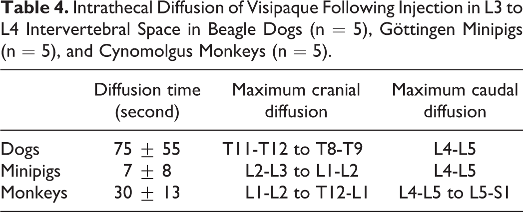
Following intrathecal injection of iodixanol, dogs showed the greatest diffusion distance; however, the longest diffusion time through the intervertebral space, followed by monkeys and minipigs (Table 4). Diffusion times after L3 to L4 injection were 75 ± 55, 30 ± 13, and 7 ± 8 seconds, respectively. Iodixanol diffused cranially up to T8 to T9 intervertebral space in dogs and T12 to L1 in monkeys. In comparison, minipigs showed minimal diffusion, with iodixanol reaching only the L1 to L2 intervertebral space in 2 of 5 animals. For 3 of the minipigs, iodixanol remained in the L3 to L4 space following injection. In all species, limited diffusion was noted toward the lumbar region, reaching from L4 to L5 to L5 to S1.
Intrathecal Diffusion of Visipaque Following Injection in L3 to L4 Intervertebral Space in Beagle Dogs (n = 5), Göttingen Minipigs (n = 5), and Cynomolgus Monkeys (n = 5).
Discussion
Safety-related drug failures remain a major concern in the drug discovery and development process. 8,9 The most frequent reason for drugs failing has been due to cardiovascular toxicity. 10 Failure due to neurotoxicity, however, has also been a leading cause of drug failure, with CNS toxicity mainly peaking in drugs intended for CNS indications. 11 Cerebrospinal fluid analysis in preclinical studies has been a valuable and a simple method for evaluating the entry of drugs into the CNS by collecting pharmacokinetic data to characterize the time course of the drug entering and exiting the CSF after a bolus injection or short-term drug infusion into the central compartment. 12 As variability in CSF characteristics across species can have an effect on drug distribution and pharmacodynamics, we aimed to assess differences in CSF characteristics and diffusion of an aqueous iodixanol solution through the intervertebral region following intrathecal injection across 3 nonrodent large animal species. As the CSF is in close contact with the nervous system, it is an excellent source of biological information reflecting alterations in this system, whether it be related to drug exposure or pathological conditions.
In order to characterize the CSF across beagle dogs, Göttingen minipigs, and cynomolgus monkeys, CSF was collected and analyzed for its clinical chemistry parameters compared to plasma. Results were fairly consistent across all 3 species with the exception of urea, which was considerably lower in minipigs (Table 2).
The composition and secretion of CSF is highly regulated and so variations in the composition can be useful for diagnostic purposes. The active physiological process of CSF production by the choroid plexus results in CSF containing a slightly higher concentration of chloride (crucial in neuronal conduction) yet significantly lower concentrations of potassium and calcium than blood plasma 13 -15 that reach statistical levels as was observed in the CSF of all 3 species. Potassium ion, which is necessary for neuronal function and neurotransmitter release, was also found at a statistically significant lower and very narrow concentration in the CSF compared to plasma as expected. 16,17 In CSF, sodium is the most abundant ion and plays an important role in transport and osmoregulation with CSF and plasma having similar sodium concentrations, 16 as was found across all 3 species in the current study.
Glucose, which is actively transported across the blood–brain barrier, is typically two-thirds 18 -20 of the fasting plasma glucose level in the CSF under normal conditions. Levels lower than this in the CSF can be indicative of a pathological condition. All glucose levels were found to be within expected physiological ranges across all animals in this study.
The integrity of the CSF–blood barrier across which diffusion and filtration of macromolecules from the blood to the CSF occurs and the CSF bulk flow plays a role in determining the level of protein in the CSF. 5,21 The elevation of CSF total protein is most frequently seen under abnormal conditions (ie, inflammation, drug-induced aseptic meningitis, tumor, subarachnoid hemorrhage). 22 The contamination of CSF with increasing blood volume during CSF collection can also result in falsely elevated CSF protein concentrations. 23 A potential limitation in our study was that blood contamination was not quantified; however, total protein levels in the CSF across all 3 species were basically devoid (<2.0 g/L). Hence, we suspect that any possible blood contamination during the procedure was at a minimal level and was not a significant confounder in our analysis. For one male monkey, CSF was contaminated by blood during the collection procedure and this was evident by the quantity of total protein present (5.3 g/L); therefore, this animal was excluded from the analysis and results not reported. Clinical chemistry parameters in the blood plasma for all species were found to be in normal in-house historical reference ranges (Table 3).
Cytological examination of the CSF is a valuable diagnostic tool for the identification of pathologies caused by infectious or noninfectious agents or adverse drug reactions associated with the CNS. 16 In a normal adult human, mononucleated cells which are predominantly seen in the CSF are small lymphocytes (∼60%-80%) and larger monocytoid cells (∼30%) with an occasional presence of a rare neutrophil, although in neonates, the percentage of monocytes and macrophages can be higher. 22 The presence of eosinophils is usually indicative of a helminthic infection 24 ; however, there have been rare occasions where CSF eosinophilia was associated with adverse drug reactions such as drug-induced (ibuprofen) eosinophilic meningitis. 25
Adverse drug effects involving the CNS have been known to have an effect on the total and differential cell counts in CSF. For example, nonsteroidal anti-inflammatory drugs have been associated with many adverse drug reactions mainly involving the gastrointestinal tract and liver, but also with drug-induced aseptic meningitis with the presence of neutrophilic or lymphocytic pleocytosis in CSF. 26 Drug-induced aseptic meningitis, although rare, has in addition been associated with antimicrobials (ie, cetuximab) 27,28 and trimethoprim/sulfamethoxazole 29,30 as well as intravenous immunoglobulins, 31 monoclonal antibodies, 32 and vaccines. 33
Across all 3 species in this study, lymphocytes and monocytoid cells represented approximately 90% of the cellular differential. In both monkeys and dogs, the percentage of lymphocytes were found to be higher than monocytoid cells as opposed to minipigs, whereby monocytoid cells represented a higher percentage of the total nucleated cell count (TNCC). For one animal, the total number of monocytoid cells represented 83.2% of the total cell count. In accordance with a previous study where CSF was collected and characterized in 20 healthy pigs, monocytoid cells also predominated and represented 77.3% ± 15.91% of the TNCC, with lymphocytes representing 20.80% ± 14.18%. 34 These results suggest that this may be a characteristic feature of the CSF in minipigs under normal conditions compared to the other species evaluated in this study. Interestingly, monocytoid cells are also the predominant cell component in the normal feline CSF, representing 69% to 100% of the TNCC. 35 -37
Intrathecal administration of drugs involves injecting into the subarachnoid space allowing diffusion into the CSF. This method of administration has been of importance for the administration of drugs such as analgesics, chemotherapeutic agents, and the specific GABA-B receptor agonist baclofen, which has commonly been used as a muscle relaxant for decades. 38 Intrathecal drug spread has been the greatest challenge in this administration technique and great interpatient variability in spread has previously been described. 39 In this study, intrathecal diffusion across dogs, minipigs, and monkeys was assessed and results showed variability in the spread of iodixanol across species, with dogs showing the longest diffusion times and greater distance from site of injection followed by monkeys and minipigs, respectively. Several factors have been known to affect drug diffusion rates via intrathecal administration. Some of these factors which are important considerations include CSF characteristics (ie, density, specific gravity, and baricity), injected drug solution characteristics (ie, baricity, volume/dose/concentration, temperature, viscosity), technique (ie, level of injection, fluid currents, positioning), and lastly characteristics such as age, weight, sex, spinal anatomy, lumbosacral CSF volume, and intra-abdominal pressure. 39 Hence, some of these factors could have contributed to the variability that was seen in the diffusion rates of iodixanol across species. In the minipig, iodixanol remained predominantly in the L3 to L4 space following injection. Previously, anatomical constraints have been identified in the pig spinal cord compared to humans when considering intrathecal drug delivery. For example, the pig epidural space is filled with abundant adipose tissue (unlike humans where it is sparse), which leads to anterior bulging of the dura mater resulting in a reduction of the intrathecal space size; hence, for a pig in a prone position, this can result in a diminished amount of CSF in the lumbar area, 10 which in the context of this study could possibly have been a contributing factor affecting the diffusion of the contrast agent. Interestingly, a previous study that aimed to characterize the distribution of baclofen and bupivacaine within CSF and spinal cord following infusion at very slow rates (20 µL/h typically used for chronic intrathecal drug delivery) into the intrathecal space in pigs showed that there was very limited distribution from the site of administration and most of the drug recovered in the CSF and spinal cord was found to be within 1 cm of the site of administration. 40 The authors suggested that the limited drug distribution was consistent with what is known about CSF motion and the existence of large permanent concentration gradients for endogenous CSF components. 40
In conclusion, results from this study showed some significant differences when CSF clinical chemistry was compared to blood plasma and these differences were consistent across all 3 species. No differences were observed in differential cell counts in CSF between monkeys and beagles; however, there were differences seen when compared to minipigs, whereby monocytoid cells were predominantly found in the TNCC. Results and previous work in the literature suggest that this may be a common characteristic seen in the normal CSF of minipigs. Finally, in this study, we saw that intrathecal diffusion rates differed significantly with the longest diffusion rate seen in the beagle, followed by the monkey and minipigs where diffusion was very limited. The limited diffusion observed in the minipig could have been a result of spinal constraints that have been previously identified in the literature; however, this would have to be explored further in future work.
Footnotes
Author’ Contributions
Cristina Ballesteros contributed to conception, contributed to analysis, and drafted the manuscript; Mylène Pouliot, Remi Froment, Mohammed Said Maghezzi, and Dominique Paquette critically revised the manuscript; Christian Li contributed to conception and critically revised the manuscript; Camille St-Jean contributed to acquisition and critically revised the manuscript; Simon Authier contributed to conception and design, contributed to acquisition, analysis, and interpretation, and critically revised the manuscript. All authors gave final approval and agree to be accountable for all aspects of work ensuring integrity and accuracy.
Declaration of Conflicting Interests
The author(s) declared no potential conflicts of interest with respect to the research, authorship, and/or publication of this article.
Funding
The author(s) received no financial support for the research, authorship, and/or publication of this article.