
Abstract
The Göttingen minipig is one of the nonrodent species recommended by various regulatory authorities for safety assessment of drugs in preclinical studies. In such studies, knowledge of background pathology is critical in order to evaluate the potential renal toxicity. In the present study, the authors report 4 cases of glomerulonephritis out of 154 microbiologically defined Göttingen minipigs microscopically evaluated in preclinical studies. One animal required early sacrifice because of general poor health, and an additional animal died spontaneously. Histopathological evaluation revealed renal lesions in all 4 animals, exhibiting membranous or membranoproliferative glomerulonephritis at different stages, accompanied by secondary tubulo-interstitial damage. The renal changes observed were considered spontaneous in origin and of unknown etiology. Development of this condition in this strain should be considered in future studies.
Introduction
Recommended by various regulatory authorities for preclinical studies (Jacobs 2006; Vodicka et al. 2005), the Göttingen minipig is commonly used as a nonrodent species in the safety assessment of drugs. In toxicity studies, differentiating between spontaneously occurring pathology from test compound–related effects is crucial. In our facility, we routinely examine a full list of organs, including kidneys, as required by the guidelines of chemical and pharmaceutical regulatory authorities. To determine the background renal pathology in the Göttingen minipig strain from Ellegaard (Dalmose, Denmark), we reanalyzed the renal pathology observed in routine preclinical studies in a total of 154 minipigs (72 males and 82 females).
Here we present 4 cases of glomerulonephritis that were observed in these animals and were considered to be of spontaneous origin. This renal change has rarely been reported in the Göttingen minipig as background pathology, and when present was usually reported to be focal and of mild severity (Madsen and Larsen 1998; Dincer and Svendsen 2006). Renal pathology evaluation was not the initial objective of our study. However, the observation of inflammatory renal changes, and in particular glomerular lesions, which are not commonly seen in this species, indicated the need for detailed studies to clarify background renal pathology in this nonrodent strain utilized in many pharmaceutical assessments.
Materials and Methods
Four microbiologically defined Göttingen minipigs, 4 to 10 months old, weighing 9 to 22 kg, received from the end of 2007 to the beginning of 2009, were used for safety evaluation in preclinical studies.
Experimental procedures concerning animal welfare were approved by our ethical committee.
Animals were housed in a limited-access animal facility. An acclimatization period of approximately 2 weeks was allowed before treatment. After arrival, each animal underwent a detailed physical examination. Any veterinary treatment was recorded in detail. Blood samples were obtained from the jugular vein of each minipig for hematological and biochemical screening 1 week before the beginning of treatment; in addition, overnight urine and fecal samples were taken. The animals were individually housed in limited indoor pens approximately 102 × 135 cm. Controls were set to maintain temperature and relative humidity at 19°C ± 3°C and 55% ± 15%, respectively; actual conditions were monitored and recorded and the records retained. There were continuous air changes, and the rooms were lit by artificial light for 12 hours each day. These enclosures were cleaned daily, and during this period, animals were exercised in the pen area. Drinking water was supplied ad libitum to each pen via an automatic valve system (Lixit Corporation, Napa, CA) or water bottles, except when urine samples were collected. Each minipig was fed twice per day with a weighed amount of diet, ranging from 120 to 220 g in relation to sex and age (SMP (E) SQC, Special Diets Services, Witham, Essex, UK) divided in two rations.
Experimental Procedure
Four minipigs were euthanized for humane reasons and then were macroscopically and microscopically examined.
Histopathology was performed on 10% neutral buffered formalin-fixed and paraffin wax-embedded tissues. Four-µm-thick sections were stained with hematoxylin and eosin (HE) for routine morphological evaluation, Periodic Acid Schiff (PAS) for glycoproteins and basal membranes, and Masson’s Trichrome (MT) for mature collagen and fibrosis.
The histological classification of glomerular diseases by Maxie and Newman (2007) was adopted.
Results
Case 1
One untreated male minipig, 5 months old and weighing 11 kg, intended for a safety evaluation study, showed pale mucous membrane, and reddening in the skin of ears, distal legs, and perineal and umbilical regions.
Hematobiochemistry revealed a slightly decreased number of white blood cells and a 10-fold increase of serum alkaline phosphatase.
At necropsy, both kidneys were hyperemic and dark and enlarged, with brown medulla and abnormal brown fluid content in the pelvis. Furthermore, the pancreas appeared enlarged and had peritoneal adhesions. Adrenal glands, mesenteric lymph nodes, lumbar skeletal muscles, gall bladder, and heart exhibited dark red areas consistent with hyperemia.
At histopathology, a moderate diffuse membranous glomerulonephritis associated with interstitial hemorrhage was observed (Figure 1A ). Bowman’s space presented proteinaceous amorphous material possibly indicative of fibrin. Glomerular tufts were found to be hypercellular. In addition, there was diffuse capillary basement membrane thickening, due to the formation of glomerular basement membrane-like and PAS-positive deposits, often encompassed by protrusions of glomerular basement membrane matrix. The interstitium was widely hemorrhagic.
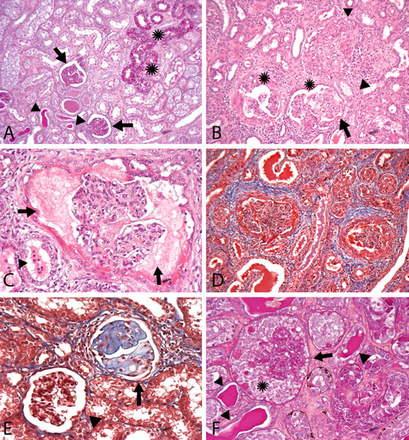
(A) Animal #1, Periodic Acid Schiff (PAS), 100X: PAS+ glomerular basal membrane proliferation (arrows), PAS+ intratubular hyaline casts (arrowheads), and PAS+ intracytoplasmic hyaline droplets of the tubular epithelial lining (asterisks). (B) Animal #2, hematoxylin and eosin, 100X: glomeruli affected at different stages by glomerulonephritis and exhibiting mesangial proliferation (arrow), membrane proliferation (arrowheads), and Bowman’s capsule thickening (asterisks). (C) Animal #2, hematoxylin and eosin, 200X: massive presence of proteinaceous material within the glomerular Bowman’s space (arrows) and neutrophils within the tubular lumen (arrowhead). (D) Animal #2, Masson’s Trichrome, 200X: periglomerular and interstitial fibrosis. (E) Animal #3, Masson’s Trichrome, 200X: focal glomerulosclerosis (arrow) surrounded by interstitial fibrosis and near a normal glomerulus (arrowhead). (F) Animal #4, Periodic Acid Shiff (PAS), 200X: PAS+ glomerular basal membrane proliferation (arrow), intratubular hyaline casts (arrowheads), and PAS-proteinaceous material within the Bowman’s space (asterisk).
Other affected organs were the heart (mild and multifocal chronic interstitial myocarditis in the context of cardiomyopathy) and the pancreas (mild and multifocal chronic pancreatitis).
Case 2
One male minipig, 4 months old and weighing 10 kg, intended for a safety evaluation study but untreated, showed asthenia and hematuria during the acclimatization period and was euthanized for humane reasons. Hematobiochemical data were unavailable.
During a postmortem examination, lesions were seen in the urinary bladder, which was uniformly dark red and whose content consisted of a wide blood clot, with irregular and raised internal surface. Dark red and variably extended discolorations were seen in the kidneys, thymus, lymph nodes, seminal vesicles, mesentery, subcutis, serosal surfaces, liver, ureters, heart, lungs, and gastrointestinal mucosa. Light brown liquid content was apparent in the pericardium. Furthermore, the gastric fundic mucosa appeared raised. Kidneys were multifocally spotted manifesting dark red small areas.
The histopathologic evaluation of the kidneys revealed a marked mesangioproliferative glomerulonephritis, characterized by thickened basal membranes associated with increased cellularity of the tuft, finely granular proteinaceous to hyaline material under the Bowman’s capsule that had hypertrophic epithelial lining, in some cases adherent to the tuft itself and creating real synechiae (Figures 1B and 1C); the hypercellularity was due to the proliferation of cells of both the capillary loops and the mesangium: therefore the lesion could have been also defined as mesangiocapillary or membranoproliferative. Periglomerular to interstitial fibrosis was evident (Figure 1D ), although the tubulo-interstitial portion was infiltrated by acute-phase inflammatory cells, such as neutrophils, and only a small number of lymphocytes and plasma cells (Figure 1C ). Mild tubulonephropathy with hyaline casts was also seen. At histopathology of the other organs, a gastric ulcer was confirmed; in addition, hyperemic areas, consistent with hemorrhages, were seen in several organs.
Case 3
One female minipig, 4 months old and weighing 9 kg, belonging to a treated group of a 2-week study testing a compound known to be safe to the urogenital tract, did not show any in vivo clinical signs or any hematobiochemical alterations; only the urinalysis showed an increase in glucose and protein content.
At necropsy no lesion was observed even though the histopathologic renal evaluation showed multifocal and moderate mesangioproliferative glomerulonephropathy. This was characterized by glomeruli with hyaline thickening of capillary basal membranes, in some cases associated with an increase in mesangial and endothelial cells; the capillary basement membrane thickening was generally seen as basement membrane-like and PAS-positive material. A few glomeruli underwent sclerotic changes, with loss of capillaries substituted by mesangial matrix (Figure 1E ). The tubulo-interstitial portion showed mild basophilia of the tubular epithelium and hyaline droplet within the tubules; in addition, aggregated neutrophils were found.
Since all other animals in the same study were unaffected by renal lesions, this observation was not considered to be treatment-related.
Case 4
One female minipig, 10 months old and weighing 22 kg, belonging to a dosed group of a 39-week study testing a compound known to be safe to the urogenital tract, appeared clinically stricken, and died spontaneously before scheduled sacrifice.
Hematobiochemistry revealed only a slightly increased level of serum alkaline phosphatase.
At necropsy, the most evident change was seen in the kidneys, which were uniformly dark red and enlarged up to 110 × 60 × 45mm. The enlargement was considered mainly related to the hyperemia. The abdominal cavity was characterized by locally extensive adhesions of fibrous tissue involving the kidneys and extending to the ureters, adrenal glands, part of the liver, gall bladder, pancreas, uterus, and ovaries. Dark red multifocal areas were apparent in the lungs, heart, diaphragm, gastrointestinal mucosa, thyroid gland, lymph nodes, and the mesentery; while the liver, pancreas, skin with subcutis, mammary area, and the plain mucosal surface of the tongue showed a diffuse yellowish color.
The histopathologic renal evaluation revealed membranous glomerulonephritis with mesangiolysis and accumulation of fibrin within the Bowman’s space. No inflammatory cell infiltrate was apparent. Associated with these glomerular lesions, severe and diffuse interstitial hemorrhage and massive tubulonephrosis with hyaline droplets (in some instances strongly PAS-positive) were seen (Figure 1F ).
Histologically, the gall bladder mucosa was necrotic, and hemorrhage was observed in the gastrointestinal tract, adrenal glands, heart, liver, lungs, thymus, thyroid, and lymph nodes. A yellowish discoloration was seen in the mammary gland, pancreas, skin, and tongue.
At the end of the study no other animals showed such findings: therefore, these were not considered to be treatment-related.
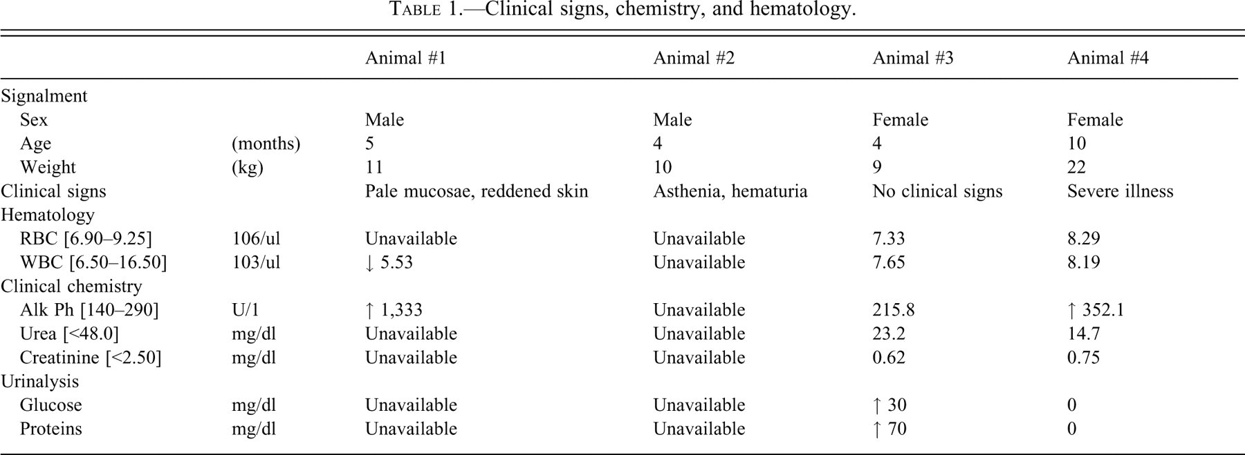
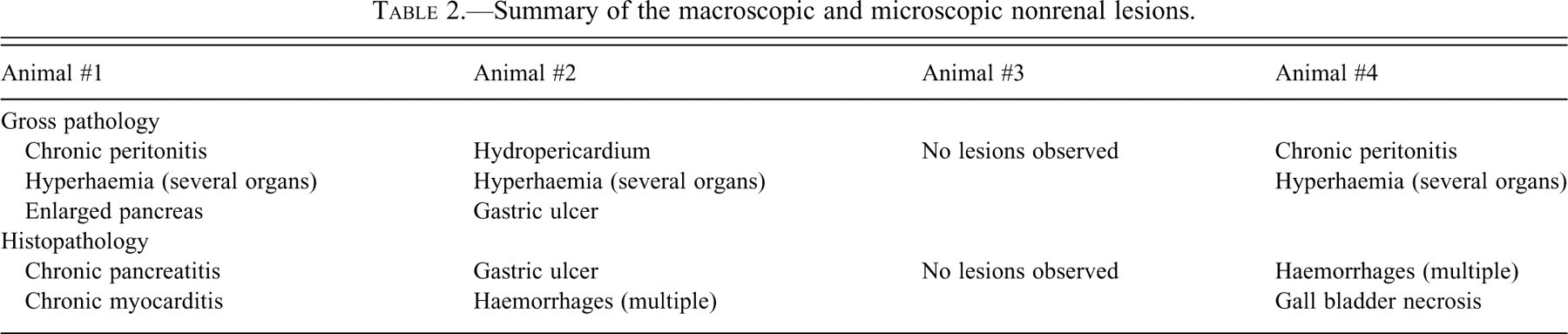
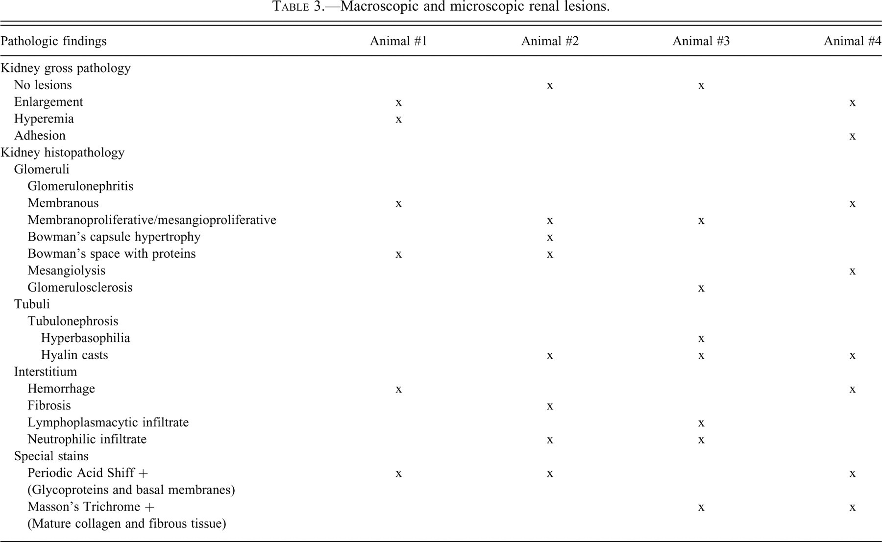
Summary of clinical chemistry and hematology data are presented in Table 1 . Non-renal lesions identified during gross and microscopical examinations are summarized in Table 2 , and the particular importance paid to the renal lesions, both macroscopic and microscopic, is shown in Table 3 .
Clinical signs, chemistry, and hematology.
Summary of the macroscopic and microscopic nonrenal lesions.
Macroscopic and microscopic renal lesions.
Discussion
Here we present four cases of spontaneously occurring glomerulonephritis in Göttingen minipigs. These changes have been rarely observed previously as background pathology in this strain, and when present they were reported to be of mild severity and focally localized. We show that these lesions can sometimes be confirmed only by detailed histopathological evaluation, without clear clinical or hematobiochemical correlation. Indeed, the minimal hematological and serum biochemical alterations revealed by blood testing were not indicative of renal pathology. However, in one case proteinuria was evident, which suggested potential glomerular pathology. The origin of these spontaneous lesions is unknown and further investigation (e.g., electron microscopy, immunohistochemistry) has not been performed as part of our study.
The glomerulonephritis observed in our cases cannot be attributed to a colony-related problem. First, the animals showing naturally occurring clinical symptoms belonged to different purchase orders and were delivered in-house at different times. Second, the renal lesions, even if mainly focused on glomeruli, were quite diverse in nature and occurred at different animal ages. Finally, the supplier rarely observed such findings, as reported in the literature (Skydsgaard 2005; Svendsen 2006).
We suggest that the renal changes observed occurred spontaneously and are not dependent on drug treatment. It has been reported that chemical-induced glomerulonephritis is known to occur after treatments with puromycin aminoglycoside, adriamycin, cyclosporine A, and histamine-receptor antagonists (Newman, Confer, and Panciera 2007). However, the test compounds administered to animals 3 and 4 were confirmed to be non-nephrotoxic, since no other animal involved in the studies exhibited such lesions. Moreover, the other two animals (1 and 2) were untreated.
The spontaneous renal lesions mainly involved the glomeruli and were classified to the membranous or proliferative pattern (Maxie and Newman 2007; Newman, Confer, and Panciera 2007) and occurred at an incidence of 2.5% (4 out of 154 minipigs examined during the past 2 years demonstrated renal lesions).
Spontaneous renal lesions in microbiologically defined Göttingen minipigs held under limited-access conditions are generally of mild and focal nature (Madsen and Larsen 1998), while in our four cases the changes were moderate to severe. Swine glomerulonephritis observed in kidneys from slaughterhouses is usually considered to be of immunological origin (Bourgault and Drolet 1995; Newman, Confer, and Panciera 2007; Zipfel et al. 2006) or related to genetic (Hegasy et al. 2002), bacterial, or viral diseases, such as hog cholera, classical and African swine fever, and cytomegalovirus or circovirus infections (Choi and Chae 2003; Maxie and Newman 2007; Segalés, Allan, and Domingo 2005). In our cases, the latter could be excluded based on the lack of microscopic dermal lesions and the absence of microscopical evidence for necrotizing vasculitis, a hallmark of porcine dermatitis and nephritis syndrome (Segalés, Allan, and Domingo 2005). Other bacterial or viral diseases usually show vascular lesions, which mainly involve the capillary vessels (Bourgault and Drolet 1995; Maxie and Newman 2007), findings that were not evident in our cases. Although blood and tissues samples in our study were not tested for potential infections, the fact that the disease did not spread to other animals excludes an infectious source. Finally, the predisposition to hereditary porcine membranoproliferative glomerulonephritis seems exclusively linked to the Norwegian Yorkshire breed (Hegasy et al. 2002).
The morphological changes in the glomerulus observed in our study could have suggested an immunological disorder resulting from the formation of antigen-antibody complexes. According to this theory, in the presence of prolonged antigenemia, formation of soluble immune complexes and deposition of antibodies against antigens within the glomerular basement membrane are possible consequences; this eventually leads to the fixation of complement, which results in leukocyte infiltration (Newman, Confer, and Panciera 2007). However, this potential mechanism was not further investigated in our cases.
Another possibility is that increased hydrostatic pressure leads to hyperfiltration, damaging capillaries and resulting in glomerulosclerosis. This was seen only in a single case as a side finding corroborating the main mesangioproliferative lesion (Newman, Confer, and Panciera 2007).
The most common histopathologic background lesions reported in the Göttingen minipig are serous atrophy of bone marrow fat cells, cholecystitis and hypoplasia of the gall bladder, as well as renal lesions characterized by mononuclear inflammatory-cell foci and minimal to mild tubular-cell basophilia and dilation (Svendsen et al. 1998; Skydsgaard, 2005). Focal chronic interstitial nephritis, commonly found in other species, may indicate a normal immunological potential. Other renal findings were subcapsular granuloma and iron deposition (Madsen and Larsen 1998), suggested to be related to prophylactic administration of colloid iron-dextran for prevention of anemia (Svedsen et al. 1998).
In a study carried out by Dincer and Svendsen (2006) from 1997 to 2001 involving 150 animals, only 1 case of focal and minimal glomerulonephritis was observed. In this study, the more common renal findings were mononuclear-cell infiltrates; mineralization; tubular basophilia; and presence of eosinophils, casts, and cysts. A recent study, which evaluated 9 cases of vasculopathy in minipigs, described 3 cases of glomerulonephritis (Maratea, Snyder, and Stevenson 2006).
Discrimination between normal variation, spontaneous changes, or compound-related changes is fundamental to evaluate the potential toxicity of putative therapeutic agents; the availability of histopathological background data is therefore essential.
Conclusions
Glomerulonephritis was reported in 4 out of 154 minipigs (2.5%). To the best of our knowledge, this renal change has been rarely reported in the Göttingen minipig as background pathology. Despite the relatively high incidence we reported, after a systematic exclusion of the main possible causes, we suggest this lesion to be spontaneous in origin and of unknown etiology. Important to note is that marked glomerulonephritis may occur in minipigs in the presence of minimal aspecific clinical signs. In our opinion, and especially in light of an increased usage of this emerging nonrodent species for toxicity studies, the report of background lesions, such as those described, is of utmost importance for the correct interpretation of preclinical studies.