
Abstract
The reproductive toxicity potential of the dental resin monomer bisphenol A glycidyl methacrylate (BisGMA; CASRN 1565-94-2) was investigated in male and female Crl: CD1(ICR) mice, 4 dosage groups, and 25 mice/sex/group. Formulations of BisGMA (0, 0.008, 0.08, or 0.8 mg/kg/d) in 0.8% ethanol in deionized water were intubated once daily beginning 28 days before cohabitation and continuing through mating (males) or through gestation day 17. The following parameters were evaluated: viability, clinical signs, body weights, estrous cyclicity, necropsy observations, organ weights, sperm concentration/motility/morphology, cesarean sectioning and litter observations, and histopathological evaluation of select tissues. No deaths or clinical signs related to BisGMA occurred. No significant changes in male and female body weights and body weight gains were recorded at any of the administered dosages of BisGMA. All mating and fertility parameters, and all litter and fetal data, were considered to be unaffected by dosages of BisGMA as high as 0.8 mg/kg/d. Gross or histopathologic tissue changes attributable to the test article were not observed. Reproductive and developmental no observed effect levels (NOAELs) for BisGMA were 0.8 mg/kg/d, the highest dose tested. Comparison of this NOAEL value to published probabilistic estimates of human BisGMA exposure from dental products suggests a margin of safety of at least 280- to nearly 2000-fold. Under the conditions of this study, BisGMA is not a reproductive toxicant.
Introduction
Dental caries (tooth decay) remains the most common chronic disease of adults and children in many parts of the world. Once established, dental caries requires treatment to prevent progression of the disease. Polymeric composite dental restorative materials, 1 option for treatment, are designed to replace biological tissue in both appearance and function. The global use of composite dental restoratives is increasing as a result of several factors, including improved esthetics, ease of handing, and potential for chemical affinity with tooth tissue as well as negative perceptions of and/or restrictions on use of mercury amalgam. 1 In 2010, polymeric composite materials were used in approximately 65% of all dental restorations performed in the United States. 2
The development of composite dental restorative materials with acceptable performance and biocompatibility is extremely challenging. The resulting product must have a stable shelf life of several years plus the ability to react rapidly in the clinical setting to form a cross-linked polymer with high modulus, high hardness, and high glass-transition temperature. 3 At the same time, the material must match the thermal expansion of the tooth, minimize extractable components and moisture uptake, be chemically and toxicologically inert, have minimal potential for shrinkage and shrinkage stress, and present an aesthetic appearance. To date, methacrylate resin formulations represent the most commercially successful approach to meeting the complex performance criteria for this category of dental materials, and contemporary methacrylate-based composites currently dominate the commercial market. 1
Development of contemporary resin composites was enabled by synthesis of bisphenol A glycidyl methacrylate (BisGMA; CASRN 1565-94-2; Figure 1) by Bowen in 1956. 4 Bisphenol A glycidyl methacrylate provides essential properties of mechanical strength, rapid polymerization, and low shrinkage that are keys to the functional performance of dental composites. Commercialization of dental composites based on BisGMA first occurred in the 1960s. 4 Although identification of alternative monomers for new product development remains an active area of research, BisGMA is estimated to be a component in more than 70% of all currently marketed composite restoratives.
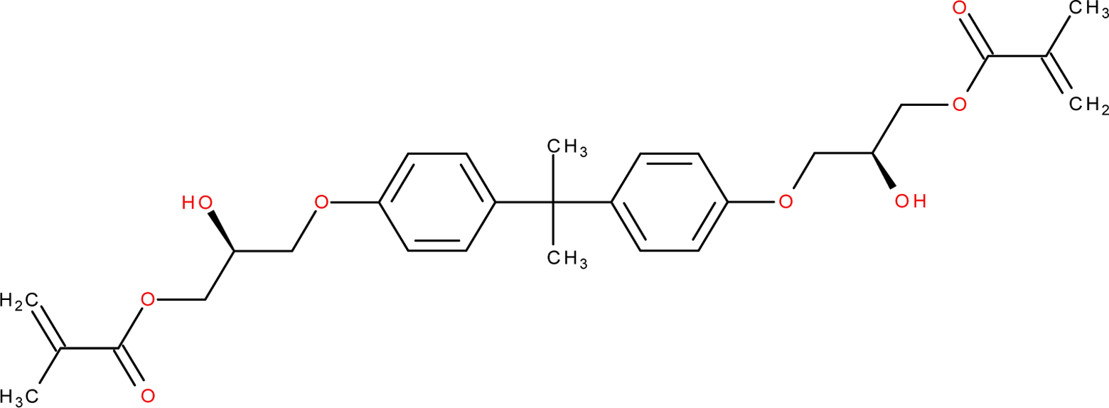
Structure of bisphenol A glycidyl methacrylate (BisGMA).
Composite restoratives are applied to teeth in an uncured form and polymerize in situ. Conversion of the component monomers is typically incomplete, and residual (unpolymerized) monomer is present in the cured product. 5 The potential for leaching of residual BisGMA monomer from polymerized composite dental materials is well documented and has been addressed in more than 20 published studies. 6 Bisphenol A glycidyl methacrylate may leach from composite dental materials into salivary fluids 7 resulting in contact with the oral mucosa and gastrointestinal tract. Alternatively, a small amount of BisGMA may leach into dentin and diffuse toward the pulp. 7 The rate and extent of leaching, and thus potential bioavailability, depend on multiple factors, including product composition, monomer properties, amount of product applied, extent of cure, and cross-linking density.
Toxicokinetic studies in guinea pigs 8 using radiolabeled BisGMA indicate that orally dosed BisGMA is rapidly and completely absorbed in the gastrointestinal tract and that the parent compound or its metabolites are widely distributed throughout the body following oral or intravenous dosing. Clearance of parent compound and metabolites appears to be essentially complete by 24 hours after dosing. Approximately 65% of the administered 14 C-radiolabel study was excreted as CO2 via the lungs, 7% was excreted in the urine, and 6% of the radiolabel remained in the body at 24 hours after dosing. Analysis of radiolabeled material excreted in the urine showed that 11.4% was parent compound, 2.2% was methacrylic acid, and 60.1% was bisphenol-A-bis(2,3-dihydroxypropyl)ether. 9 The peak equivalent levels of BisGMA in all examined tissues were reported to be at least 1000-fold less than known toxic levels determined in in vitro cytotoxicity assays using cultured cells. 8
Limited information is available on the reproductive effects of BisGMA. Because BisGMA is synthesized from bisphenol A (BPA), an estrogenic compound, a variety of studies have evaluated BisGMA for potential estrogenicity. Mariotti et al 10 evaluated the estrogenicity of subcutaneously injected BisGMA in ovariectomized mice. Treatment with 100 μg of BisGMA 3 times/week for 3 weeks resulted in modest but statistically significant increase in normalized uterine wet weight and uterine collagen content but had no effect on cell number, cell size, or RNA content in comparison to ovariectomized control animals. When tested elsewhere using in vitro systems, BisGMA was not estrogenic in yeast 2 hybrid assays, 11,12 MCF 7 cells, 13,14 or an estrogen receptor competition binding assay. 11 Thus, the weight of evidence suggests that BisGMA is not estrogenic. Although breakdown of BisGMA to BPA has been postulated, such breakdown appears unlikely based on the results of studies that investigated potential degradation via enzymatic or salivary action, 15 extreme pH values, 13,15 and high temperature. 16
Recently, 2 published studies 17,18 reported that BisGMA produced adverse effects on the fertility and reproductive systems of male and female mice, respectively. In a study of male reproductive toxicity, Al-Hiyasat and Darmani 17 reported that BisGMA, administered in an alcoholic water solution to male mice for 28 days at 25 and 100 μg/kg, caused significant dose-related reductions in body weight, absolute weight of preputial glands, testis weight, and total sperm per epididymis. Dose-related effects at 100 μg/kg included significant reductions in relative testes weight and daily sperm production and increased relative testes weight. Untreated female mice mated to 25 or 100 μg/kg males had dose-related, significant reductions in pregnancy rate and number of viable fetuses. In a study of female reproductive toxicity by the same authors, 18 female mice exposed to daily oral doses of 25 or 100 μg/kg BisGMA showed significant dose-related increases in the total number of resorptions, resorptions per implantation site, and ovary weight. Females exposed to 100 μg/kg had significantly reduced body weight and pregnancy rate.
Interpretation of these study results was limited by several factors including the use of small group sizes for this type of study (10 animal/sex/dose), lack of information on preparation and delivery of dosing solutions, and choice of statistical methods. Uncertainty regarding the above-mentioned results and lack of a full reproductive and developmental toxicity study in mice prompted the initiation of the present study. Reproductive studies are usually conducted primarily in rats, but due to the frequent use of the mouse in the published literature, verification of results in this species was considered essential.
Materials and Methods
Regulatory Compliance
A modified 1-generation reproductive toxicity study was designed and conducted following recommendations in Organisation for Economic Co-operation and Development (OECD) Test Guideline 415. 19 In a 1-generation study, the test material is administered to male and female animals for a period of time covering sperm development in the males and at least 1 or 2 ovulations in the females, through mating, gestation, lactation, and growth of the next generation. The work was considered 1 study. The male and female mice were administered the vehicle or test material during the same time period. At the end of the administration period, nonexposed female mice were brought in-house to mate with the treated male mice, and breeder male mice were used to mate with the treated female mice. The study conformed to Good Laboratory Practice (GLP) regulations promulgated by the US Food and Drug Administration 20 and OECD. 21
Materials
The test substance was BisGMA, CAS registry number 1565-92-4, lot # PK, a clear viscous gel. The test substance was obtained from the 3M ESPE Dental Products Division (St Paul, Minnesota) and stored at room temperature protected from light.
Bisphenol A glycidyl methacrylate formulations (solutions) in 0.8% ethanol in reverse osmosis (RO) processed deionized water were prepared weekly at the Testing Facility throughout the study and were stored refrigerated (2-8°C) and protected from light. Prepared formulations were stirred continuously during sample collection and dosage administration. Each batch was analyzed for concentration. Verification of nominal concentrations is essential for BisGMA, because this compound can adsorb to glass and other surfaces, potentially affecting the dosing concentration.
The control article (vehicle) for the BisGMA formulations was 0.8% ethanol in reverse osmosis (RO) processed deionized water, prepared using 200 proof, United States Pharmacopeia/National Formulary ethanol.
Animals
Male and female Crl: CD1(ICR) mice (Charles River Laboratories, Inc, Raleigh, North Carolina, and St Constant, Quebec, Canada [Males, first group]), were used in the study because (1) they are one mammalian species accepted for use in toxicity studies; (2) historical data and experience exist at the Testing Facility; and (3) they are generally comparable to the mice used in 2 existing studies of this compound. 17,18
Mice arrived at the Testing Facility in 4 shipments and were assigned to individual housing on the basis of computer-generated random units to 4 replicates to accommodate Testing Facility scheduling. After acclimation (14 days), male mice were selected for study on the basis of physical appearance and body weights. Female mice were selected after acclimation for 12 days, on the basis of an estrous cycle evaluation, appearance, and body weights. The mice were then assigned to groups based on computer-generated (weight-ordered) randomization procedures. The weight variation of mice used on study did not exceed ±20% of the mean body weight of each sex. Male mice weighed 27.8 to 31.2 g and female mice weighed 19.4 to 29.8 g at study assignment.
Study rooms were maintained under conditions of positive airflow relative to a hallway and independently supplied with a minimum of 10 changes per hour of 100% fresh air that had been passed through 99.97% high-efficiency particulate air filters. Room temperature and relative humidity were targeted at 64°F to 79°F (18°C-26°C) and 30% to 70%, respectively. An automatically controlled 12-hour light:12-hour dark fluorescent light cycle was maintained. Mice were individually housed in stainless steel, wire-bottomed cages, except during cohabitation period when each pair of male and female mice was housed in the male’s cage. All cage sizes and housing conditions were in compliance with the Guide for the Care and Use of Laboratory Animals. 22
Mice were given ad libitum access to Certified Rodent Diet #5002 (PMI Nutrition International, Inc., St. Louis, MO) in individual feeders. Reverse osmosis processed water was available via an automatic watering access system to which chlorine was added as a bacteriostat.
Methods
Dosage Justification and Administration
The test substance, BisGMA, is a leached component of approved and marketed dental composites. The oral (gavage) route was selected for testing because (1) it is the primary route of human dental patient exposure; (2) exact dosage can be accurately administered; and (3) 2 published studies 17,18 claimed that oral dosages of 25 and 100 μg/kg (0.025 and 0.100 mg/kg, respectively) BisGMA had caused adverse fertility and reproductive effects in male and female mice.
Relevant dosages were also investigated in a 14-day dosage range-finding study in which 0.008, 0.08, or 0.80 mg/kg of BisGMA were intubated daily into male and female mice. In the dosage-range study in mice no mortality or clinical observations related to BisGMA occurred. Body weights and body weight gains during the overall dosage period were increased in the 0.8 mg/kg/d dosage group male mice in comparison to the control group values. Body weight gains in the female mice were 87%, 87%. and 84% of the control group value in the 0.008, 0.08, and 0.8 mg/kg/d dosage groups, respectively, during the overall dosage period. The absolute weight of the right and left testes were increased (15% and 19%, respectively) in male mice in the 0.8 mg/kg/d dosage group, in comparison to the control group value. All other absolute organ weights for the male mice, as well as the ratios of organ weights to terminal body weights and brain weights, were generally comparable among the groups. Absolute organ weights and ratios of organ weights to terminal body weights and brain weights for the female mice were generally comparable among the groups. Histopathology was performed on both testes from all male mice assigned to the control and high dosage groups. Minimal or mild test article-related multifocal spermatid degeneration was observed in both testes of 4 of 5 male mice treated with 0.8 mg/kg/d. This effect consisted of an increased amount of large, densely basophilic spermatids, usually on the luminal surface or in the lumen of scattered (multifocal) seminiferous tubules. The cells were enlarged and appeared to have a dense basophilic nucleus, which occasionally appeared to be pyknotic and were considered to be spermatogenic cells (spermatids). Associated with this spermatid degeneration was an increased amount of large, anuclear, densely basophilic structures considered to be residual bodies. In some of the affected seminiferous tubules, there also appeared to be disorganization of the layers of spermatogenic cells. These changes in the seminiferous tubules did not appear to be stage specific. An occasional basophilic spermatid and/or residual body occurred in the testes of a few control mice of this study and were considered to be within normal histologic limits. However, the amount and size of affected spermatids/residual bodies were notably increased in the mice treated with 0.8 mg/kg/d and were considered to be related to the test article. On the basis of this information, dosages of 0 (Vehicle), 0.008, 0.08, and 0.8 mg/kg/d of BisGMA were selected for the definitive study in male and female mice.
In the present study, the treatment period for male mice began 28 days before cohabitation (maximum 14 days) and continued through the day before euthanasia at completion of the cohabitation period. Treatment of female mice also began 28 days before cohabitation, but continued through gestation day 17 (GD 17). Dosages were adjusted daily for body weight changes and intubated at approximately the same time each day. Specific information on the dosing regimen is summarized in Table 1.
Dosage Regimen for Male and Female Mice Intubated With BisGMA.a
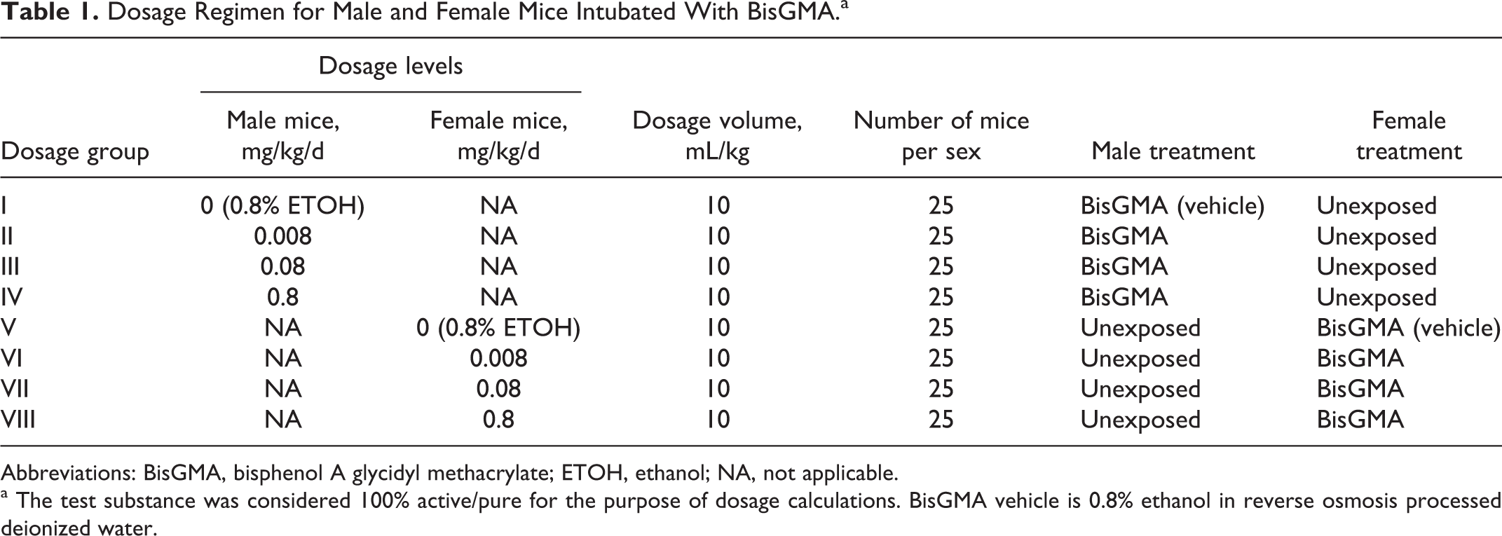
Abbreviations: BisGMA, bisphenol A glycidyl methacrylate; ETOH, ethanol; NA, not applicable.
a The test substance was considered 100% active/pure for the purpose of dosage calculations. BisGMA vehicle is 0.8% ethanol in reverse osmosis processed deionized water.
Animals were observed twice daily for viability and examined for abnormal clinical signs, abortions, or premature deliveries before dosage administration and approximately 2 hours later. Body weights of treated mice were recorded weekly prior to the start of the study and daily during the dosage period and at euthanasia. Non-BisGMA-exposed (“unexposed”) male mice were weighed weekly; unexposed females were weighed weekly during the acclimation and precohabitation periods and on GDs 0, 6, 9, 12, 15, and 18. Estrous cycling was evaluated by examination of vaginal cytology for 9 days before initiation of treatment, for 27 days beginning with the day after the first treatment, and then until a copulatory plug was observed in situ during the cohabitation period. Female mice with a copulatory plug in situ were considered to be at GD 0 and assigned to individual housing as were female mice not mated within the 14-day cohabitation period.
All mice were euthanized by inhalation of carbon dioxide. Male mice were subjected to a gross necropsy of the thoracic, abdominal, and pelvic viscera, and the following reproductive organs were weighed: right and left testis, left epididymis (whole and cauda), right epididymis, and seminal vesicles (with coagulating gland and with and without fluid). With the exception of the pituitary, which was weighed after fixation, brain, kidneys, and liver were also weighed prior to fixation in all treated and vehicle control male mice. Sperm concentration and motility were evaluated using computer-assisted sperm analysis for all male mice treated with vehicle or a test article. Motility and sperm concentration (sperm/g tissue weight) were also evaluated via the Hamilton Thorne IVOS by collection of samples from the left cauda epididymis, while the remaining portion of the left cauda was used to manually evaluate sperm morphology. Brain, epididymides, kidneys, liver, pituitary, prostate, and seminal vesicles (with coagulating gland) were retained in 10% neutral-buffered formalin (NBF) for histological examination. Testes were initially fixed in Bouin solution for up to 72 hours before being transferred to 10% NBF.
Female mice were cesarean sectioned on day 18 of gestation based on day 0 pending the day mating was confirmed, and uteri of nonpregnant mice were examined while pressed between glass plates to confirm the absence of implantation sites. Uteri from pregnant mice were excised and examined for number and distribution of implantations, live and dead fetuses, and resorptions. Numbers of corpora lutea on each ovary were also recorded. Brain, kidneys, liver, ovaries, pituitary (fixed weight), and uterus were weighed and retained in 10% NBF.
Fetuses were removed from the uterus, weighed, and examined for gender and gross external alterations. Live fetuses were then euthanized by an intraperitoneal injection of pentobarbital before undergoing further examination. Approximately half of the fetuses in each litter were fixed in Bouin solution for possible future soft tissue evaluation; the remaining fetuses were retained in alcohol for possible future skeletal evaluation.
Histopathological examination was performed on tissues from 10 randomly selected male and female mice from the control and highest dosage group (0.8 mg/kg/d). The tissues were routinely processed, embedded in paraffin, sectioned at 5 μm, and stained with hematoxylin and eosin. All gross lesions were also examined. Testes were fixed in Bouin fixative prior to being transferred to formalin. This procedure assures good fixation of the testes to allow for an adequate evaluation of all stages of sperm development and good visualization of the tubules in the testes. The Periodic Acid Schiff and hematoxylin staining was used for evaluation of the male reproductive organs.
Data generated during the course of study were recorded either by hand or by using the Argus Automated Data Collection and Management System, the Vivarium Temperature and Relative Humidity Monitoring System and the Hamilton Thorne IVOS. Data were tabulated, summarized, and/or statistically analyzed using the above-mentioned systems in conjunction with Microsoft Excel (Microsoft Office 97/2000/XP) and/or The SAS System (version 6.12). Clinical observation and other proportional data were analyzed using the Variance Test for Homogeneity of the Binomial Distribution. 23 Continuous data were analyzed using the Bartlett Test of Homogeneity of Variances 24 and Analysis of Variance. 25 The Dunnett test 26 was used to identify statistical significance of individual groups. If the analysis of variance was not appropriate, the Kruskal-Wallis test 27 or Dunn method of multiple comparisons 28 was used to identify the statistical significance of the individual groups. If there were greater than 75% ties, Fisher exact test 29 was used to analyze the data.
Results
Analytical
Concentrations and purity of the bulk BisGMA and dosing solutions were analyzed by high-performance liquid chromatography and/or ultraviolet methodology. Chromatograms from bulk test articles at the beginning and end of the study did not differ. Weekly concentration analyses (12 weeks of dosing) for BisGMA were all within ±20% of calculated concentrations except for the 0.008 and 0.08 mg/mL samples (−35% and −47%, respectively) taken from dosing preparations used during week 3 and the 0.008 mg/mL sample (+59%) taken during week 9. Homogeneity for all preparations was within the acceptable range of ≤5% relative standard deviation. Stability for low-dose formulations was confirmed for 15 days of refrigeration at 5°C.
Treated Male Mice
No deaths or clinical signs occurred that were related to treatment with BisGMA. One male mouse in the 0.008 mg/kg/d BisGMA dosage group was euthanized due to an intubation error.
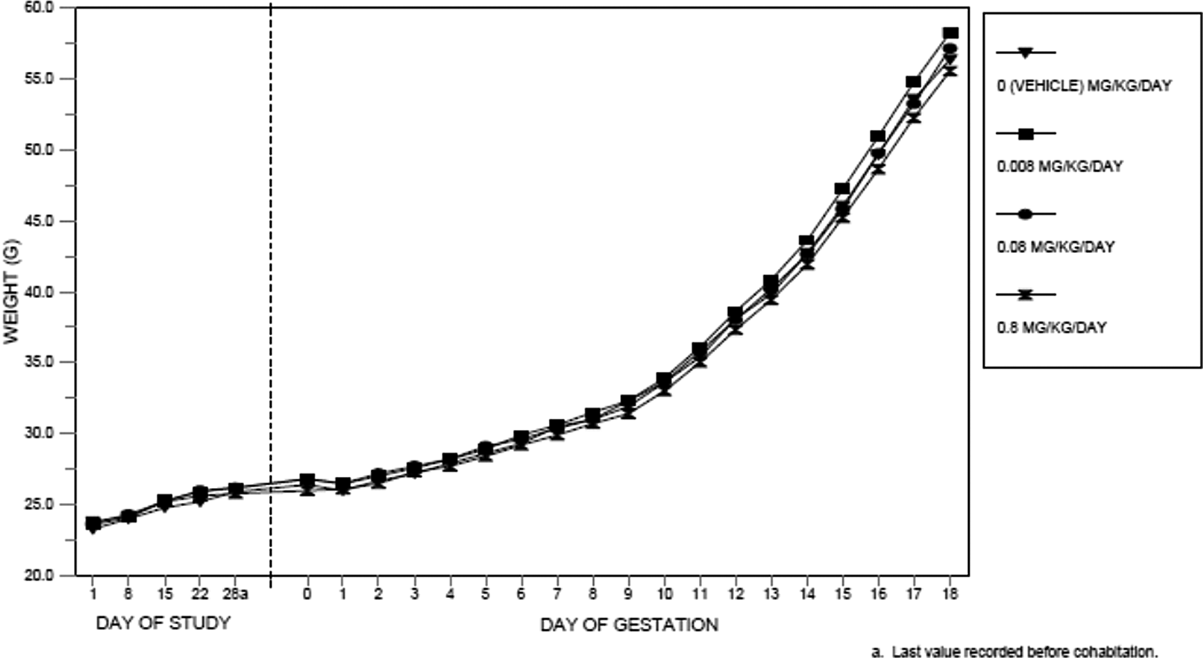
Body weights, body weight gains, and terminal body weights were not significantly affected by dosages of BisGMA as high as 0.8 mg/kg/d. All values were comparable among the dosage groups and did not significantly differ (Figure 2).
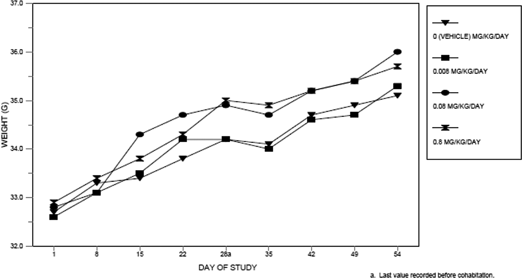
Body weights of treated male mice.
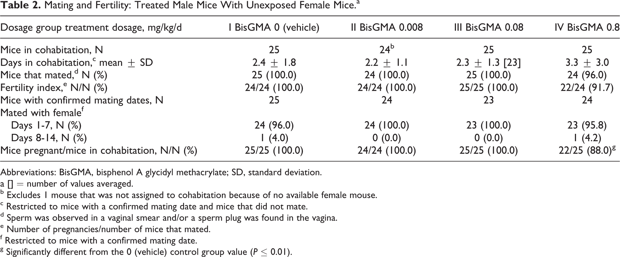
All mating and fertility parameters (days in cohabitation, mice that mated, Fertility Index [pregnancies per mated mice], mice with confirmed mating dates during the first week of cohabitation and number of pregnancies per number of mice in cohabitation) were comparable among control and treatment groups (Table 2). The number of mice pregnant per number of male mice in cohabitation was significantly reduced (P ≤ 0.01) in the 0.8 mg/kg/d dosage group. This reduction was not considered related to the test article because the Test Facility’s historical range for this parameter is 80% to 100%.
Mating and Fertility: Treated Male Mice With Unexposed Female Mice.a
Abbreviations: BisGMA, bisphenol A glycidyl methacrylate; SD, standard deviation.
a [] = number of values averaged.
b Excludes 1 mouse that was not assigned to cohabitation because of no available female mouse.
c Restricted to mice with a confirmed mating date and mice that did not mate.
d Sperm was observed in a vaginal smear and/or a sperm plug was found in the vagina.
e Number of pregnancies/number of mice that mated.
f Restricted to mice with a confirmed mating date.
g Significantly different from the 0 (vehicle) control group value (P ≤ 0.01).
Litter averages for implantations, corpora lutea, litter size, live fetuses, dead fetuses, early and late resorptions, fetal weights, sex ratio, and percentage of dead and resorbed fetuses per litter were unaffected by paternal dosages as high as 0.8 mg/kg/d of BisGMA (cesarean data from the untreated female mice not shown).
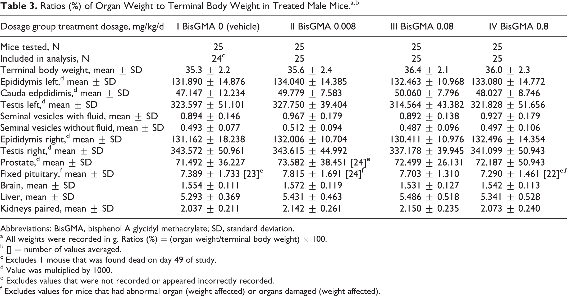
There were no test article-related gross morphological tissue changes observed at necropsy on Study Day 54. The weights of the pituitary, brain, liver, kidneys, epididymides, caudal epididymis, testes, seminal vesicles (with and without fluid) and prostate, and the ratios of these organ weights to terminal body weight and brain weight were unaffected by dosages of the test article as high as 0.8 mg/kg/d. There were no statistically significant differences among the groups (Table 3).
Ratios (%) of Organ Weight to Terminal Body Weight in Treated Male Mice.a,b
Abbreviations: BisGMA, bisphenol A glycidyl methacrylate; SD, standard deviation.
a All weights were recorded in g. Ratios (%) = (organ weight/terminal body weight) × 100.
b [] = number of values averaged.
c Excludes 1 mouse that was found dead on day 49 of study.
d Value was multiplied by 1000.
e Excludes values that were not recorded or appeared incorrectly recorded.
f Excludes values for mice that had abnormal organ (weight affected) or organs damaged (weight affected).
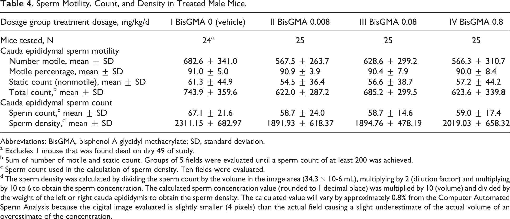
There were no statistically significant differences in sperm parameters, including sperm morphology (not shown), between control groups and treatment groups intubated with dosages as high as 0.8 mg/kg/d of BisGMA (Table 4). Test article-associated histopathological changes were not observed in any organs mentioned earlier from 10 randomly chosen male mice the BisGMA high-dose groups. The lack of effect on sperm parameters and histopathology in this study versus the dosage-range study can be attributed to the differences in group size (8/group verses 25/group) between the 2 studies.
Sperm Motility, Count, and Density in Treated Male Mice.
Abbreviations: BisGMA, bisphenol A glycidyl methacrylate; SD, standard deviation.
a Excludes 1 mouse that was found dead on day 49 of study.
b Sum of number of motile and static count. Groups of 5 fields were evaluated until a sperm count of at least 200 was achieved.
c Sperm count used in the calculation of sperm density. Ten fields were evaluated.
d The sperm density was calculated by dividing the sperm count by the volume in the image area (34.3 × 10⁻⁶ mL), multiplying by 2 (dilution factor) and multiplying by 10 to 6 to obtain the sperm concentration. The calculated sperm concentration value (rounded to 1 decimal place) was multiplied by 10 (volume) and divided by the weight of the left or right cauda epididymis to obtain the sperm density. The calculated value will vary by approximately 0.8% from the Computer Automated Sperm Analysis because the digital image evaluated is slightly smaller (4 pixels) than the actual field causing a slight underestimate of the actual volume of an overestimate of the concentration.
Treated Female Mice
No deaths or clinical signs related to BisGMA were observed.
Body weights and body weight gains during the precohabitation and gestation periods were unaffected by dosages of the test article as high as 0.8 mg/kg/d. All values were comparable among the 4 dosage groups and did not significantly differ (Figure 3). Reductions in body weight that occurred in the dosage-range study were not repeated probably due to the difference in group sizes between the dosage-range study and this study, 8/group versus 25/group.
Body weights of treated female mice.
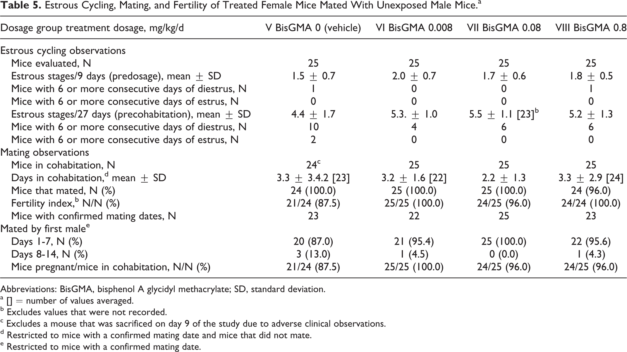
Estrous cycling and all mating and fertility parameters (days in cohabitation, mice that mated, Fertility Index [pregnancies per mated mice], mice with confirmed mating dates during the first week of cohabitation and number of pregnancies per number of mice in cohabitation) were unaffected by the maternal administration of BisGMA (Table 5). Cesarean-sectioning and litter parameters were also devoid of adverse effects and were unaffected by any of the maternal dosages of BisGMA (Tables 6 and 7).
Estrous Cycling, Mating, and Fertility of Treated Female Mice Mated With Unexposed Male Mice.a
Abbreviations: BisGMA, bisphenol A glycidyl methacrylate; SD, standard deviation.
a [] = number of values averaged.
b Excludes values that were not recorded.
c Excludes a mouse that was sacrificed on day 9 of the study due to adverse clinical observations.
d Restricted to mice with a confirmed mating date and mice that did not mate.
e Restricted to mice with a confirmed mating date.
Cesarean-Sectioning Observations in Treated Female Mice.
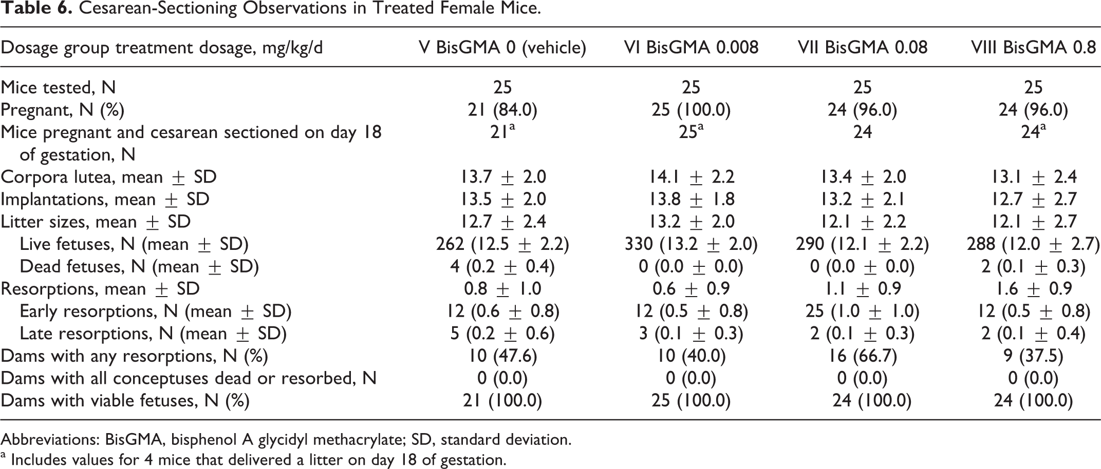
Abbreviations: BisGMA, bisphenol A glycidyl methacrylate; SD, standard deviation.
a Includes values for 4 mice that delivered a litter on day 18 of gestation.
Litter Observations (Cesarean-Delivered Fetuses) in Treated Female Mice.a
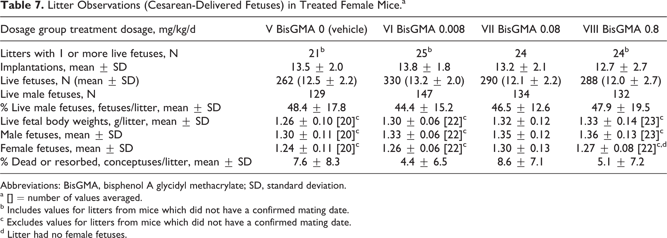
Abbreviations: BisGMA, bisphenol A glycidyl methacrylate; SD, standard deviation.
a [] = number of values averaged.
b Includes values for litters from mice which did not have a confirmed mating date.
c Excludes values for litters from mice which did not have a confirmed mating date.
d Litter had no female fetuses.
There were no gross morphological tissue changes at necropsy related to treatment with BisGMA. Absolute weights of the pituitary, brain, liver, kidneys, ovaries, and nongravid uterus with cervix, and the ratios of these organ weights to terminal body weight and brain weight, were unaffected by the administered dosages of BisGMA (Table 8). Significant (P ≤ 0.05) increases in the ratio of the paired ovarian weight to the terminal body weight in the 0.008 and 0.8 mg/kg/d dosage groups were not considered related to BisGMA because the increase was not dosage dependent considering the 100X difference between the low- (0.008 mg/kg/d) and high-dose (0.8 mg/kg/d) dosage group. Test article-associated histopathological changes also were not observed in any of the tissues listed earlier which were examined microscopically from 10 randomly chosen female mice in the BisGMA high-dose group.
Ratios (%) of Organ Weight to Terminal Body Weight for Treated Female Mice.a,b
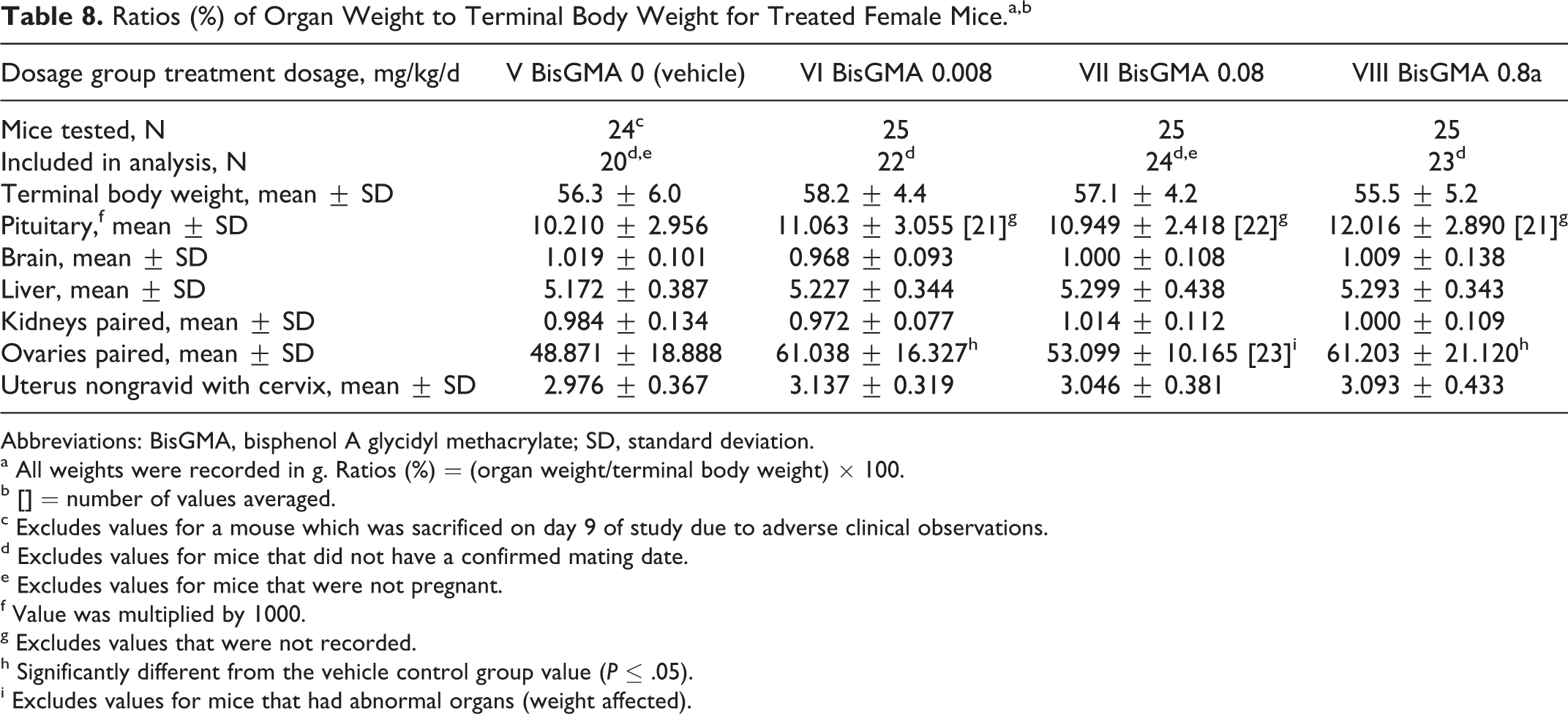
Abbreviations: BisGMA, bisphenol A glycidyl methacrylate; SD, standard deviation.
a All weights were recorded in g. Ratios (%) = (organ weight/terminal body weight) × 100.
b [] = number of values averaged.
c Excludes values for a mouse which was sacrificed on day 9 of study due to adverse clinical observations.
d Excludes values for mice that did not have a confirmed mating date.
e Excludes values for mice that were not pregnant.
f Value was multiplied by 1000.
g Excludes values that were not recorded.
h Significantly different from the vehicle control group value (P ≤ .05).
i Excludes values for mice that had abnormal organs (weight affected).
No cesarean-sectioning or litter parameters were affected by dosages of the test article as high as 0.8 mg/kg/d. The litter averages for corpora lutea, implantations, litter size, live fetuses, dead fetuses, early and late resorptions, fetal weights, sex ratio, and percentage of dead and resorbed fetuses per litter were comparable among the 4 dosage groups and did not significantly differ. No dam had a litter consisting of only nonviable embryos.
Dosages of BisGMA as high as 0.8 mg/kg/d administered to female mice prior to and during gestation did not cause any fetal gross external alterations.
Discussion
The widespread use of composite resins in dentistry and the observed leaching of components from dental resins have prompted discussion of the safety of this category of dental products. The present reproductive and developmental toxicity study was initiated to provide GLP- and guideline-compliant basic animal reproductive information, which has been lacking, on the widely used resin component BisGMA. The study was conducted in mice, rather than rats, in order to try to replicate and verify some of the data published by Darmani and Al-Hiyasat. 17,18
The results from the present extensive mouse study indicate that BisGMA does not produce any remarkable untoward effects in adult male or female Crl: CD1(ICR) mice, or in the F1 offspring, when administered in dosages as described in the study. The BisGMA did not produce any decrease in pregnancy rate, increase in ovarian weights, increase in resorptions, decrease in sperm counts, or testicular weights, even at dosages that were 8x higher than reportedly administered in the previously published studies. 17,18 Routine microscopic examination of reproductive organs from high-dose male and female mice failed to find any histological changes that might indicate any untoward male or female reproductive effects. Based on these data, it was concluded that the reproductive and the developmental no observed effect level (NOAEL) was 0.8 mg/kg d, the highest dosage tested in this study.
The results of this definitive study are in contrast to the previous studies in mice 17,18 that report BisGMA-induced impairment of reproduction and fertility in male and female mice at doses as low as 25 μg/kg/d (0.025 mg/kg/d). A direct comparison of study methodology and results between this study and the previously published studies is difficult because the published descriptions of these studies do not fully report methodological details and data. Nevertheless, it is important to note that there are numerous departures in the published studies from the standard approaches used for conducting reproductive and developmental toxicology studies for risk assessment and regulatory purposes. These departures include small group size for this type of study (10/sex/dose); lack of information on measured concentration, homogeneity, stability, or administered volume of the dosing solutions; failure to report confirmation of mating in the evaluation of effects on male reproductive toxicity; and sacrifice of males shortly after the mating period, which may have confounded measurement of sperm counts in the epididymis. In addition, data for pregnancy rate are presented for all mated females, without reference to the results for individual, test article-treated breeding males. Finally, it is not clear that appropriate statistical methods were used for all end points. These departures from standard methodology significantly limit the use of the previously published data on BisGMA for human health risk assessment purposes.
The results of the current study address the concerns mentioned earlier and provide a reliable basis for screening level risk assessment calculations. For example, estimates of human BisGMA exposure resulting from its use in composite dental products are available from a probabilistic assessment sponsored by the International Academy of Oral Medicine and Toxicology. 30 This assessment reported estimated mean adult exposure to BisGMA of 0.41 μg/kg/d (0.00041 mg/kg d) and maximum (100th percentile) adult exposure of 2.83 μg/kg/d (0.00283 mg/kg/d). Comparison of these estimated exposure values with the NOAEL value of 0.8 mg/kg/d for reproductive effects, the highest dose tested in this study, suggests margins of safety for reproductive effects of at least 1950- and 280-fold, respectively. Because a LOAEL was not identified in this study, the actual margins of safety may be significantly higher than those calculated here.
The development of polymeric dental restorative materials capable of replacing biological tissue in appearance and function is a challenging process. Methacrylate resin formulations that use BisGMA remain among the most successful approaches to meeting the complex design requirements for development of such products. Under the conditions of this study, BisGMA did not cause reproductive toxicity; these results support the acceptable use of BisGMA in dental products applied according to the manufacturers’ instructions.
Footnotes
Declaration of Conflicting Interests
The author(s) declared a potential conflict of interest (eg, a financial relationship with the commercial organizations or products discussed in this article) as follows: Lori H. Moilanen and Janelle K. Dahms are employees of 3M, a manufacturer of dental products that contain the dental resin monomer analyzed in this study. Alan M. Hoberman is an employee of the laboratory contracted to perform the research.
Funding
The author(s) disclosed receipt of the following financial support for the research, authorship and/or publication of this article: The study described in this publication was sponsored by 3M.