
Abstract
Recent chronic toxicity studies performed on green tea extracts in fasted dogs have revealed some unique dose-limiting lethal liver, gastrointestinal, and renal toxicities. Key findings included necrosis of hepatic cells, gastrointestinal epithelia and renal tubules, atrophy of reproductive organs, atrophy and necrosis of hematopoietic tissues, and associated hematological changes. The polyphenol cachetins (a mixture of primarily epigallocatechin gallate [≥55%]; plus up to 10% each of epigallocatechin, epicatechin, and epigallocatechin gallate) appeared to be the causative agents for the observed toxicities because they are the active ingredients of green tea extract studied. Conduct of the study in nonfasted dogs under the same testing conditions and dose levels showed unremarkable results. Assuming both studies were valid, at the identified no observed adverse effect levels (NOAEL) of each study, systemic exposures (based on area under the curve [AUC]) were actually lower in fasted than nonfasted dogs, suggesting that fasting may have rendered the target organ systems potentially more vulnerable to the effects of green tea extract. The toxicity mechanisms that produced lethality are not known, but the results are scientifically intriguing. Because tea drinking has become more popular in the United States and abroad, the mode of action and site of action of green tea extract-induced lethal toxicities during fasting and the role of other phytochemical components of Folia Camellia sinensis (including nonpolyphenol fractions, which are often consumed when whole-leaf products are presented) warrant further investigation.
Commentary
Tea [Folia Camellia sinensis (L) Kuntze, Family Theaceae] has been historically used as nutrition or food in Asia for thousands of years. 1 Tea-drinking has been traditionally believed to be associated with various health benefits. 2 Safety information, including animal toxicology studies in particular, on the tea and its extracts are widely available. 3 –5 However, several recent National Cancer Institute Division of Cancer Prevention (NCI DCP) and others sponsored 13-week and 9-month dog studies (the latter terminated on day 17 because of morbidity/moribundity) have revealed some unique dose-limiting liver, gastrointestinal, and renal toxicities produced by green tea extract given orally to fasted dogs. 6,7 Key findings included necrosis of hepatic cells, gastrointestinal epithelia and renal tubules, atrophy of reproductive organs, atrophy and necrosis of hematopoietic tissues, and associated hematological changes. The polyphenol cachetins (a mixture of primarily epigallocatechin gallate [≥55%]; plus up to 10% each of epigallocatechin, epicatechin, and epigallocatechin gallate) appeared to be the causative agents for the observed toxicities because they are the active ingredients of green tea extract studied. Conduct of the 13-week study in nonfasting dogs under the same testing conditions at the same dose levels (50-500 mg/kg per day, same test article) showed unremarkable results and none of the above-mentioned toxicities were elicited. The underlying reasons for such a difference in toxicity (lethality vs relatively unremarkable findings) may be due to higher systemic exposures (3- to 10-fold) to the active ingredients under fasting conditions. 6,7 Toxicologically, a review of those studies revealed that systemic drug exposures, which might have directly or indirectly induced certain organ/system failure, may not be the sole cause for the lethal toxicity observed during fasting conditions, that is, the difference in systemic exposures between the 2 studies at the identified no observed adverse effect levels (NOAEL) may be only a partial explanation for the observed discrepancies in toxicity findings reported in these studies 7 :
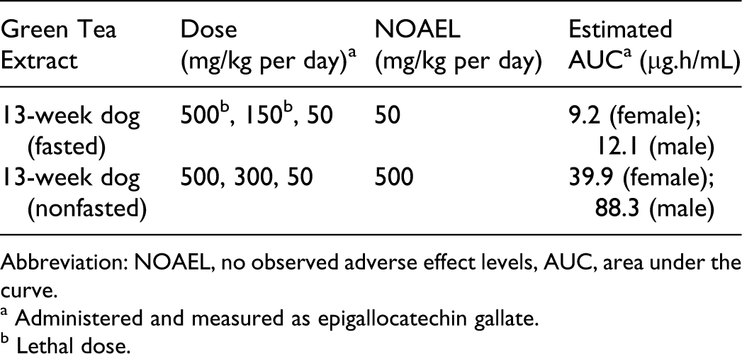
Abbreviation: NOAEL, no observed adverse effect levels, AUC, area under the curve.
a Administered and measured as epigallocatechin gallate.
b Lethal dose.
It is apparent that there was an approximately 10-fold difference in maximum tolerated dose between fasted and nonfasted states. Toxicological principle denotes that if drug exposure is to be the primary factor for eliciting the toxicity, the NOAEL exposure levels obtained from different studies conducted in the same animal species should be similar to one another. According to the studies reported above and data cited, the NOAEL systemic exposures were actually lower in fasted than nonfasted state, leading to the speculation that fasting had rendered the target organ systems potentially more vulnerable to the effects of green tea extract.
Green tea extract-induced severe toxicities during fasting state were complex and had multifactorial components that led to the final toxicity expression. Although sufficient human safety margins exist and that peak cachetin levels at NOAELs (Cmax) in fasted dogs were 4-10 times higher than those achieved in humans consuming an equivalence of approximately 10-16 cups of tea (personal communication – NCI DCP), the toxicity mechanism is clearly intriguing. In addition, these findings seem to be consistent with historical precautions in Asian countries that one should not drink tea with an empty stomach. 8 Because tea drinking has become more popular in the United States and abroad, the mode of action and site of action of green tea extract-induced lethal toxicities during fasting and the role of other phytochemical components of Folia Camellia sinensis (including nonpolyphenol fractions, 9 which are often consumed when whole-leaf products are presented) warrant further investigation.
Footnotes
Acknowledgment
The authors wish to thank Drs William Taylor, Gary Bond, Susan Walker, Ke Zhang of CDER FDA and Hao Zhang of NIAID NIH for their valuable inputs and discussions.
The author(s) declared a potential conflict of interest as follows: This article is not an official FDA or NCI guidance or policy statement, and no official endorsement by the FDA or NCI is intended.
The author(s) received no financial support for the research and/or authorship of this article.