
Abstract
Antiapoptotic Bcl-2 family proteins are validated cancer targets composed of six related proteins. From a drug discovery perspective, these are challenging targets that exert their cellular functions through protein-protein interactions (PPIs). Although several isoform-selective inhibitors have been developed using structure-based design or high-throughput screening (HTS) of synthetic chemical libraries, no large-scale screen of natural product collections has been reported. A competitive displacement fluorescence polarization (FP) screen of nearly 150,000 natural product extracts was conducted against all six antiapoptotic Bcl-2 family proteins using fluorochrome-conjugated peptide ligands that mimic functionally relevant PPIs. The screens were conducted in 1536-well format and displayed satisfactory overall HTS statistics, with Z′-factor values ranging from 0.72 to 0.83 and a hit confirmation rate between 16% and 64%. Confirmed active extracts were orthogonally tested in a luminescent assay for caspase-3/7 activation in tumor cells. Active extracts were resupplied, and effort toward the isolation of pure active components was initiated through iterative bioassay-guided fractionation. Several previously described altertoxins were isolated from a microbial source, and the pure compounds demonstrate activity in both Bcl-2 FP and caspase cellular assays. The studies demonstrate the feasibility of ultra-high-throughput screening using natural product sources and highlight some of the challenges associated with this approach.
Keywords
Introduction
Natural product extracts (NPEs) represent a tremendous resource for novel chemical matter with potential application toward therapeutic development. In fact, 50% of drugs approved in the United States from 1981 to 2010 were either natural products or their derivatives. 1 Despite this strong track record, most pharmaceutical firms have stopped natural product research in the past decade, which signifies a 30% drop in activity between 2001 and 2008, as a consequence of changes in the drug discovery paradigm and other technical limitations. 2 This decline represents an opportunity for government and noncommercial entities to drive this important area of drug discovery research.
Several academic organizations maintain natural product libraries of considerable scale and diversity, including the Spanish Fundación Medina, the plant extract bank of the Korean Institute of Bioscience and Biotechnology, and the Australian Eskitis Institute. 3 However, industrial-scale high-throughput screening of these collections has been limited. Recently, the National Cancer Institute (NCI) assembled the Chemical Biology Consortium (CBC) to conduct drug discovery within the academic and nonprofit sectors. The CBC’s directive includes screening to identify novel natural products with potential utility as anticancer agents. A large-scale pilot project was undertaken to screen the NCI NPE collection of approximately 150,000 unique samples against a panel of 10 antiapoptosis cancer targets, including six members of the Bcl-2 family proteins and the BIR2 and BIR3 domains of cIAP1 and cIAP2. This report describes the screening and hit follow-up studies for the Bcl-2 family targets (Bcl-2, Bcl-W, Bcl-XL, Bcl-B, Mcl-1, and Bfl-1).
Overexpression of antiapoptotic proteins is a common feature in the pathogenesis and progression of malignancies, making these proteins promising anticancer targets.4,5 BCL-2 was the first antideath gene discovered and constitutes a new anticancer target class with far-reaching implications for tumor biology. 6 Multiple members of the human Bcl-2 family proteins have since been identified, including six antiapoptotic members. The Bcl-2 proteins bind and sequester the proapoptotic BH3-only proteins such as Bim, thus blocking cell death. 7 Bcl-2 family proteins are regulated through myriad posttranslational modifications and interactions with other proteins, but most compellingly, Bcl-2 family proteins regulate all major types of cell death, including apoptosis, necrosis, and autophagy. Therefore, these proteins operate as nodal points at the convergence of multiple pathways with a broad and profound relevance to oncology.
Small-molecule BH3 mimetics that antagonize the interaction between antiapoptotic Bcl-2 proteins and proapoptotic BH3-only proteins represent potential anticancer therapeutics. 8 The rationale for screening natural product collections for compounds targeting the Bcl-2 family proteins has both a biological and a biophysical foundation. From a biological standpoint, it is noteworthy that Bcl-2 family genes are conserved throughout the animal kingdom and are found in insects, nematodes, and simple marine organisms.9,10 Plants and microbes have evolved chemical biosynthetic pathways that use natural products to defend themselves against predatory or pathological attack by competing animal species, and thus gene products required for cell survival are ideal targets of such agents. Indeed, examples of natural products targeting Bcl-2 have been found, including those with known anticancer activity. The most advanced of these is gossypol, a Bcl-2 inhibitory natural product from cottonseeds with a history of use in Chinese herbal medicine. Gossypol has advanced into phase III clinical trials for cancer; however, three other phase II trials were either suspended or terminated, casting doubt on the future development of this agent. 11 From a biophysical standpoint, natural products are attractive as candidate inhibitors of Bcl-2 family proteins because strategies for neutralizing these proteins are predicated on mimicking protein-protein interactions, a task for which more complex chiral molecules found in nature are best suited.
A limited number of synthetic small-molecule inhibitors of Bcl-2 family proteins have been described and are in various stages of preclinical and clinical development, the most advanced of which is ABT-199 (GDC-0199), currently in phase III clinical trials (Abbvie/Roche). ABT-199 is a highly selective and potent inhibitor of Bcl-2, which was generated using nuclear magnetic resonance (NMR)–based chemical fragment screening and structure-based drug design technologies.12–14 There remains a need for potent agents that act on other members of Bcl-2 family proteins—including Bfl-1 and Mcl-1, which are upregulated in many cancers—but are not blocked by existing compounds. In addition, a potent broad-spectrum Bcl-2 family inhibitor could be superior to chemical entities that target only one member of the family due to the simultaneous overexpression of several members in many tumors. To enable the identification of agents functioning as broad-spectrum and isoform-specific inhibitors of these key antiapoptotic proteins, we took a multitarget parallel HTS approach.
This report describes the large-scale crude extract library reformatting, assay optimization, multitarget parallel ultra-high-throughput screening (uHTS), and bioassay-guided fractionation of active extracts of interest. These efforts have led to the identification of several known, as well as novel, natural products with potential anti–Bcl-2 family activity. Despite efforts to minimize potential interference from nuisance compounds, it was noted that many of the compounds isolated contain structural features that could cause assay interference. Nonetheless, several of the known compounds that were identified have been described previously to possess anticancer activity via unknown mechanisms. Moreover, the purified altertoxins identified from this screen demonstrated broad-spectrum activity against Bcl-2 family proteins and activated caspase-3/7 in cells, providing the intriguing possibility that their apoptosis-inducing properties may in part be due to the direct inhibition of Bcl-2 family proteins.
Materials and Methods
Natural Product Library Reformatting
The collection of NPEs shipped from the NCI Natural Products Branch contained a total of 148,250 extracts in 1685 microplates of 96 wells at 0.5 mg/well. This collection was composed of 825 organic NPE plates and 860 aqueous NPE plates. Reformatting protocols (96 to 384 and 384 to 1536 plate formats) were optimized with a Biomek FX liquid handler (Beckman Coulter, Brea, CA). Additional information on library reformatting is available in the supplemental materials (
Protein Purification
GST-tagged proteins containing Bcl-Xl, Bcl-2, Bcl-W, Bcl-B, Bfl-1, and Mcl-1 lacking their C-terminal transmembrane domains (last 20 amino acids; ΔTM) were expressed from pGEX 4T-1 plasmid in XL-1 Blue cells (Stratagene, La Jolla, CA), as previously reported. 15 Additional details of protein expression and purification are available in the supplemental materials.
Peptide Synthesis and Purification
N-terminal rhodamine, fluorescein isothiocyanate (FITC), and Cy5-labeled Bim BH3 peptides were synthesized on Rink Amide-MBHA resin using Fmoc synthesis and DIC/HOBt coupling with an Advanced Chem Tech 396 multiple peptide synthesizer. Mass spectrometry of dye-labeled peptides was performed using an AutoFlex II MALDI-TOF (Bruker Daltonics, Billerica, MA), employing α-cyano-4-hydroxycinnamic acid as matrix. High-performance liquid chromatography (HPLC) of the same sample was accomplished using a C18 column with a gradient of 0.1% trifluoroacetic acid (TFA) in water to 0.1% TFA in acetonitrile for 20 min at a flow rate of 1.5 mL/min.
Competitive Peptide Displacement Fluorescence Polarization Assays
Methods for competitive peptide displacement using various Bcl-2 family proteins and FITC assays were similar to those described in a previous publication. 15 The Kd for each assay was estimated by varying the protein concentrations by factors of 2 while maintaining the concentration of the N-terminally labeled FITC-Bim or Cy-5-Bim peptide at 10 nM. Determinations were carried out in phosphate-buffered saline buffer plus 0.005% Tween 20. Fluorescence polarization (FP) was measured after a 20 min incubation. Reactions were run in a total volume of 6 µL per well in 1536-well black solid-bottom microplates (Corning No. 3724, Corning, NY). Additional information is available in the supplementary materials.
High-Throughput Screen
For high-throughput screening (HTS), an acoustic dispenser (Echo 555, Labcyte) was used to transfer organic extracts, and a pintool device (High Resolution Biosciences) was used to transfer aqueous extracts from 1536-well Echo-qualified NPE plates into 1536-well assay plates (Corning No. 3724). The Multidrop Combi bulk dispenser (Thermo Fisher Scientific, Hudson, NH) was then used to dispense buffer, protein, and tracer solutions. A volume of 10 nL of organic NPEs (5 mg/mL stock) or 20 nL of aqueous NPEs (5 mg/mL stock) was dispensed into the assay plates predispensed with 2 µL of 0.15% gamma-globulin (Sigma, St. Louis, MO) followed by the addition of 2 µL of GST-Bcl-2 proteins, then followed by the addition of 2 µL of a mixture of FITC or Cy5-Bim BH3 peptide (each at 10 nM in 25 mM Bis-Tris, pH 7.0, containing 0.005% Tween-20) with a final volume of 6 µL. Each well thus contained 8.3 µg/mL of organic NPEs and 16.7 µg/mL of aqueous NPEs and various concentrations of GST-Bcl-2 proteins and 10 nM each of FITC- and Cy5-Bim BH3 peptide. After 20 min of incubation, FP was measured using the Envision Microplate Reader for FITC channel and the PHERAstarPlus Microplate Reader (for Cy5 channel) with an excitation filter (485 nm) and an emission filter (530 nm) for the FITC channel and an excitation filter (590 nm) and an emission filter (675 nm) for the Cy5 channel. Data analysis was performed using CBIS software (ChemInnovations Software, Inc.).
Hit Confirmation
Hits were cherry-picked with a Biomek FX liquid handler (Beckman Coulter) from 384-well source NPE plates into 384-well polypropylene microplates (Greiner No. 781280, Monroe, NC). To use acoustic dispensing (Echo 555) for transfering hits into 1536-well assay plates, one set of Echo-certified hit plates (8 µL/well) was generated. The remaining polypropylene hit plates were kept at −80 °C for secondary assays of confirmed hits. For confirmation fluorescence polarization assays (FPAs), an acoustic dispenser (Echo 555, Labcyte) was used to transfer both organic NPE hits and aqueous NPE hits from 384-well Echo-certified hit plates into 1536-well assay plates (Corning No. 3724) in quadruplicate. The FPAs were performed as described above.
Cell Culture and Caspase-3/7 Assay
Reconfirmed NPE hits (30 µL each) were cherry-picked again from the hit plates (384-well polypropylene plates; see the Hit Confirmation section) into 384-well polypropylene plates (Greiner No. 781280) to generate hit dose-response (DR) plates at five concentrations (5, 2.5, 1.25, 0.625, and 0.3125 mg/mL). Cells were seeded into 384-well microplates (Corning No. 3570) at a cell density of 10,000 cells/well for RS11846 or 5000 cells/well for Caco-2 in RPMI or MEM medium (25 µL/well), respectively. Cells were cultured at 37 °C with 5% CO2 overnight prior to addition of NPE hits from DR plates in duplicate using a Biomek FX liquid handler (Beckman Coulter). Final NPE concentrations tested were 50, 16.7, 5.6, 1.9, and 0.6 µg/mL. After incubation at 37 °C with 5% CO2, 25 µL of ApoLive-Glo reagent (Promega, Madison, WI) was added, and luminescent signals were measured with an Envision microplate reader. The cell viability and caspase-3/7 activation were simultaneously determined according to the manufacturer’s instructions.
Solid Phase Extraction Filtration and Bioassay-Guided Fractionation
Approximately 20 mg of the resupplied crude extracts was suspended in an appropriate solvent to a known concentration. An aliquot of the original crude extract was transferred to a preweighed, bar-coded vial for evaporation as the assay validation sample. The remaining solution was filtered using an appropriate solid phase extraction (SPE) cartridge and eluted with additional solvent. All samples were filtered using commercially available SPE cartridges and accessories. The filtrate was then collected and placed into a preweighed, bar-coded vial. The solvent from the vials was removed under reduced pressure using a Genevac EZ 2 centrifugal evaporator with mild convection heating. All final vial content weights were determined on a FlexiWeigh automated vial-weighing workstation. Vials 1 (crude retest) and 2 (SPE filtered) were then tested in the appropriate FPAs after resuspension in appropriate solvents. FPAs were conducted as described above.
Extracts resupplied at 100 mg were filtered through the appropriate SPE cartridges as established for the corresponding 20 mg samples. The post-SPE filter eluent was evaporated to dryness to obtain an accurate sample weight. The residue was resuspended in a solvent compatible with the HPLC mobile phase (DMSO, MeOH, or aqueous mixtures of these solvents) and at a known concentration for injection onto a Waters AutoPurification preparative liquid chromatography–mass spectrometry instrument with ultraviolet detection. The final step of the sample preparation for HPLC injection was particulate filtration using a 0.4 µm PTFE cartridge. Fraction collection started at 0 min, stopped at 12 min, and was timed at 0.5 min intervals, generating a total of 24 fractions. Fractions (as well as crude and post-SPE samples) were retested at three concentrations in the appropriate FPAs (4, 12, and 25 µg/mL) in triplicate. The Echo 555 was used to transfer both organic and aqueous samples from source plates to assay plates. Additional information on the processing of 100 mg samples is described in the supplemental materials.
Isolation and Characterization of Altertoxins
A detailed summary of the isolation and purification of altertoxins will be described elsewhere (manuscript in preparation). Briefly, the crude extract of the fungus was fractionated by C18 vacuum chromatography (Discovery DSC18 SPE) with a step gradient of H2O/MeOH to give fractions 2 (100% H2O), 3 (50 % H2O/MeOH), and 4 (100% MeOH). Fractions 2 and 3 were combined and subjected to Sephadex LH-20 chromatography (MeOH) to give altertoxin I. Additional purification of adjacent LH-20 fractions with semipreparative HPLC resulted in isolation of alterpherylenol. Fraction 4 was purified using semipreparative C18 HPLC to give altertoxin II. Compounds were identified by comparison of one- and two-dimensional NMR spectra and mass spectrometric data to published reports. 16
Results and Discussion
NPE Library Reformatting
The Natural Products Branch of the NCI has amassed one of the largest and most diverse collections of NPEs in the world, totaling more than 180,000 samples from more than 50,000 source organisms. The library is composed of solvent-extracted samples obtained from terrestrial and marine biomass resources. The library samples were generated by taking a portion of each source material and dissolving it either with aqueous (milliQ water) or organic (typically 50:50 methylene chloride:methanol) extraction solvents to yield two extract solute classes, referred to as aqueous NPE and organic NPE, respectively. The majority of test samples are derived from terrestrial plants (approximately 100,000), with the remainder derived from marine plant, marine invertebrate, and microbial sources, and they are predominantly crude extracts (
A series of experiments was conducted to determine the feasibility of solubilizing, replating, and transferring NPEs from 1536-well source plates to 1536-well assay plates. Methods were established to accurately and reproducibly transfer the NPEs in this high-density format using both pintool and acoustic transfer technologies (supplemental materials). Once transfer methods were validated, the dry-film NPE 96-well plates received from the NCI repository were solubilized in bulk and stamped into Labcyte Echo-certified 1536-well plates for future screening efforts. When not in use, all crude extract plates were maintained at −80 °C, and samples were thawed and visually assessed for solubility immediately prior to use. Efforts were also taken to limit freeze-thawing to two cycles prior to testing in bioassays.
Assay Optimization and Miniaturization
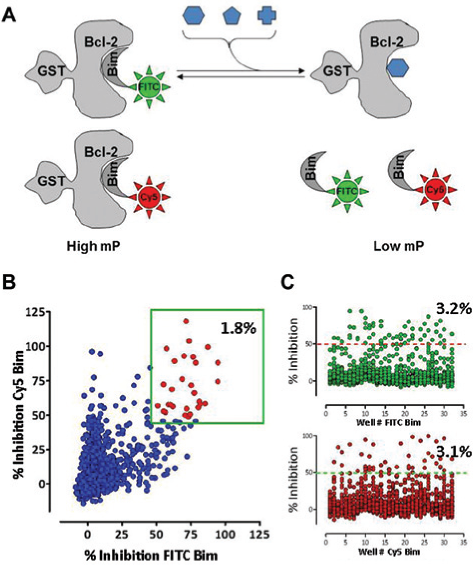
The antiapoptotic Bcl-2 family proteins function by sequestering various proapoptotic BH3-only proteins such as Bim and Bid, thereby modulating processes that control permeability of mitochondrial membranes and thus determine cell viability. 7 FP 384-well assays were developed previously to monitor the protein-protein interaction between antiapoptotic Bcl-2 proteins and the BH3-only proteins by using FITC-labeled Bim-derived peptides. 17 Assay windows were based on “no protein” controls, but these matched well with the positive control small molecule, ABT-737, a potent BH3 mimetic and inhibitor of certain Bcl-2 family proteins (Bcl-2, Bcl-XL, and, to a lesser extent, Bcl-W). Assay optimization was conducted to ensure that the FPAs could be miniaturized to 1536-well format with suitable assay statistics to ensure quality data sets from screening.
Despite the advantage of unparalleled chemical diversity offered by natural product mixtures, the presence of potential assay-interfering substances makes HTS of these collections challenging. Therefore, we took several steps to mitigate the interference of nuisance compounds during screening and later downstream assays. Two assay buffer components were included to limit the potential for interference from nonspecific binding and aggregation,18,19 including a non-ionic detergent (0.005% Tween 20) and an inert protein “scrubber,” gamma globulin (0.05%).
Published reports have suggested NPEs interfere with blue-shifted fluorescence wavelengths.20,21 To minimize interference by substances causing fluorescence or light scattering, we generated an N-terminal–conjugated Bim peptide coupled to the red-shifted Cy5 dye. Protein titration studies indicated similar apparent Kd values for FITC-conjugated and Cy5-conjugated Bim peptides against the six Bcl-2 family targets. Titration curves were similar for both Bim peptides when tested alone or in combination, demonstrating the feasibility of testing samples with both peptides simultaneously (dual-color FPA;
Fig. 1A
;
(
In addition to incorporating dual-color probes in all Bcl-2 family assays in the primary screen, we monitored potential fluorescence interference by determining a fluorescence ratio (F_ratio) for each test well. The F_ratio is a comparison of the intrinsic fluorescence intensity of any given test well relative to the average fluorescence of the vehicle controls (F-ratio = F-total/MAX [positive control, negative control]). This calculation allows an assessment of the likelihood that a given NPE is either autofluorescent or, alternatively, is acting as a fluorescence quencher in the assay. Overall, Z′ values were between 0.71 and 0.91 for all targets using final optimized assay conditions (
HTS and Hit Confirmation
HTS of the NCI NPE library was conducted batchwise on a total of 106 source plates against 10 FPAs, resulting in the generation of HTS data on 1060 plates of 1536 wells. Assay execution was directly coupled with compound plate production to minimize the time crude extracts were kept at ambient temperature. Briefly, up to 14 source plates—each batch consisting of equal numbers of organic extract plates (transferred using acoustic dispensing) and aqueous extract plates (transferred using pintool)—were replicated into 10 assay plates for a total of up to 140 assay plates per batch. One replicate from each source was then screened against one of the target FPAs. All assays were assembled and conducted in a similar fashion on identical equipment, with the only differences centering on the target protein and target protein concentrations.
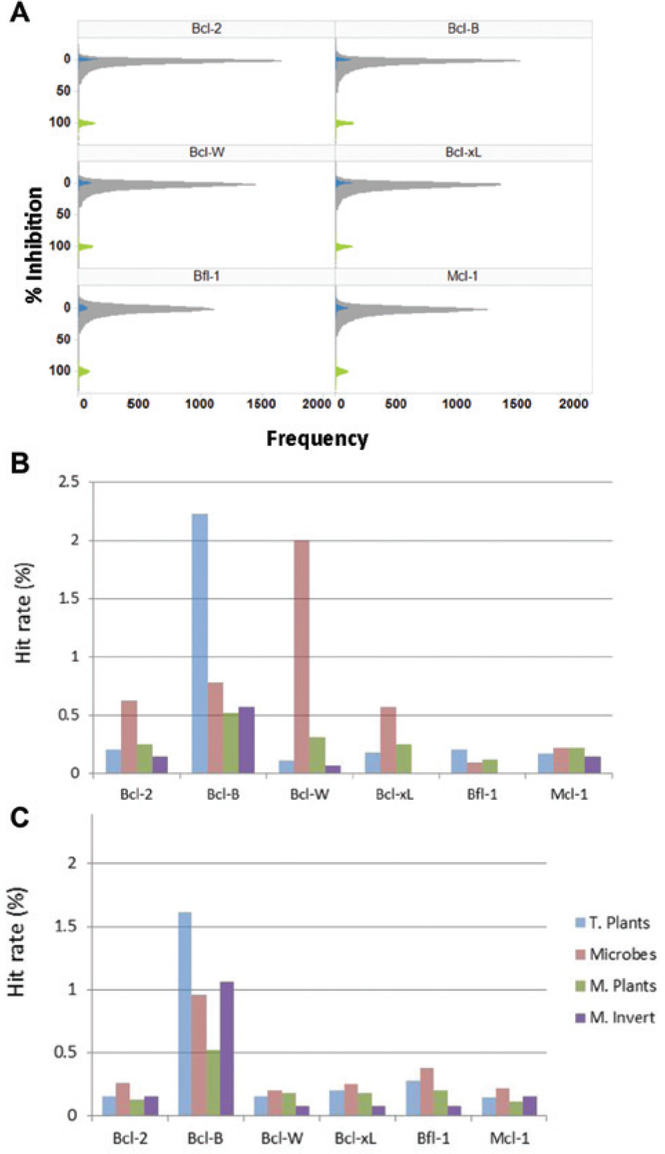
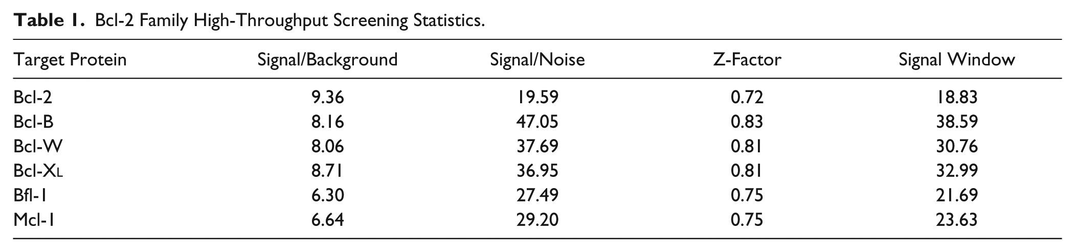
All screening data were uploaded and processed in the CBIS HTS data management system (ChemInnovation Software, Inc.). All of the 1060 screened plates met the Z-factor ≥0.5 criteria. A histogram distribution of extract activity relative to controls for all Bcl-2 targets is shown in Figure 2A . The majority of extracts were inactive against all targets, although the average activity of the negative controls is not centered with the peak of inactive NPEs, suggesting a weak background inhibition by a common component present in the extracts. The overall HTS statistics are further summarized in Table 1 . In general, all assays demonstrated good signal windows ranging from 18.8-fold to 38.6-fold, with robust signal to background. The average Z-factor ranged from 0.72 to 0.83. After data processing, results were generated for the 1,482,580 NPE wells screened. By applying hit criterion of inhibition ≥50% in the FITC channel and an F_ratio between 0.5 and 1.5, we identified a total of 4037 hits for the 10 assay targets. Activity in the Cy5 channel was not included in the hit cutoff criteria, thus reducing the possibility of missing false-negatives and allowing retrospective comparison of dual-color versus single-color hit confirmation rates. By running all targets simultaneously, an initial assessment of target selectivity for extracts was possible. Many of the hits register as active on more than one target. As a result, the number of unique Bcl-2 family active hits was 2962. Of these hits, 2312 NPEs were active against only one Bcl-2 family target assay. Interestingly 524 NPEs hit more than one target but were restricted to members of the Bcl-2 family over the cIAP targets (not shown).
(
Bcl-2 Family High-Throughput Screening Statistics.
To identify trends, screening data were analyzed in several ways to assess hit distribution across the targets, extract solvents, and extract sources. Among all targets screened, Bcl-B has a significantly higher number of hits, approximately three times higher than the next highest target, Bcl-W. Interestingly, among the antiapoptotic Bcl-2 family proteins, Bcl-B displays differences in its BH3 binding domain and binding partner preference relative to other family members. This may be related to its apparent promiscuity.22,23 With respect to extract solvent, the hit rate was higher for aqueous samples versus organic (ranging from 20% to 45% higher) for all targets except Bfl-1. For technical reasons, aqueous samples were tested at twice the concentration relative to organic samples, which may in part account for the relative hit distribution.
Figures 2B
Cherry-pick confirmation studies were conducted in quadruplicate on all hits identified in the primary HTS. Data from confirmation assays on 2962 Bcl-2 family active NPEs were uploaded into the CBIS database to identify confirmed NPEs and determine the confirmation rate and other screening statistics. Data for all test plates were of high quality (Z′ factor ≥0.5) with an average Z′ factor of 0.7. Final confirmation criteria were chosen to be more inclusive with respect to confirmed hits, specifically, using the criteria in which two of the four replicates satisfied inhibition of ≥40% and an F_ratio between 0.5 and 1.5 resulted in 994 unique NPE to follow up in orthogonal assays. Surprisingly, the confirmation rate for the dual positive extracts was not consistently higher than that for the FITC channel alone, with only Bcl-2 showing a higher confirmation rate and Bcl-W, Mcl-1, and Bfl-1 showing notably lower confirmation rates ( Table 2 ). The confirmation rate was not uniform across targets, with Bfl-1 and Mcl-1 having a lower hit confirmation rate (16% and 17%, respectively) relative to other targets. This was also true for these targets when applying the dual positive confirmation criteria. Reasons for the very low confirmation rates of these targets/assays are not clear, although the signal to background for these two assays was somewhat lower than the other assays screened, potentially giving rise to a higher false-positive rate. Overall, the FITC hit confirmation rate ranged from 16% to 60% across the targets.
High-Throughput Screening Hit Confirmation Statistics.
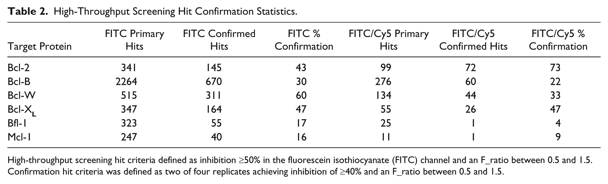
High-throughput screening hit criteria defined as inhibition ≥50% in the fluorescein isothiocyanate (FITC) channel and an F_ratio between 0.5 and 1.5. Confirmation hit criteria was defined as two of four replicates achieving inhibition of ≥40% and an F_ratio between 0.5 and 1.5.
Cell-Based Orthogonal Assays
After confirming the activity of crude extracts in the FPAs, active samples were re–cherry-picked and tested in a functional cellular assay to further validate them as containing proapoptotic Bcl-2 family inhibitory activity. Inhibitors of the Bcl-2 family are reported to induce caspase-dependent cell death by liberating the proapoptotic BH3-only proteins (e.g., Bim, Bid, Bad) from their antiapoptotic partners, leading to mitochondrial outer membrane permeabilization (MOMP) via Bax and Bak. MOMP results in the release of cytochrome-c and other proapoptotic factors in the assembly of the apoptosome. This in turn activates caspase-9 and subsequent downstream executioner caspases (caspases-3 and -7).7,24 Therefore, we tested confirmed hits for their ability to activate caspase-3/7 in tumor cells. We simultaneously monitored cell viability and caspase-3/7 activation using a single multiplex assay (ApoLive-Glo, Promega) to determine the ability of extracts to induce programmed cell death. Importantly, in addition to being an orthogonal cell-based assay, the caspase-3/7 activity was monitored using a luminescent readout, thereby providing a non–fluorescent-based measurement of extract bioactivity.
Previous studies have demonstrated that human leukemia and lymphoma-derived cancer cell lines overexpress a diversity of antiapoptotic Bcl-2 family members, which likely contributes to their resistance to apoptosis. 25 As such, the lymphoma cell line RS11846 was used to evaluate NPE-induced apoptosis via interference with Bcl-2, Bcl-XL, Bcl-W, Mcl-1, and Bfl-1, because these cells have been shown to express detectable levels of these antiapoptotic genes. 25 Caco-2 cells express higher levels of Bcl-B relative to other cell lines and were therefore selected as a second cell line to assess cellular activity against this target.
We confirmed the ability of the potent nanomolar Bcl-2 inhibitors, ABT-737 and ABT-263, to induce caspase-3/7 activity in RS11846 cells within 6 h of treatment (
The highest NPE concentration tested was 50 µg/mL, which is approximately fivefold higher than the NPE concentrations used in primary screening. All cell-based assays were performed in 384-well format, and confirmed hits were directly cherry-picked from intermediate plates in five-point concentration response in triplicate. Effects on viability in this assay using potent target selective inhibitors was modest at both the 6 h and 24 h time points. Moreover, relatively few NPEs tested showed significant effects on cell viability under the conditions tested. As a consequence, hit validation criteria focused on caspase activation wherein an extract registered as a hit if the average caspase induction (measured as an increase in luminescence) was greater than the average induction of the negative control plus three times the standard deviation of the sample and negative control. The range of caspase induction achieved by NPEs (2- to 10-fold) was generally less than that observed for 10 µM ABT-737 (15-fold induction) but was similar or superior to effects observed using gossypol and related analogs at ~20 µM final concentration. In fact, some NPEs actually demonstrated greater than 10-fold induction of caspase activity, even at concentrations below 10 µg/mL. A total of 167/994 NPEs met validation criteria, representing a hit validation rate of approximately 17%. This hit validation rate demonstrates a considerable level of enrichment given that only 1 of 128 (<1%) randomly selected NPEs was active in the caspase-3/7 assay (data not shown). Figure 3 shows the distribution of caspase-3/7 activation across both cell lines for NPEs meeting threshold criteria. Caspase activation was observed to similar extents in both Caco-2 ( Fig. 3A ) and RS11846 cells ( Fig. 3B ). Maximal caspase induction was not necessarily observed at the highest concentration tested; in fact, some of the most effective extracts induced maximal activation between 1.9 µg/mL and 5.6 µg/mL. Although the majority of NPEs induced modest caspase activation (2- to 5-fold), some induced caspases up to 15-fold. In general, caspase induction was more dramatic at 6 h versus 24 h, particularly for organic extracts. The decline in caspase activity at 24 h may be a reflection of reduced cell health or extract stability at later time points.
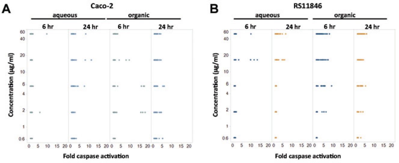
Summary of caspase activity of confirmed natural product extract (NPE) hits. Fold caspase-3/7 activation of all confirmed extract hits tested in (
The majority of cell-active NPEs were derived from organic extracts, possibly as a consequence of better cell permeability properties intrinsic to these agents. Nonetheless, several potent actives were identified from aqueous sources. Terrestrial plant extracts represented the source with the most abundant cell-active samples, 108 in total. This was followed by microbial samples, with 34 total extracts. Both single-target and broad-spectrum active NPEs were represented in the validated hit set. At this stage, validated hit extracts demonstrated reproducible activity in the biochemical assay as well as activity in the cellular caspase assay. The cell-based caspase-3/7 induction data—in conjunction with source extraction information and resupply availability—was used to guide the selection of extracts for bioassay-guided fractionation.
SPE and Bioassay-Guided Fractionation of Validated Hits
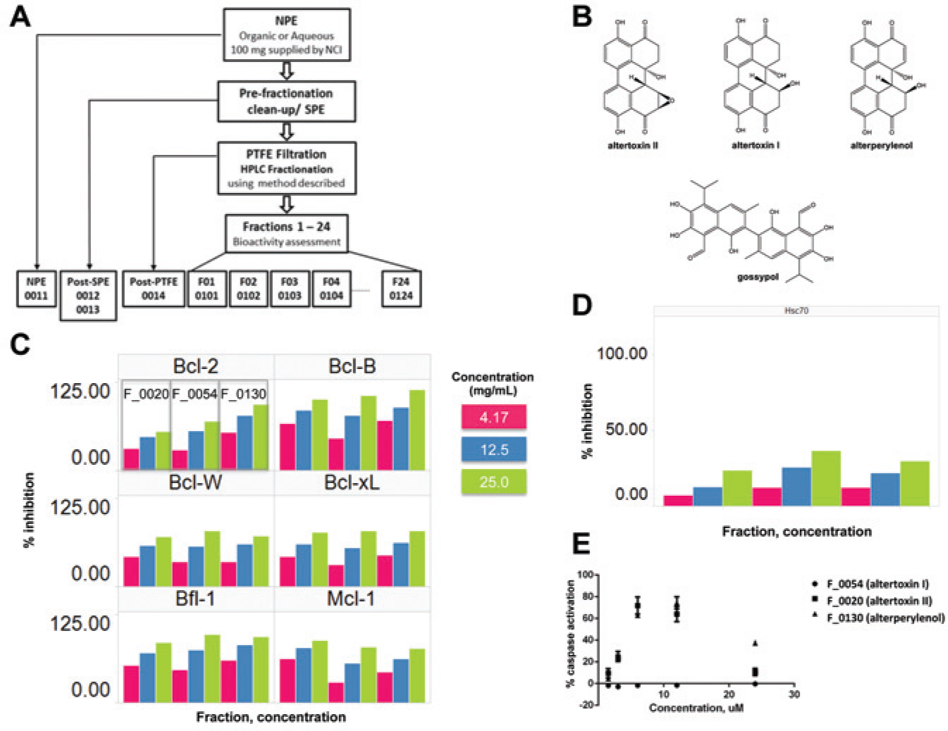
Once validated cell-active extracts were identified, the purification and isolation of pure active natural products was initiated. All subsequent steps required independent resupply of crude material from NCI. As a first step toward purification, SPE was applied to a 20 mg resupply of all active crudes to remove potential materials that might interfere with biochemical assays or with subsequent HPLC-based fractionation steps (e.g., salts, tannins, saponins, polymers). Following removal of nuisance and interfering components, the samples were retested for activity in the primary assays. If bioactivity was retained, or preferably enriched, the NPE was selected for column chromatography. Active samples for HPLC chromatography were prepared from a second independent 100 mg resupply of crude extract and processed ( Fig. 4A ). Following SPE filtration, the samples were loaded onto the appropriate HPLC column, and fractions were collected and processed for bioassay. Approximately one-third (56/157) of the resupplied crude NPEs retained bioactivity in the respective biochemical FPAs following SPE and first-round HPLC fractionation, further increasing confidence that tractable bioactive components were present in these mixtures.
(
Subsequent to the initial round of HPLC fractionation and bioassay, a comprehensive review of fractionated bioactivity profiles, as well as an assessment of extract complexity (analytical and structural data) and potential for resupply, was conducted for all extracts. Following this review, extracts were prioritized and selected for iterative bioassay-guided purification and identification of discrete pure substances of biological and potential drug development interest. To date, these efforts have led to the identification of several purified or nearly pure natural products, of which three represent novel structures. A more detailed description of these activities and results will be described elsewhere (manuscript in preparation).
Among the compounds isolated are three related perylene quinone mycotoxins ( Fig. 4B ). Altertoxins I and II and alterperylenol (x-y) were isolated from the fungus Alternaria alternata after several rounds of chromatography and display robust broad-spectrum activity against all Bcl-2 family targets, with limited activity against an unrelated target, Hsc-70 ( Fig. 4C , D ). These purified fractions inhibit the Bcl-2 Bim interaction between 80% and 100% at 25 µg/mL and even show significant inhibition (>40%) at the lowest concentrations tested, whereas they display only weak activity against the unrelated protein (Hsc70) at the highest concentration tested. Previous reports suggest these agents may be genotoxic and mutagenic.26,27 Alterpyrelenol was shown to inhibit telomerase with an IC50 of 30 µM. 28 We tested the purified compounds (fractions F_0020, F_0054, and F_0130) in the cellular caspase-3/7 assay against RS11846 lymphoma cells using a similar protocol to that implemented during hit validation. As seen in Figure 4E , both F_0020 (altertoxin II) and F_0130 (alterperylenol) robustly activate caspase-3/7 at 6 h in cells at concentrations as low as 3.16 µg/mL. The level of caspase activation was more than 70% that of ABT-737 at 10 µM, indicating fairly robust and rapid apoptosis following treatment with these compounds. Caspase induction increased with compound concentration up to the second highest dose (12.5 µg/mL). The reduced activation at the highest concentration (25 µg/mL) may be a consequence of reduced cell count or compound solubility. The original crude extract containing the altertoxins required higher concentrations (50 µg/mL) to induce significant caspase activity (not shown). These results are consistent with the enriched apoptotic activity being derived from the purified compounds. Curiously, altertoxin I failed to activate caspases under these conditions, possibly due to reduced cell permeability or solubility in cellular media, although further studies are necessary to confirm these hypotheses. Caspase activation has not been reported previously for any of the altertoxin compounds. The altertoxins share some structural similarity to the known Bcl-2 family inhibitor gossypol, suggesting a potentially related mechanism of inhibition. Like the altertoxins and gossypol, many of the samples isolated thus far contain polyphenolic substructures, likely reflecting a combination of the inherent sensitivity of the assays and/or targets to this class of compounds and the abundance of these compounds within the extract collection.
In summary, we have completed a parallel, multitarget HTS campaign against nearly 150,000 NPEs in high-density 1536-well format. The antiapoptosis Bcl-2 family proteins screened in this project represent oncology targets that garner significant clinical interest. A total of 994 NPEs were confirmed as active against one or more Bcl-2 family targets, with 17% of these also inducing caspase-3/7 activation in tumor cells. Reconfirmation and orthogonal testing in cell-based assays enabled the identification of NPEs for bioassay-guided fractionation and isolation. However, the complex nature of crude extract mixtures and the potential for isolating nuisance compounds make this screening approach challenging. Further studies are required to elucidate the mechanisms of action of the pure natural products identified. Notwithstanding these limitations, our results demonstrate the feasibility of uHTS of natural product libraries to discover leads for therapeutically active natural product substances.
Footnotes
Acknowledgements
We thank Antimone Dewing for technical assistance with cellular assays and Drs. Barbara Mroczkowski, Shizuko Sei, Neal Green, and Bill Moore for scientific discussions.
Declaration of Conflicting Interests
The authors declared no potential conflicts of interest with respect to the research, authorship, and/or publication of this article.
Funding
The authors disclosed receipt of the following financial support for the research, authorship, and/or publication of this article: This project has been funded in whole or in part with federal funds from the National Cancer Institute NExT Program, National Institutes of Health, under contract No. HHSN261200800001E. The content of this publication does not necessarily reflect the views or policies of the Department of Health and Human Services, nor does mention of trade names, commercial products, or organizations imply endorsement by the U.S. government.
References
Supplementary Material
Please find the following supplemental material available below.
For Open Access articles published under a Creative Commons License, all supplemental material carries the same license as the article it is associated with.
For non-Open Access articles published, all supplemental material carries a non-exclusive license, and permission requests for re-use of supplemental material or any part of supplemental material shall be sent directly to the copyright owner as specified in the copyright notice associated with the article.