
Abstract
Antibodies are powerful research tools that can be used in many areas of biology to probe, measure, and perturb various biological structures. Successful drug discovery is dependent on the correct identification of a target implicated in disease, coupled with the successful selection, optimization, and development of a candidate drug. Because of their specific binding characteristics, with regard to specificity, affinity, and avidity, coupled with their amenability to protein engineering, antibodies have become a key tool in drug discovery, enabling the quantification, localization, and modulation of proteins of interest. This review summarizes the application of antibodies and other protein affinity reagents as specific research tools within the drug discovery process.
Keywords
Introduction
Antibodies are invaluable tools in research and are applied throughout the drug discovery process from target identification to target validation and in the identification and optimization of lead compounds ( Fig. 1 ).
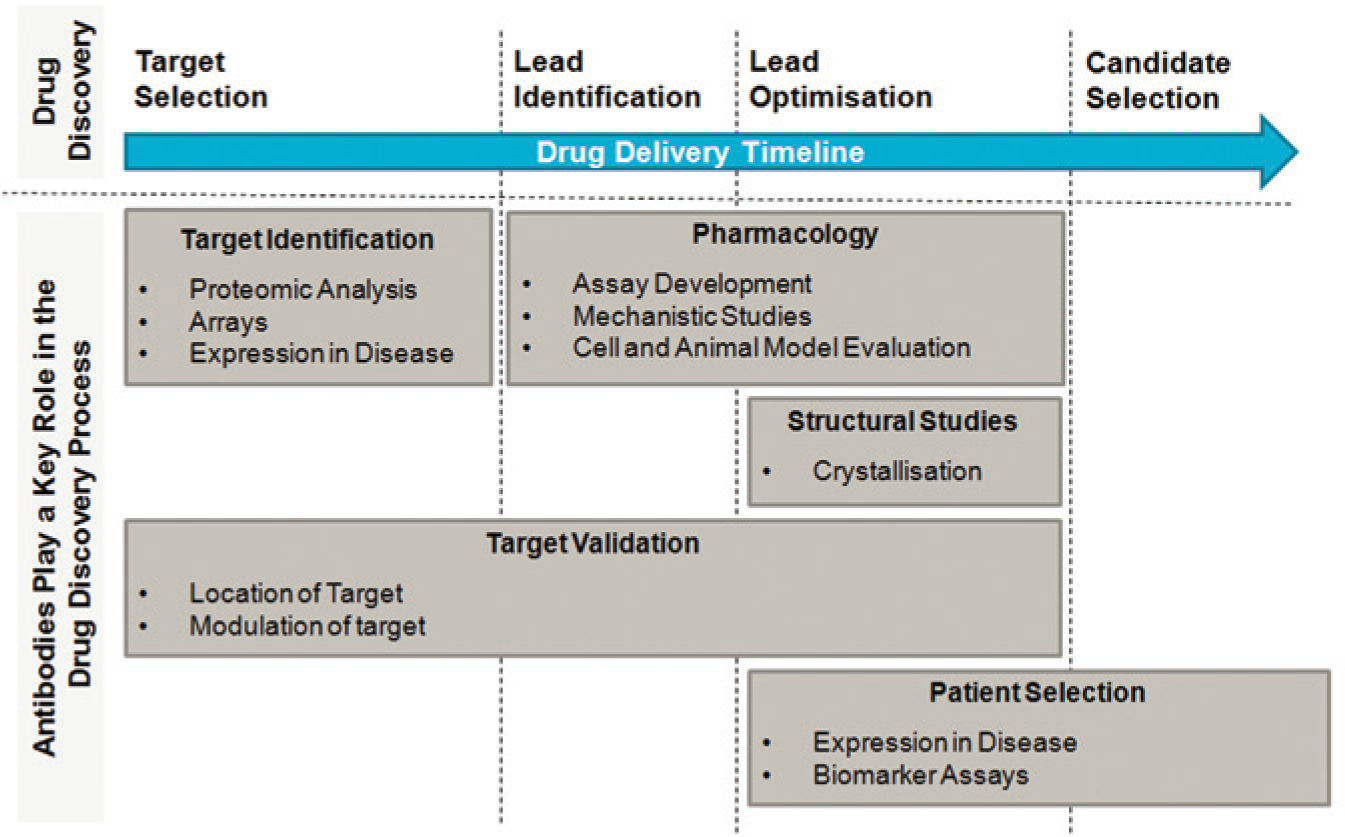
Antibodies are invaluable tools in research and are applied throughout the drug discovery process.
Antibodies play a key role in target identification technologies, including analysis of proteomic changes in disease 1 that may be causative in pathology, and more recently, both antibodies and antibody fragments have been applied as tools in phenotypic screening. 2 Throughout the drug discovery process, from target selection to patient stratification, target validation is essential, and affinity reagents such as antibodies are powerful tools to investigate target distribution, subcellular localization and function and, importantly, how these factors might play a role in disease. In addition, functional antibody tools play a key role in building a platform of evidence for modulating a target in the context of disease. Once a lead small-molecule drug candidate has been identified, affinity reagents can be applied to aid crystallography of the target protein, providing structural and ligand interaction data to drive lead optimization. 3
Some specific aspects of the use of antibody tools have recently been described in more detail, including their use in the stabilization and sensing of specific protein conformations. 4 This review provides a broad commentary on the technical use of antibodies and other protein affinity reagents. It focuses exclusively on antibodies as tools in all aspects of small-molecule drug discovery, summarizing their current application and emerging technologies in these areas.
Antibody Tools in Target Identification: Arrays and Proteomics
The human genome has approximately 20,000 protein-encoding genes, 5 of which only a few hundred proteins have been explored as drug targets 6 ; there is therefore great potential in applying proteomics platforms to identify disease-related proteins.
Traditionally, proteomic analysis including protein separation via two-dimensional (2D) gel electrophoresis coupled with mass spectrometry has been the method of choice for large-scale analysis of proteins; however, this “shotgun” proteomics approach is limited by its sensitivity and throughput, particularly as many plasma proteins are present at or below the ng/mL range.7,8
The current favored method for quantification of peptides/proteins is selected reaction monitoring 9 or a multiplexed version termed multiple reaction monitoring mass spectrometry (MRM-MS) coupled with stable isotope dilution. This targeted mass spectrometry (MS) analysis enhances the lower detection limit for peptides by up to 100-fold by allowing rapid and continuous monitoring of the specific ions of interest. However, its sensitivity is constrained by sample complexity including posttranslationally modified forms and splice variants. Because of this, to gain less than ng/mL sensitivity, plasma processing prior to MRM-MS is required to reduce the level of abundant proteins. This is carried out using immunoaffinity depletion followed by multidimensional protein/peptide fractionation, increasing the proteome depth and detection. 8
Although MS-based methods avoid the lead time and cost required to generate high-quality target-specific antibodies, there remains a need for approaches that deliver high-throughput parallel analysis of proteins. The specificity and affinity of antibodies make them a natural choice for proteomics analysis, 10 and antibody arrays can be applied to the parallel detection of low-abundance proteins in a range of samples including tissues, cells, and plasma. 11
Antibody arrays have recently been reported in multiple different formats, ranging from commercially available planar arrays, through qualitative multiplexed bead-based arrays and array-based surface plasmon resonance systems (which enable rapid, label-free, high-throughput analysis in low sample volume), to the greatly enhanced sensitivity of nanostructured immunoassays.12–15 These technologies have been successfully applied in identifying global protein changes associated with a range of diseases including cancer16–19 and neurodegenerative disorders. 20 The detailed technical aspects of these methodologies including some of the different technological characteristics have been reviewed previously by Borrebaeck and Wingren. 21
The quality of data generated from the different antibody microarray technologies is determined by the specificity and sensitivity of the antibodies generated. It is clear, therefore, that any assay that is based on detection with a single-antibody binder of unproven specificity may be limited. This limitation has recently been addressed by several technologies, including the development of size exclusion chromatography-resolved microsphere-based affinity proteomics (Size-MAP) in which labeled sample proteins are separated by size exclusion chromatography before contact with a microsphere-based antibody array, 22 effectively producing an “antibody array Western blot.” Paired antibody approaches have also been applied in which two independent antibodies targeting different and nonoverlapping epitopes on the same protein are used, thus allowing improved specificity of detection. 23
As described above, there remain a number of drawbacks with both antibody-based technologies and MS platforms. These might be overcome by combining the two technologies, for example, the use of stable isotope standards and capture by antipeptide antibodies method, 24 which has been described as providing a multiplexed, specific, and standardized alternative to conventional immunoassays. Using this method, antipeptide antibodies immobilized on nanoaffinity columns are used to enrich specific peptides and stable-isotope–labeled internal standards of the same sequence before elution and quantitation using electrospray MS. 24 Further development of this method has included improvement in peptide enrichment and the use of combinations of multiple antibodies to provide a flexible multiplex capability. 25
It is apparent from the diverse methods described above that there have been many technology improvements taking place in the field of antibody-based proteomic research. There is a clear requirement for proteomic approaches that discriminate between complex and diverse proteins, including their splice variants and posttranslationally modified derivatives, offering improved sensitivity and throughput; a major challenge remains in deconvolution of data generated, including the analysis of off-target effects. 26 For target identification, antibody arrays might perhaps be viewed as an initial screen of large numbers of proteins, for example, in normal versus diseased tissues, to identify proteins or protein complexes on which to focus further research. Indeed, once a protein with a potentially interesting expression pattern has been identified, validation can be supported by the use of other platforms, such as tissue microarrays, that allow for the simultaneous analysis of both expression patterns and subcellular localization in hundreds of tissue samples with a directed antibody. 27
Antibody Tools in Phenotypic Screening
In recent decades, drug discovery in the hit and lead identification stages has relied heavily on in vitro biochemical assays to power high-throughput screening (HTS) and hit identification, 27 with engineered-cell assays used for post-HTS lead generation and secondary assays. 28 Although this approach has been widely applied in drug discovery, arguably the HTS paradigm has proved challenging in terms of successful translation into the clinic, with skepticism evident for more than a decade. 29 Phenotypic screening has contributed to the discovery of first-in-class drugs and is an alternative drug discovery approach to complement target-based screening. 30 The pharmaceutical industry is therefore increasingly focusing on cell-based phenotypic screening, as well as primary and native-like cell systems, to discover drugs with efficacy in an environment that is physiologically relevant to the disease state.28,31 The increased use of nonengineered cells for screening, rather than target overexpression systems, requires high-quality antibodies to establish endogenous target expression. In addition, high-quality tool antibodies are required for high-content assays, 32 to provide high-throughput, multiparametric readouts of protein expression and cell-signaling events as a measure of compound effects.
The most common approach to phenotypic screening to identify novel targets involves applying small-molecule collections in disease-relevant cell-based assays. This approach has been applied successfully to identify new molecules that inhibit pathways involved in pathology, including infection 33 and neurodegenerative disease.34,35 The application of antibodies as pharmacologic tools in phenotypic screens offers great potential to complement a small-molecule approach and to identify antibody tools that can be used to deconvolute novel targets.
The potential to identify cell surface antigens involved in, for example, tumorigenesis and hematological malignancy with antibodies that mediate receptor function and cell signaling demonstrates that this area is an excellent opportunity to apply antibody-based phenotypic screening. To identify antigens associated with cancer, a common approach is to screen antibodies against tumor cells or cell lines. 36 Subsequent cell-based assays and in vivo studies confirm antibody-mediated cytotoxicity and antitumor activity. A recent study by Rust et al 2 demonstrates that screening of a cancer cell line with antibodies derived from hybridomas or phage display libraries can identify antigens previously cited as potential therapeutic cancer targets.
The use of antibodies in phenotypic screening for target identification is not limited to extracellular targets. The intracellular application of antibodies or intrabodies can be used to probe cellular function and phenotypic modulation. A recent study has applied the intracellular expression of an antibody library in a eukaryotic cell using lentivirus, such that antibodies were expressed within the cell and secreted. 37 Using the erythropoetin receptor as proof of principle, a combination of expressed antibodies that gave potent erythropoietin agonism was identified. This work has been extended by the further use of antibody libraries in lentiviruses, infecting eukaryotic cells containing a fluorescent reporter coupled to a receptor. This has allowed the selection of single cells in which receptor signaling is activated by the expressed antibody. 38 The phenotype of the cell and the genotype of the antibody are linked, allowing cells with a signaling response to be selected and the sequence of the antibody inducing the response to be identified. This system could be applied to any phenotypic screen in which responding cells can be selected and has the potential to identify both intra- and extracellular targets.
Target identification has previously focused on the use of RNA interference and target-gene knock-down in cells.39,40 Applying antibodies to modulate protein function may prove to be a more effective approach for target identification, more closely mimicking the effect of a small molecule than modulation of RNA. The availability of combinatorial antibody libraries and the application of antibodies both intra- and extracellularly and in vivo provide an excellent opportunity for the application of antibodies in phenotypic screening and target identification.
Antibody Tools in Target Validation
In a target-based drug-discovery program, a thorough understanding of whether a protein is the right target for a given disease process is paramount to success. Recent failures in the clinic 41 have highlighted the importance of early target validation and therefore the need for proteomic approaches and highly specific, fully characterized affinity tools. There are two key target validation questions that antibodies play a key role in addressing. First, is the target expressed in a relevant tissue and cell type and associated with disease? Second, if the target activity is modulated, does this result in a phenotype consistent with involvement in the disease process? Here we review the application of tool antibodies in answering these questions.
Target Protein Expression
Expression profiling at the RNA level is an essential component of target validation, and a range of techniques are available for quantitative gene expression, which are reviewed elsewhere. 42 Selective and sensitive antibodies are applied to assess changes in target protein expression levels and posttranslational modifications in a range of immunoassay platforms. Well-validated antibodies that have demonstrated specificity and assay platform compatibility are essential in delivering data regarding disease linkage and for understanding patterns of expression in normal and diseased tissue. For example, antibodies have been critical in understanding the distribution, subcellular localization, and composition of ion channel complexes, challenging targets due to their heteromultimeric subunit assembly, electrophysiological diversity, and the number of different cell types and channel compositions involved. Subunit-specific antibodies have also led to a greater understanding of the localization of individual ion-channel subunits to specific cell types, allowing construction of heterologous cell lines expressing combinations of channel subunits for use in screening for molecules with specific channel-modulating activity. This has been successfully demonstrated with both Kv1 channels and in the identification of small-molecule inhibitors of the interaction of KChIP1 with Kv4 channels. 43
The final step in target validation is demonstration of efficacy in patients, and a key goal is to establish target protein expression in diseased tissue. The application of antibodies to localize protein expression levels and distribution at the cellular level is valuable in building a hypothesis that modulating the target may have therapeutic value. However, immunohistological analysis is often complicated by off-target and nonspecific binding of antibodies. One approach to overcome this problem is the application of in situ proximity ligation assays (PLAs). 44 A PLA uses two antibodies, each covalently linked to an oligonucleotide probe, that recognize different epitopes in the same target protein. 45 When the probes are in close proximity, the oligonucleotides ligate and can be amplified to generate a reporter signal. This method provides a highly sensitive and specific method for localization and quantification of protein at the cellular level, and the precision of the readout can be greatly enhanced by the application of next-generation sequencing. 46 PLA can also be carried out by using two antibodies to different target proteins, providing a detailed evaluation of protein co-localization and interaction changes. For example, using an in situ PLA, antibodies to MARK2 and tau have been applied in brain tissues from Alzheimer’s patients, demonstrating an increased interaction between the two proteins associated with disease. 47 Enhanced phosphorylation of tau is associated with Alzheimer’s disease, and the association with MARK2 raises the possibility that this kinase might be a therapeutic target.
Validation of Extracellular Target Proteins
Investigation of the effects of target modulation at both the cellular and in vivo level is important in building validation of a novel drug target. Modulation of the target can be carried out at the level of the gene, RNA, or protein. There are numerous examples of transgenic animal and siRNA studies that have provided validation of drug targets. However, modification of gene or RNA expression levels may not predict the effects of inhibition of protein activity, and therefore intervention at the protein level is important in target validation. In addition, modulation at the protein level also enables targeting of specific protein domains or protein-protein interaction sites. This level of validation can be obtained with the availability of potent and selective tool compounds, although such compounds are often unavailable until the lead optimization phase of the drug discovery process. This represents a clear challenge for target-based drug discovery, and the application of antibody reagents represents an opportunity to drive the understanding of disease biology prior to the availability of small molecules. Antibodies with high sensitivity and selectivity can be generated relatively rapidly using technologies such as phage display 48 and used to target proteins in the natural cell environment, to mimic and predict the effects of functional interference.
Potent agonistic and antagonistic antibodies are highly valuable and ideally suited to the functional validation of extracellular drug targets, including both secreted and cell surface proteins, which have been implicated in many pathophysiological processes. Extracellular proteases are important targets in drug discovery because of their differential expression in many disease states, including cancer, cardiovascular conditions, and inflammatory, pulmonary, and periodontal diseases. 49 For example, about 50% of proteases are extracellular and thus available to targeting via traditional antibody technologies using either active site-directed or allosteric inhibitors. Allosteric inhibitors isolated from phage display libraries have been used to determine the mechanism for the regulation of the proteolytic activity of hepatocyte growth factor activator. 50 Allostery was confirmed by epitope mapping, with antibody-protease complex structural studies showing the conformational changes and structural basis for the functional link between the epitope and active site.
Plasma membrane proteins, such as ion channels, G-protein–coupled receptors (GPCRs), and tyrosine receptor kinases, form 60% of current drug targets 51 and can be targeted via their extracellular regions. Naylor and Beech 52 have described the generation of extracellular ion channel inhibitor antibodies for multiple targets by targeting the third extracellular loop. Although inhibition has been shown to be partial (perhaps due to partial occlusion of the channel, allosteric modulation, or channel internalization), the antibodies have demonstrated target specificity, allowing them to be used to determine their function in native tissues. 52 Antibodies that bind the second extracellular loop of the heterodimeric protein channel connexin have recently been demonstrated to specifically prevent the opening of the Cx43 hemichannel, without affecting gap junction channel function, 53 and are being used to study their function in normal and cancer cells, highlighting the potential of antibodies in the functional validation of multimeric transmembrane channels.
Whereas antibodies to the epitopes of soluble proteins often have neutralizing effects, those to plasma membrane proteins can induce or sense conformational changes, resulting in specific activation or inhibition of the target and its cellular responses. 54 The ability of antibodies to recognize activity-mediated changes has been successfully harnessed in drug screening. 55 Antibodies to regions within the N-terminus of G-protein–coupled opioid receptors have been generated and used to probe their differential conformation states. Data suggest that the N-terminal region of these receptors undergoes conformational changes following receptor activation that can be selectively detected by region-specific antibodies. 56 Ullmer et al 57 have described a monoclonal antibody that binds to the metabotropic glutamate receptor 7 (mGluR7), a GPCR involved in modulating the release of excitatory and inhibitory neurotransmitters. Antibody binding was shown to lead to a novel mechanism of induction of receptor internalization, leading to the potent antagonism of both orthosteric and allosteric agonist-induced inhibition of cAMP accumulation, thus providing a valuable tool to increase the understanding of mGluR7 function.
The generation of specific binding and functional antibodies to membrane proteins such as GPCRs and ion channels is particularly challenging because of their structure, consisting of large hydrophobic regions and often rather short exposed surface loops. 58 Even if purification of the whole membrane protein is achieved, they can be unstable outside the membrane and may not be in a biologically relevant conformation, making them unsuitable for use as antigens for the generation of useful antibodies. Because of this, much recent work has focused on novel methods of antigen generation and subsequent presentation, aimed at stabilizing the antigen in a more native conformation. These methods include the generation and use of soluble ectodomains or chimeric fusions as antigens, or alternatively, the addition of neutralizing antibodies to endogenous binding partners and in vivo selection of overexpressed receptor. 59 Using these antigen presentation techniques, antibody phage display has identified membrane receptor modulators with agonistic and antagonistic activities covering a range of targets and disease indications. 60
Validation of Intracellular Target Proteins
In contrast to extracellular and cell surface targets, antibodies have a number of limitations in validating intracellular targets. The use of traditionally generated whole antibody molecules (IgG) in target validation is well established and has been the mainstay when characterizing the localization, activity, and function of a wide range of intracellular targets, from the interaction and regulation of transcription factors to the activation state of the preceding kinase-driven signaling pathways. The major limitation of using whole antibody molecules for target validation is the limited access that these reagents have to intracellular targets. Although studies of the subcellular localization of targets or their activation states can be carried out using lysed cell assays or fixed and permeabilized cells, 61 the application of antibody reagents to functional studies within intact living systems is more challenging. The difficulties in delivering antibodies across the cell membrane have been addressed using recombinant intracellular antibodies, so-called intrabodies. 62 Two key limitations persist when recombinantly expressing antibodies intracellularly: first, the tendency of whole antibody molecules to be secreted from cells, and second, the reducing environment of the cell, which leads to destabilization of disulphide bonds and poor folding and aggregation. A number of recombinant strategies have therefore been developed to enhance the expression and retention of antibody fragments allowing functional studies to be carried out in intact cellular systems.63,64
Intrabodies modified for intracellular localization have been used for a number of years to characterize potential drug targets or pathways. 65 Intrabodies that are highly specific and mechanistically similar to small molecules are powerful tools in drug discovery, especially against targets that are not amenable to tool compound generation. Intrabodies can be used to validate targets based on isoform or conformational form. For example, Haque et al. 66 have used a phage display approach to isolate single-chain variable fragment (scFv) antibody to selectively stabilize the inactive oxidized form of protein-tyrosine phosphatase 1B (PTP1B) phosphatase within cells, thereby inhibiting its activity and suggesting a new approach to the design of PTP-directed inhibitors. The ability to produce antibody fragments, specific to the conformation of a potential drug target, opens up new ways to understand and validate a target’s function and druggability. It has been demonstrated, however, that scFv molecules, like many nonendogenous proteins, show relatively low stability in the cytoplasm. 67 It is possible to overcome this problem, for example, by engineering the scFv molecules to contain the entire kappa light chain constant region with or without a nuclear targeting sequence (SV40), which leads to scFv molecules that can selectively target to the cytoplasm or the nucleus of the cell 68 or by the fusion of the Escherichia coli maltose-binding protein at the C-terminus. 63
The intrinsic limitation of antibody fragments in terms of both half-life within the cell and stability of correctly folded functional molecules has led to the use of other antibody formats and alternative affinity tools to validate intracellular targets, including nanobodies and designed ankyrin repeat proteins (DARPins; Fig. 2).69,70 Nanobodies, single-domain antibodies (VHH) engineered from heavy chain antibodies that occur naturally in camelids, 69 are highly soluble and fold correctly even in the reducing environment of the cytoplasm. In addition, VHH antibodies have longer CDR3 loops than conventional antibodies, which can reach protein regions beyond the antigen-binding surface. Nanobodies have been applied as protein conformation–specific intracellular binders, and this approach has been used to probe the mechanism of action of an adrenergic receptor agonist, 71 revealing a new understanding of intracellular localization and signaling for this receptor. This type of detailed intracellular mechanistic information of a target pathway has the potential to fill the knowledge gap in how the modulation of the target might affect cell function.
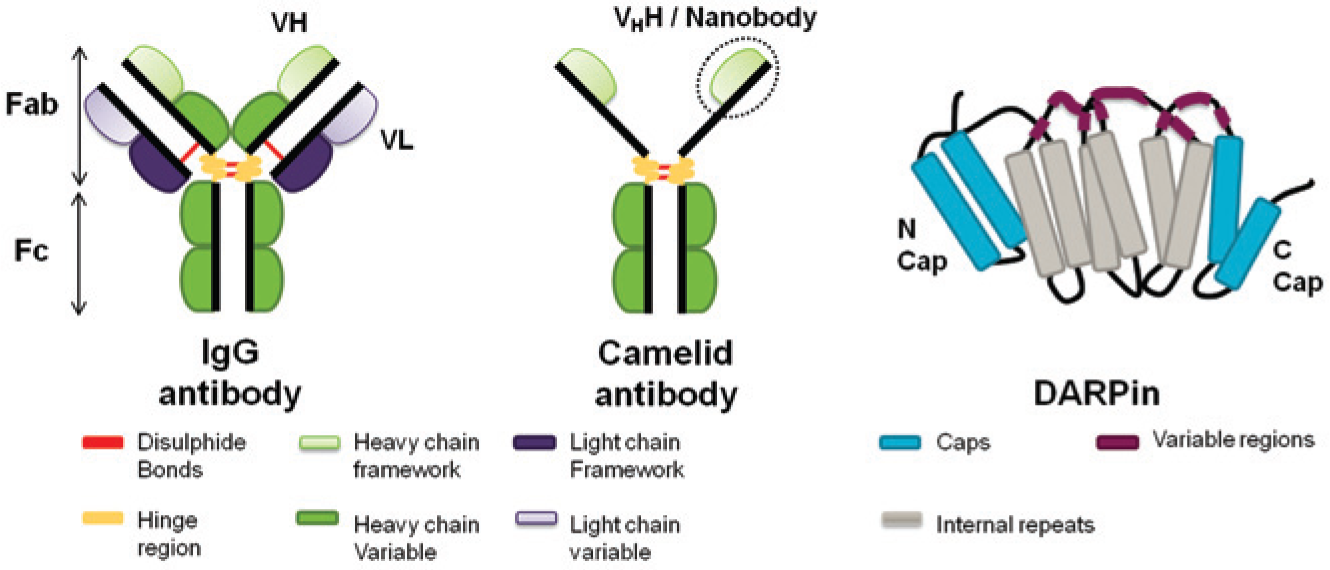
Schematic cartoons of affinity reagents used as tools in the drug discovery process. (
Antibodies have many attributes that make them very effective affinity reagents for target validation, as described above. However, interest in nonantibody affinity tools, for example DARPins, has increased considerably in recent years for use as target validation tools.72–74 DARPins consist of three or more ankyrin repeats within a constant framework. 75 This modular design provides a large surface area that can be modified by the addition of repeats, to produce an alternative binding surface without compromising the 3D structure of the tool. DARPins do not have disulphide bonds and are stable and soluble when expressed in the cytosol, making them ideal tools for intracellular protein modification, and as such, DARPins have demonstrated their use in target validation strategies for potential therapeutic targets. 70 Thus, it is clear that although antibody affinity tools still play a dominant role in the validation of intracellular drug targets, the use of alternative protein binders may, in the future, allow greater flexibility in the design and application of affinity tools for target validation.
Antibody Tools in Structural Studies
With the rapid rise of rational drug design, biophysical techniques are now routinely used to identify and study potential active sites before a drug is even conceptualized.
X-ray crystallography is an important technique for structural studies, as it has the highest possible resolving capability for deducing biological structures and is often used for lead site identification in pharmacology. In addition, knowledge of the 3D structure of a lead compound bound to its target protein generates structural information that can be used to optimize these leads efficiently. 76 The generation of crystals suitable for interrogation is, however, the limiting factor for X-ray crystallography. There are few well-known underlying principles, 77 and as such, crystallizing a target protein can prove to be extremely difficult. A method that has been successfully applied to enhance protein crystal formation is the use of chaperones, including antibody fragments or other specific binding proteins. The bound antibody fragment stabilizes the protein conformation and reduces structural heterogeneity, as well as provides additional crystal contacts. 3
The use of antibody tools to aid crystallization of “uncrystallizable” targets has been well characterized using both scFvs 78 and antigen-binding fragments (Fabs). 79 Alongside other protein affinity tools including DARPins, 80 work has also been carried out using nanobodies that are more stable than heterodimeric antibodies. 81
Wu et al. 82 have described the successful use of the VHH scaffold ( Fig. 2 ) as a crystallization chaperone for the KREPA6 protein, an important interaction protein unique to trypanosomatids, parasites that cause diseases such as sleeping sickness and Chagas disease. Many different methods had been tried previously to generate crystal structures, including the use of homologous proteins from different species, various protein lengths, mutating residues, and the use of RNA-based chaperones, as well as thousands of different crystallization conditions. However, it was not until single-domain (VHH) antibodies were used as crystal chaperones that a crystal with a 2.1 Å resolution, a sufficiently high resolution for facilitating pharmacologic drug design, was obtained.
GPCRs are highly dynamic structures and undergo a wide range of complex conformational changes. Low expression levels and instability on purification from their native membrane environment present problems in obtaining pure protein for crystallization and antigen production. 83 These problems lead to great difficulty obtaining crystals of the specific state of interest. Addressing these issues, Rasmussen et al. 84 have described the use of camelid antibody VHH fragments, selected from a fragment library generated by immunizing a llama with the β2AR protein displayed in a phospholipid vesicle, to stabilize the active conformation of the β2 adrenoceptor, generating an agonist-bound crystal structure. The resulting fragment demonstrated the ability to bind the β2AR structure in a manner analogous to the GS protein. This is an important insight as GPCRs are attractive targets for drug development because of their role in cell-signaling cascades. 85 However, comparatively little is known about the structural changes associated with agonist or antagonist binding, as of the GPCRs, only rhodopsin has been crystallized in the multiple conformations required to understand the complex structural processes associated with ligand binding. 84
Alongside acting as structural chaperones, the use of antibodies as crystallization aids has been extended to provide vital mechanistic understanding in addition to structural information. Domanska et al. 86 have reported the structure of β2M, a protein implicated in amyloidosis, using a camelid antibody bound to the truncated ΔN6 version (the version found in amyloids that aggregates easily at neutral pH 87 ). As the antibody recognized this switched dimer configuration, it suggests the mechanism by which the fibrous protein is formed with the swapped dimer acting as a template for amyloidogenesis.
In addition to antibodies acting as chaperones to aid crystallogenesis, they also have a role in supporting structure determination. Labeling antibodies with selenium-methione (SeMet), the heavy atoms required to phase X-ray scattering, allows the introduction of labels without inducing conformational changes associated with labeling proteins directly. 80 Tereshko et al. 88 have used a yeast display system to incorporate additional methionine groups into a VHH scaffold. Once bound to the antigen, the SeMet-labeled antibodies provided sufficient phasing information to assemble the structure without requiring further labeling of the protein or use of complex and expensive techniques, such as multiple anomalous dispersion.
It is important to note that the production of affinity tools to specific targets is not always trivial. An advantage of traditional crystallography solvents is the high throughput that is possible with screening kits when automated, and this speed can be lost when custom antibodies are required. This suggests that the use of antibodies as co-chaperones should be limited to previously intractable targets. As an alternative to proteinaceous affinity tools, small molecules can be used to stabilize dynamic protein structures via “tethering” (as discussed in a recent communication by Wang et al. 89 ). These approaches are more amenable to screening but do little to address the issue of nondynamic but difficult to crystallize proteins such as those cases discussed above.
It is clear that antibodies are effective tools to aid crystallogenesis, and although they are at times difficult to generate, they may provide secondary information in terms of protein-protein interactions and mechanistic pathways. Developing antibody-mediated protein crystal structures at an atomic resolution will provide new targets and mechanistic information to facilitate drug design for previously intractable targets and inform the optimization of lead compounds.
Concluding Remarks
The ability to measure and monitor the expression and function of ubiquitously expressed proteins in cells and tissues is a fundamental requirement in drug discovery, and antibodies remain the most effective tools for this purpose. The exquisite sensitivity and selectivity that antibodies provide, combined with the now advanced molecular technologies available for generation, make them ideal tools to apply in a range of assay platforms. Furthermore, advances in modern molecular biology have led to the generation of a number of proteinacious framework affinity molecules as alternatives to classical antibodies, all of which are based on a scaffold and one or more variable domains conveying the binding characteristics. The conclusion, however, still remains that beyond some limited generalizations owing to the biophysical properties of the affinity molecules themselves, most have the intrinsic property to bind a very large number of epitopes, and any favored framework will be due to a combination of the specific target in question and the intended application.
As the number of commercial organizations providing antibodies has increased dramatically, there is the potential to obtain antibodies for an unprecedented spectrum of targets; with this increase in availability comes the challenge of ensuring that antibody tools are fit for purpose in terms of specificity and selectivity. Evaluation of commercial antibodies has demonstrated that lack of selectivity is an issue,22,58,90 highlighting the need for establishing best practice in antibody validation.43,91,92 A platform for rigorous antibody validation to build a high-quality collection of antibody tools is essential, and a number of initiatives are under way to build standardized antibody collections against all human proteins (for example, AFFINOMICS, 93 NCI Antibody Characterisation Programme, 94 NIH Protein Capture, 95 Human Protein Atlas, 96 Human Proteome Organisation, 97 Recombinant Antibody Network 98 ). Significant effort is also applied within the pharmaceutical industry to meet the challenge of obtaining specific, platform-compatible tool antibodies in sufficient quantities to support drug discovery campaigns.
Footnotes
Declaration of Conflicting Interests
The authors declared no potential conflicts of interest with respect to the research, authorship, and/or publication of this article.
Funding
The authors received no financial support for the research, authorship, and/or publication of this article.