
Abstract
In this work, the authors developed a new screening approach using multiplexed immunization and immunogen array analysis to improve the efficiency of antibody screening for high-throughput antibody generation. The immunogen array is based on a 96-well format in which different immunogens and negative as well as positive controls are immobilized in each well, thus making it possible to screen hundreds of antibody candidates simultaneously. To demonstrate this approach, a model of 4 mixed immunogens immunization was employed. In total, 675 antibody candidates were screened before and after established antibody hybridomas in parallel with immunogen arrays and enzyme-linked immunosorbent assay. The signal intensity, specificity, and cross-reactivity of produced antibody candidates were analyzed using a hierarchical cluster algorithm to track the characteristics of antibody candidates during antibody generation, which might reduce the number of false-positive and false-negative binding of antibodies. Moreover, 4 monoclonal antibodies that were produced successfully recognized their corresponding target antigens.
Introduction
T
Protein array technology that is high throughput, is highly sensitive, and requires a low amount of reagents is a promising solution. 9-14 For example, De Masi et al. 15 decreased antibody screening time using multiplexed immunization and antigen microarrays. In their work, the antigen microarrays were fabricated by coating the aminosilane-modified slide with a layer of antigen. High-throughput screening (HTS) was performed by spotting hundreds of hybridoma supernatants on the surface of the antigen-coated slide, which significantly improved the efficiency of antibody screening. However, the specificity and cross-reactivity of an antibody cannot be not well characterized because only 1 antigen is used in the multiplexed assay. Furthermore, the long spotting process may impair protein activity.
To address these limitations, we developed a new antibody screening approach by using multiplexed immunization and immunogen array analysis. The difference from the approach by De Masi et al. 15 is that the immunogen array described here is based on a 96-well format where immunogens and negative and positive controls are spotted in each well. This type of multiplexing allows the specificity and cross-reactivity of antibody candidates to be characterized during the antibody generation. This is especially useful for HTS of hundreds of samples within a short time. Moreover, the amount of work saved is proportional to the number of immunogens. Finally, only minute amounts of immunogens and antibodies are employed. In this work, a model of 4 mixed immunogens immunization was employed. In total, 675 antibody candidates produced before and after established antibody hybridomas were screened in parallel with immunogen arrays and ELISA. The signal intensity and specificity as well as cross-reactivity of these antibody candidates were characterized using a hierarchical cluster algorithm.
Materials and Methods
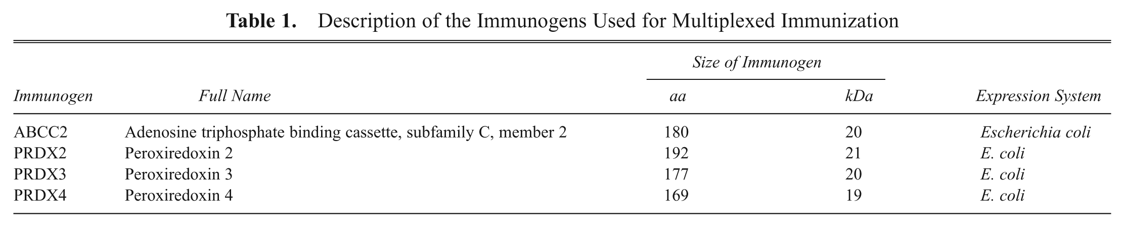
Generation of immunogens
Three proteins (PRDX2, PRDX3, and PRDX4) sharing 72% amino acids and an unrelated ABCC2 were selected as immunogens (
Description of the Immunogens Used for Multiplexed Immunization
Immunization
BALB/c mice were immunized with 4 mixed immunogens (ABCC2, PRDX2, PRDX3, and PRDX4) with the HIS tag at the N-terminus. BALB/c mice were boosted at 14-day intervals with 20 µg of each protein, with a total of 80 µg each time. The serum titer levels were monitored by ELISA every 10 days after each boost to 1:8000 dilutions. Then the antibody hybridomas were generated by fusing the spleen cells with myeloma partner SP2/0 cells based on standard protocols. 4
Fabrication of immunogen arrays
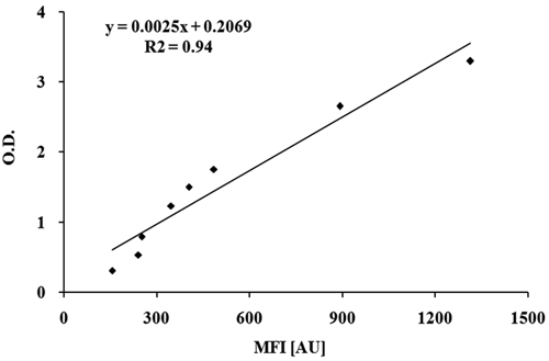
To perform HTS of the produced antibody candidates, an immunogen array based on a 96-well format was developed using 6 aldehyde-modified slides (CEL Associates, Pearland, TX) assembled into a microarray holder. Each slide consisted of 3 × 6 arrays with the array spacing identical to that of a 96-well plate. This makes this technique compatible with multichannel pipettes or robotics. For each individual array, 4 immunogens (ABCC2, PRDX2, PRDX3, and PRDX4) with a GST tag were spotted in triplicate with the Smart Arrayer-48 microarrayer (CapitalBio, Beijing, China). Bovine serum albumin (BSA) and biotinylated goat antimouse IgG antibody (Sino-Am Biotech Co., Luoyang, China) served as negative and positive controls, respectively. The biotin groups on the surface of goat antimouse IgG could be recognized by streptavidin-Cy3 (Sigma, St. Louis, MO) to produce fluorescent signal. Humidity and temperature were maintained at 60% and 20°C throughout the spotting process. All prepared immunogen arrays were blocked in 10% BSA solution at 4°C overnight.
Detection and image analysis
Before screening, the immunogen arrays were rinsed with 1× PBST (phosphate-buffered saline with 0.5% Tween-20, pH 7.4) 3 times and ddH2O once for 5 min per rinse. Then, 25 µL of culture supernatants containing hybridoma fusion cells was transferred to each array surface and incubated for 1 h at room temperature. The arrays were rinsed with PBST again following the procedure described above, and then the immunogen arrays were further incubated with biotinylated goat antimouse IgG solution (1:100) (Sino-Am Biotech Co.) and streptavidin-Cy3 solution (1:800) for 1 h, respectively. Finally, the immunogen arrays were rinsed in PBST 3 times and air-dried.
After the reaction, each slide was scanned with the Luxscan-10K/A microarray scanner (CapitalBio, Beijing, China) at 532 nm. The fluorescent image analysis was performed with Spot Data pro 3.0 software (CapitalBio). The local background intensity was subtracted from the median fluorescent intensity of each spot.
Hierarchical cluster analysis
The specificity and signal intensity of antibody candidates based on immunogen array screening was analyzed, and a heat map was drawn with MultiExperiment Viewer 4.1 software (www.tigr.org). 18 To perform hierarchical cluster analysis, the signal-to-noise (S/N) ratio was first calculated by dividing the signal of each antibody candidate by its negative control. Then the data were transformed by using the following equation:
where X is the S/N ratio, i is the sequence of antibody candidate, and M is the median S/N ratio of all antibody candidates.
Western blot detection
Total protein concentrations were determined with a bicinchoninic acid (BCA) assay (Pierce, Rockford, Illinois) using BSA to create the standard linear curve. Then, 1 µg of recombinant protein with a HIS or GST tag was separated on a 10% reduced sodium dodecyl sulfate–polyacrylamide gel electrophoresis (SDS-PAGE) and transferred to a nitrocellulose membrane. The membrane was then blocked with PBST containing 5% nonfat dry milk powder at 4°C overnight. The following morning, the membrane was incubated with 5 mL of culture supernatant containing the hybridomas at room temperature for 1 h followed by an addition of horseradish peroxidase (HRP)–conjugated antimouse IgG (Amersham Pharmacia Biotech, Buckinghamshire, UK). Immunoreactive blots were identified by using a chemiluminescence detection kit (GE Healthcare Biosciences, Piscataway, NJ).
Indirect immunofluorescence staining detection
Immunofluorecent staining of liver carcinoma cells (HepG2) was performed. Monolayer cells were washed with 1× PBS (pH 7.2) twice at room temperature and fixed with 1% Triton in 4% polyformaldehyde/PBS for 15 min. The fixed cells were washed 3 times with PBS and blocked with 1% BSA for 30 min and incubated with the primary antibody overnight at 4°C. The cells were then incubated with the secondary antibody, FITC-conjugated goat antimouse IgG, for 30 min at room temperature. Finally, the cells were incubated with Hoechst (Zhongshan Chemical Co., Beijing, China) in PBS (1:5000), and the slides were observed under fluorescence microscopy (Olympus, Tokyo, Japan).
Results And Discussion
Multiplexed immunization
With the development of hybridoma technology, cell fusion-based hybridoma technology has been the most important resource for the generation of mAbs. However, this traditional 1-immunogen/1-antibody approach has become the bottleneck in current proteomics research. 19 The development of a high-throughput antibody generation methodology will promote advancements in proteomics and associated biological research. One solution is to produce mAbs in a multiplex format, such that a single mouse can be immunized with several mixed immunogens so that their corresponding mAbs are produced at one time. The antibodies produced would specifically recognize their corresponding target or several targets. 4,15,20 Although antibody generation has improved, the accompanying high-throughput antibody screening methods are still not well established. In this report, we describe an antibody screening approach based on an immunogen array and hierarchical cluster analysis. To demonstrate our HTS approach, we immunized mice with 4 immunogens. Moreover, to see whether the screened mAbs could tell the difference between proteins sharing similar sequences, 3 proteins (PRDX2, PRDX3, and PRDX4) sharing 72% amino acids and an unrelated ABCC2 were selected. Mice were immunized with these 4 mixed immunogens, and the hybridomas secreting antibodies were isolated. Cultures of all of the 96-well sterile plates plated with fusion cells were subjected to immunogen array and ELISA analyses in parallel. To prevent potential cross-reaction between antibody and tag epitope, the recombinant proteins were expressed with 2 expression tags such that the HIS tag protein was used for BAlb/C mice immunization, and the GST tag protein was used for antibody screening.
Screening of mAbs using immunogen array and hierarchical cluster analysis
Comparison of assay performance between immunogen array and ELISA
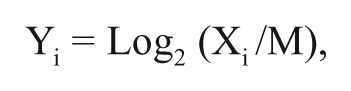
To demonstrate the ability of our immunogen array for the screening of antibody candidates, an albumin-antialbumin antibody pair was employed. Albumin was immobilized on aldehyde-modified slides and in ELISA plates, as well as incubated with mouse antialbumin antibody in parallel. Antialbumin antibody concentrations from 1 to 30 ng/mL were tested, as shown in
Comparison of the assay performance between immunogen array and enzyme-linked immunosorbent assay (ELISA). The concentration of antialbumin antibody tested is from 1 to 30 ng/mL.
Specificity and affinity analysis of antibody candidates
To screen the produced antibody candidates in high throughput, an immunogen array based on 96-well format was developed. A 5 × 5 array was fabricated in each well consisting of 4 GST tag recombinant proteins (ABCC2, PRDX2, PRDX3, and PRDX4) in triplicate, 7 negative controls (BSA), and 6 positive controls (biotinylated goat antimouse IgG;
The fluorescent image of antibody screening using an immunogen array based on the 96-well format. (
To characterize these antibodies, 22 candidates and their subclones produced before and after established hybridomas were selected to perform specificity and cross-reactivity analysis. The S/N of each antibody candidate was calculated, normalized, and used to draw a heat map. In
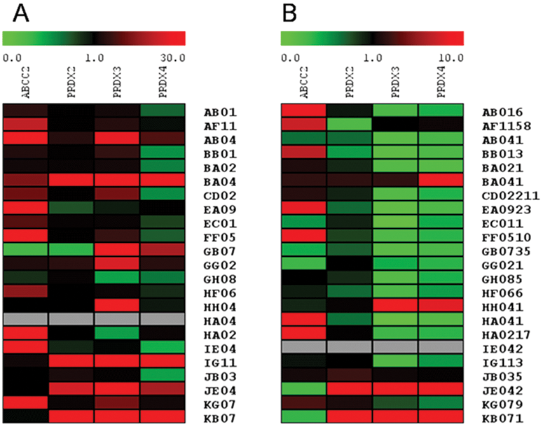
The heat map shows the specificity and signal intensity of antibody candidates based on immunogen array analysis (
Moreover, a comparison of these antibody candidates and their subclones (
Validation of antibodies using ELISA, Western blot, and Immunofluorescence staining
To validate the antibodies produced, 16 candidates were examined using immunogen arrays and ELISA, 7 antibodies were evaluated via Western blot, and 4 antibodies were tested with immunofluorescence staining (
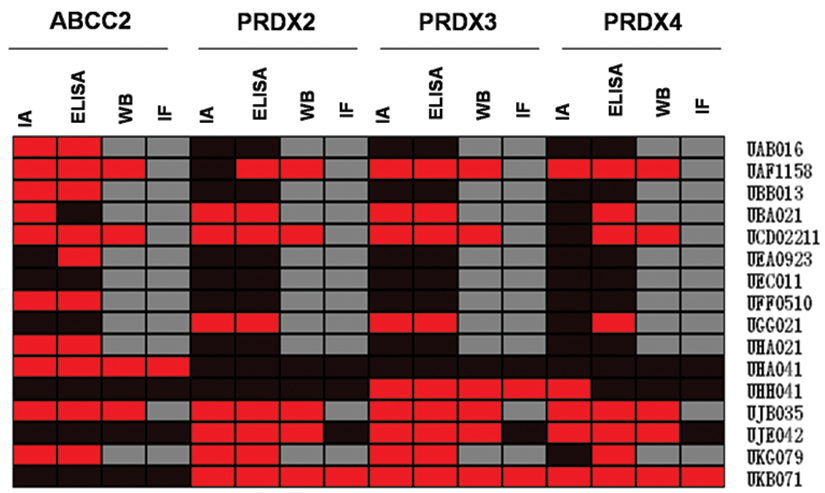
Validation of produced antibody candidates using enzyme-linked immunosorbent assay (ELISA), Western blot (WB), and immunofluorescence staining (IF) techniques. The red color is positive. The black color is negative. The gray color is no value, in which case the antibody candidate was not tested. IA, immunogen array.
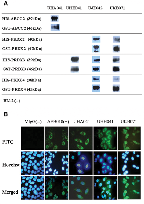
Representative images of produced mAbs from (
List of Antibodies Produced Using a Multiplexed Immunization and Immunogen Array Screening Approach
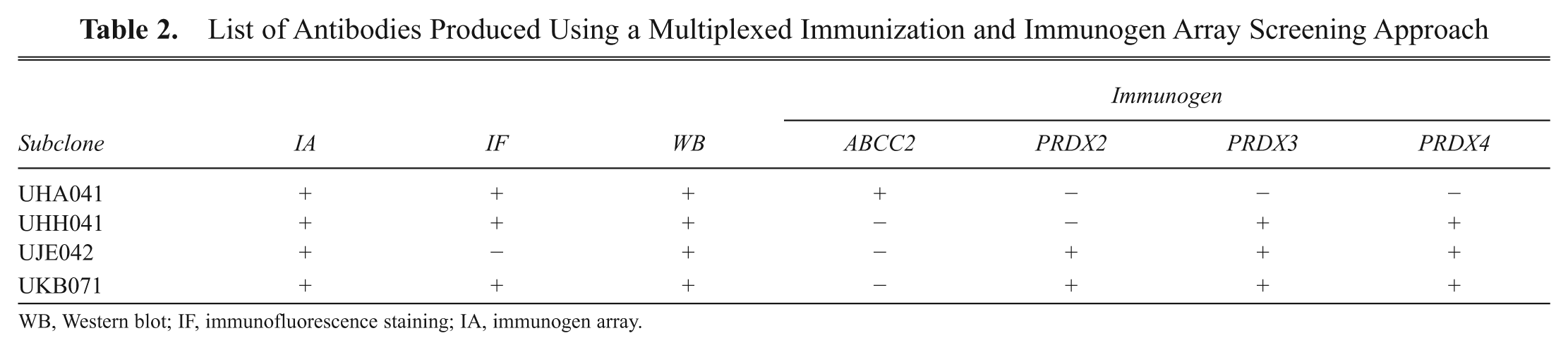
WB, Western blot; IF, immunofluorescence staining; IA, immunogen array.
Conclusion
In this work, we developed an antibody screening approach based on immunogen array and bioinformatics analysis for high-throughput mAbs generation. The immunogen array based on the 96-well format is compatible with standard laboratory equipment, which enables the rapid screening of hundreds of samples. Furthermore, using hierarchical cluster analysis, the signal intensity and specificity as well as cross-reactivity of candidate antibodies corresponding to their immunogens can be simply characterized simultaneously. This enables us to track the mAb characteristics during the mAb generation process and might prevent the potential false-positive or false-negative antibodies being produced. These results show that this new antibody screening approach has promise to push antibody generation in high throughput for proteomics and related biological research.
Footnotes
Acknowledgements
We acknowledge Dr. Thomas Joos (NMI Natural and Medical Sciences Institute, University of Tuebingen, Reutlingen, Germany) for his valuable comments and discussion for manuscript preparation. We also acknowledge financial support from the National Natural Foundation of China (grant no. 20975050) and National Basic Research Program of China (973 Program, no. 2006CB910803).