
Abstract
We report the first confirmed case of Physalia physalis (Portuguese man-o’-war) envenomation in Malaysia, involving a 9-y-old boy who sustained a sting while swimming off Labuan Island. He experienced sudden, intense burning pain in his right forearm. Visible tentacles were removed by the father, who also irrigated the area with vinegar before bringing the boy to the emergency department. The child arrived in visible distress, crying, with a linear erythematous papular rash and transiently elevated blood pressure (135/85 mm Hg) and heart rate (120 beats/min), but otherwise the boy remained hemodynamically stable. Intravenous morphine (0.1 mg·kg–1) provided effective analgesia, and the boy was admitted for observation. A photograph of the marine organism confirmed characteristic features of P physalis. Nematocysts were identified on a skin sample using the cellophane tape method and light microscopy. This case highlights gaps in region-specific guidelines and emphasizes the need for public education on marine envenomation in tropical settings.
Introduction
The Portuguese man-o’-war (Physalia physalis) is a venomous siphonophore often mistaken for a jellyfish due to its gas-filled float and trailing tentacles. It delivers painful and medically significant stings through specialized nematocysts, which can cause intense local pain and, in some cases, systemic effects.1,2 Unlike true jellyfish, P physalis is a colonial organism composed of specialized zooids functioning as a single unit.
Envenomations are well documented in Australia, the Atlantic, and the Mediterranean. 3 However, such encounters remain undocumented in Malaysia, with no previously confirmed or published case reports, despite anecdotal media accounts. 4 Labuan Island, a federal territory off the coast of Borneo in East Malaysia, is known for marine biodiversity and recreational water activities. Its warm waters and diverse marine life may attract venomous species. 5 As ocean temperatures and currents shift due to climate change, sightings of Physalia spp are expanding into previously unaffected geographic zones. 6
Despite growing marine tourism, envenomation events are often underreported in Malaysia, and standard diagnostic pathways are seldom used. 4 This case presents the first confirmed P physalis sting in Malaysia—diagnosed clinically and confirmed via nematocyst microscopy—thereby highlighting significant gaps in preparedness and clinical guidance for marine stings.
Recent genomic and morphologic studies have challenged the traditional view that P physalis represents a single cosmopolitan species. Church et al. performed whole-genome sequencing of 151 specimens and analyzed >4000 citizen-science images, revealing at least 4 distinct Physalia spp: P physalis, P utriculus, P megalista, and a newly described P minuta. These lineages exhibit strong reproductive isolation, high genomic divergence (fixation index FST up to 0.61), and region-specific population structures shaped by prevailing ocean currents and winds. 7 The study highlights that even highly mobile neustonic organisms such as Physalia are genetically structured rather than globally panmictic. These findings underscore the need for accurate species identification because regional differences may influence venom composition, envenomation severity, and clinical outcomes in Physalia-related stings. In Malaysian waters, where P physalis and P utriculus are both likely present, genomic diversity may account for variations in sting severity and clinical responses reported along different coasts. Incorporating species-level identification into future envenomation reports could enhance understanding of toxin variability and improve targeted management strategies. Despite recent genomic evidence revealing multiple Physalia spp with distinct regional distributions, clinical reports from Southeast Asia rarely differentiate stings by species, leaving gaps in understanding species-specific venom effects and patient outcomes.
Case Report
A 9-y-old boy from the Czech Republic sustained a marine sting while swimming near Labuan Island, Malaysia. He experienced a sudden, intense burning sensation in his right forearm. His father observed a gas-filled, blue-tinted marine organism floating nearby and quickly removed visible tentacles from the child's arm. The affected area was rinsed with vinegar, and the child was brought to the nearest emergency department within 30 min of envenomation.
On arrival, the child presented in visible distress, crying, with a reported pain score of 10/10. His initial vital signs were blood pressure of 135/85 mm Hg, heart rate of 120 beats/min, respiratory rate of 24 breaths/min, temperature of 36.8°C, and SpO2 of 97%. After administration of intravenous morphine (0.1 mg·kg–1) for pain control, the child’s blood pressure decreased to 105/70 mm Hg, heart rate decreased to 105 beats/min, respiratory rate decreased to 20 breaths/min, and SpO2 improved to 99%. The boy remained hemodynamically stable throughout his observation in the ward. Physical examination revealed a linear erythematous papular lesion (∼20 cm) over the dorsum of the right forearm (Figure 1). No systemic symptoms such as dyspnea, nausea, or hypotension were present.
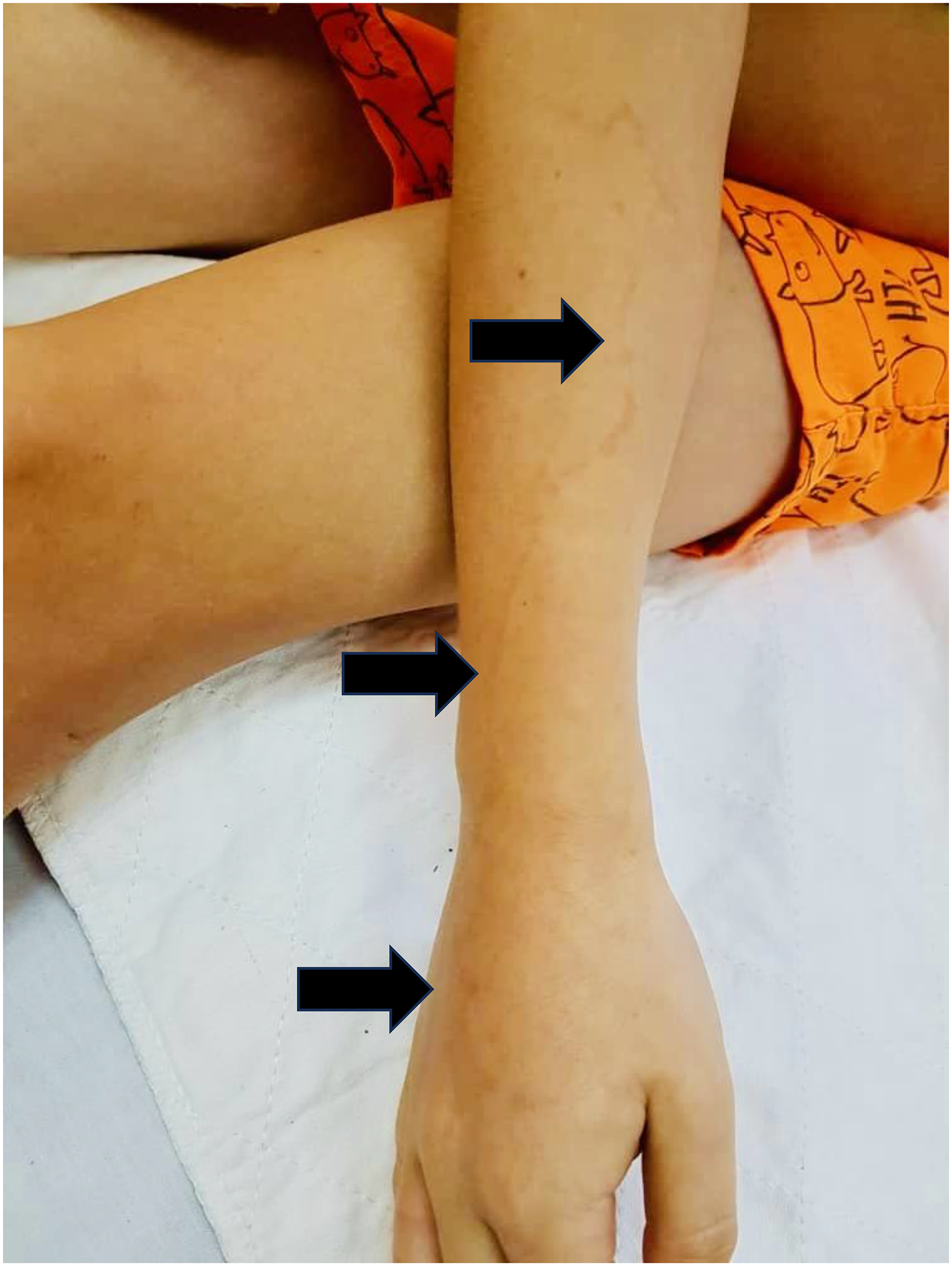
Dorsal view of the patient's right forearm showing a linear serpiginous erythematous lesion extending from the proximal forearm to the wrist. The lesion follows a whiplike pattern consistent with tentacle contact from Physalia physalis. There was no evidence of blistering, necrosis, or systemic involvement at the time of presentation.
Dr Muhamad Na’im Ab Razak, of the Emergency and Trauma Department, Hospital Lahad Datu, Sabah, Malaysia (a member of the Remote Envenomation Consultation Service [RECS] Malaysia), was consulted, and later, marine biologist Dr B. A. Venmathi Maran (Borneo Marine Research Institute, Universiti Malaysia Sabah) confirmed the findings. A photograph of the marine organism, captured by the father on a mobile phone (though slightly unclear), was reviewed and confirmed to be P physalis 8 (Figure 2).
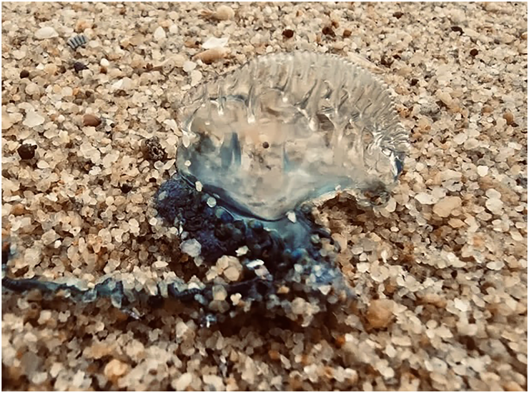
Photograph of the Portuguese man-o'-war (Physalia physalis) captured by the patient's father at the site of envenomation.
To confirm the diagnosis, a skin sample was collected using the cellophane tape method, a noninvasive technique for nematocyst detection. 9 Tape was applied to the lesion and transferred to a clean glass slide. Microscopic analysis at 10× and 40× magnifications revealed oval capsules with coiled filamentous structures consistent with Physalia nematocysts.9,10 We were unable to obtain a photograph of the nematocysts because the sample was processed for diagnostic purposes only, and the preserved material was not of sufficient quality for imaging.
Discussion
This report describes the first laboratory-confirmed P physalis envenomation in Malaysia. The genus Physalia includes several siphonophore species, with P physalis (Portuguese man-o’-war) predominantly found in the Atlantic Ocean, whereas P utriculus (commonly called the Indo-Pacific bluebottle) is widely distributed across the Indian and Pacific Oceans, including Southeast Asia. 8 P physalis typically has a larger, elongated pneumatophore (up to 30 cm), with multiple long fishing tentacles that can extend over 10 m. In contrast, P utriculus has a smaller, oval or spherical pneumatophore (rarely exceeding 10 cm) and usually possesses a single, shorter tentacle, generally <3 m in length.
Clinically, stings from both species produce painful, whiplike linear lesions, but systemic symptoms (eg, cardiovascular or neurologic effects) are reported to be more frequent and severe with P physalis, likely due to differences in venom composition. 2 Although P utriculus is more commonly reported in Southeast Asia and Australia, the morphology of the organism in this case—marked by an elongated purple–blue pneumatophore and multiple tentacles—was consistent with P physalis, as confirmed by expert review. 8
First aid for Physalia stings remains controversial. In this case, vinegar was applied on site as the initial first aid measure. Although vinegar is widely used for box jellyfish stings, it may cause nematocyst discharge in Physalia and is therefore not recommended.3,6 Instead, current guidelines favor seawater rinsing followed by hot water immersion (40–45°C) for up to 20 min to inactivate venom proteins. 6 The proposed mechanism of action—protein denaturation—remains hypothetical, with vasodilation and local dilution of venom toxins also considered potential contributing factors.
The cellophane tape method was applied successfully in this case to confirm the presence of nematocysts. It offers a simple, cost-effective diagnostic tool for use in remote or low-resource settings. 9 However, its sensitivity can be limited by sample quality or delayed application. No nematocyst photograph was obtained because the sample was processed solely for diagnostic purposes, and the preserved material was of limited quality for imaging.
From an environmental standpoint, the expanded distribution of Physalia beyond traditional habitats has been linked to shifting sea temperatures and currents. 5 A recent analysis by Kumar and Prasad (2021) highlighted increasing stings in the Indo-Pacific, reinforcing the need for clinician preparedness in newly affected regions. 5 Despite these trends, Malaysia still lacks comprehensive reporting systems for marine stings, and awareness of updated first-aid practices remains low. 4
The recent genomic delineation of Physalia spp by Church et al. strengthens the clinical significance of accurate species identification in envenomation cases. Our case aligns with these findings because the observed morphology and clinical presentation are consistent with P physalis, one of the 4 genetically distinct species now recognized. 7 Because venom composition and sting severity may vary between Physalia lineages, integrating genomic and morphologic data into regional envenomation reports can enhance both diagnostic precision and the development of evidence-based treatment protocols.
Conclusion
This first confirmed case of P physalis envenomation in Malaysia underscores critical gaps in public health preparedness, clinical awareness, and response to marine envenomations in Southeast Asia. Accurate species identification is essential, particularly in differentiating P physalis from P utriculus (Indo-Pacific bluebottle), because differences in morphology, venom potency, and clinical effects can influence management and outcomes. Clinician vigilance, rapid pain management, and simple bedside diagnostic approaches such as the cellophane tape test can facilitate timely recognition and care.
As marine biodiversity shifts due to climate change and altered ocean currents, region-specific guidelines, community education, and emergency preparedness systems must evolve to effectively address emerging threats. The recent genomic studies of Physalia spp provide a scientific basis for recognizing species diversity and potential venom variability, underscoring the need for regional envenomation reports to include both morphologic and genomic identification for accurate clinical and epidemiologic understanding. 7
Footnotes
Patient Consent
Written informed consent for the publication of this case report and accompanying clinical images was obtained from the patient's parents.