
Abstract
A panel convened to develop an evidence-based set of guidelines for the recognition and treatment of eye injuries and illnesses that may occur in the wilderness. These guidelines are meant to serve as a tool to help wilderness providers accurately identify and subsequently treat or evacuate for a variety of ophthalmologic complaints. Recommendations are graded based on the quality of their supporting evidence and the balance between risks and benefits according to criteria developed by the American College of Chest Physicians.
Introduction
Although eye complaints constitute approximately 1.5% of visits to emergency departments in North America each year, their incidence in the wilderness is unknown. 1 Eye problems in the wilderness represent a challenging group of complaints for several reasons: access to proper equipment is limited, access to proper medications may be limited, and most practitioners in the wilderness are not specially trained. 2 The Wilderness Medical Society (WMS) published a set of guidelines on eye injuries in the wilderness in 2001. A panel was convened at the WMS Mountain Medicine meeting in Park City, Utah, in February 2011 to update those guidelines with the most relevant evidence-based information. These guidelines were again updated in 2014 and in 2023.
Methods
The panel was initially convened at the 2011 Wilderness and Mountain Medicine WMS conference in Park City, Utah. The WMS members were selected based on clinical interest, research experience, and ophthalmologic expertise. Relevant articles were identified through the PubMed and Cochrane Collaboration databases using keyword searches with the appropriate terms corresponding to each topic. Studies were reviewed, and the level of evidence was assessed. Data regarding specific wilderness treatment of eye injuries are sparse; therefore, we evaluated the data regarding eye injuries and illness in general and adapted these to the wilderness setting. The panel used a consensus approach to develop recommendations regarding diagnosis, treatment, and prevention of ocular injuries in the wilderness. These recommendations have been graded based on clinical strength as outlined by the American College of Chest Physicians (ACCP) updated 2018 GRADE methodology (see Supplemental Table) and in conjunction with other WMS practice guidelines. 3 This is an updated version of the original guidelines published in WEM (Wilderness Environ Med. 2014 Dec; 25(4):S19–29).
Examination and Equipment
Examination of the eye in the wilderness may be difficult owing to the lack of specialized equipment, but many basic elements of the eye examination can be completed in austere settings. Table 1 provides a list of equipment and medications that should be included in both a basic and an advanced eye kit. A basic kit is appropriate for short excursions, whereas the basic plus the advanced kit could be used for prolonged expeditions, especially to remote locations. Many items may have a dual purpose and could also treat other conditions in the wilderness setting.
Basic and advanced equipment and medications.
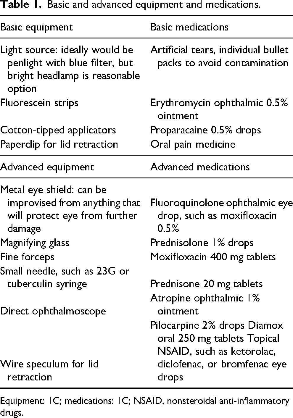
Equipment: 1C; medications: 1C; NSAID, nonsteroidal anti-inflammatory drugs.
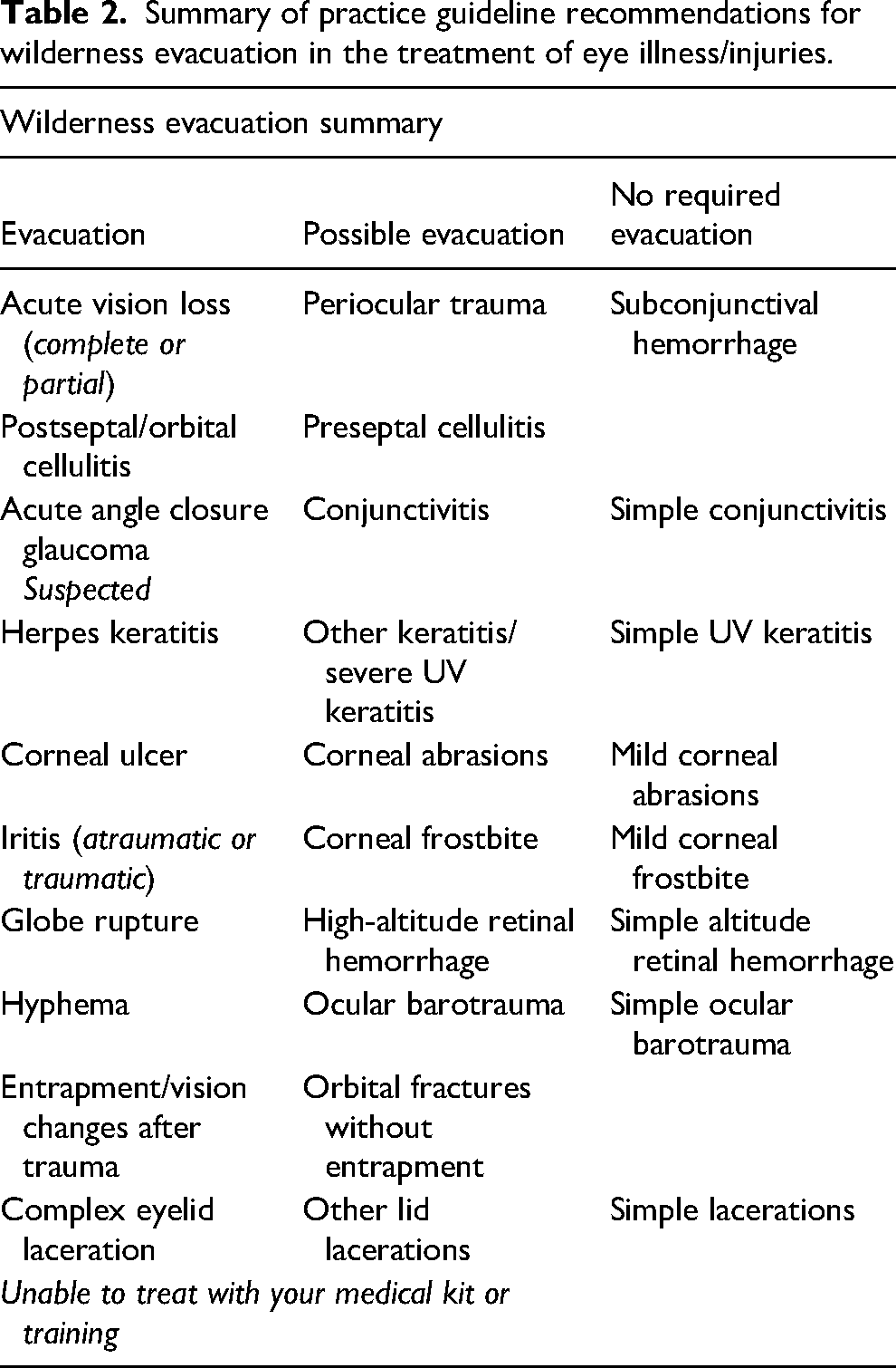
Summary of practice guideline recommendations for wilderness evacuation in the treatment of eye illness/injuries.
Pretrip Planning and Prevention
We recommend people with a history of ophthalmologic problems who are participating in wilderness activities have a complete eye examination within 3 mo of any extended trip. Participants should be encouraged to bring all of their own equipment and medications, including glasses, goggles, sunglasses, contacts, lens solution, and any specific medications that they are currently taking or anticipate requiring. Additionally, we recommend participants bring a copy of their lens prescription to aid in the acquisition of new corrective wear if needed or simply bring extra glasses or contacts if they will be in austere settings.
Prevention of eye illness and injuries is paramount. Many eye illnesses are a result of accidents and thus may not be preventable, yet adequate sun and protective eyewear, good hygiene, and proper hand washing can prevent many of the conditions addressed in these guidelines.
Medical Eye Complaints
Acute Vision Loss in the White Eye
Vision loss can occur in both the red and white eye. As these are quite rare, data on these illnesses in the wilderness are very limited; therefore, wilderness management is adapted from clinic or hospital treatments to the wilderness setting as appropriate. There are numerous causes of acute vision loss; regardless of the cause, any vision loss should be considered an emergency, and we recommend that all patients with vision loss be considered for emergent evacuation.
Central Retinal Artery Occlusion
Central retinal artery occlusion (CRAO) is an ischemic stroke of the retina, usually due to a thrombotic or embolic event, that causes abrupt vision loss. 4 Treatment options in the wilderness are limited, but there are case reports to suggest improvement in visual acuity after providing high-flow oxygen. 5 There are also several case studies that report improvement with hyperbaric oxygen therapy. Central retinal artery occlusion is currently an approved indication for hyperbaric oxygen therapy by the Hyperbaric Oxygen Committee of the Undersea and Hyperbaric Medicine Society. 5 If oxygen or hyperbaric therapies are available, we strongly recommend they be considered for patients with suspected CRAO. A 2009 Cochrane Review found 2 randomized controlled trials evaluating 2 potential treatments for CRAO: pentoxifylline tablets and enhanced external counterpulsation. Both trials showed improved retinal artery flow with their treatments, but neither showed improvement in vision. 6 More recently, interest in the treatment of CRAO with thrombolytic therapy has garnered observational support, but randomized trials have not demonstrated efficacy in the recovery of vision. 7 While CRAO is a disease that does not have a specific treatment, management should follow the treatment of other vascular end-organ ischemic diseases. 7 These treatments are not available in the wilderness. We recommend emergent evacuation of these patients.
Oxygen: Strong recommendation, low-quality evidence
Hyperbaric oxygen therapy: Strong recommendation, low-quality evidence
External counterpulsation: Weak recommendation, moderate-quality evidence
Pentoxifylline: Weak recommendation, moderate-quality evidence
Emergent evacuation: Strong recommendation, low-quality evidence
Central Retinal Vein Occlusion
Central retinal vein occlusion (CRVO) is a blockage of the venous outflow from the eye, leading to profound, painless vision loss. It can be a result of a thrombus, external compression, or vasculopathy and usually occurs in people aged more than 50 years who have hypertension. 8 Patients with CRVO will usually have an afferent pupillary defect 4 : when a light is shone in the normal eye, both pupils will constrict, but when the light is then quickly shone in the abnormal eye, both pupils will dilate. It should be noted that many diseases that cause unilateral optic neuropathy or retinal problems (such as ischemic optic neuropathy, retinal detachment, and CRVO) can lead to an afferent pupillary defect. It is not necessarily important in the wilderness setting to differentiate these conditions but to recognize this abnormal finding and move toward evacuation.
Although the treatments of CRVO in the hospital are complicated and involve laser treatments and intravitreal injections with antivascular endothelial growth factor (anti-VEGF) or steroid administration, it is nevertheless reasonable to start treatment in the wilderness with topical steroids, if available (prednisolone 1%, 2 drops in the affected eye 4 times daily). 8 We recommend emergent evacuation, as these patients need specialist care and treatments.
Topical steroid: Strong recommendation, low-quality evidence
Emergent evacuation: Strong recommendation, low-quality evidence
Retinal Detachment
A retinal detachment is characterized by vision loss or floaters and flashes of bright light in a patient's visual field. The vision loss is often described as a fixed cloudy or curtainlike defect in the visual field. This condition is usually painless. Visual loss is variable, depending on where the tear has occurred on the retina. 4 We recommend emergent evacuation, as surgical management may be necessary, and there is no effective field treatment.
Emergent evacuation: Strong recommendation, low-quality evidence
Periocular Inflammation
Inflammation of the periocular region includes preseptal cellulitis, orbital cellulitis, and dacrocystitis. Preseptal cellulitis is an infection limited to the space anterior to the orbital septum, which includes the superficial tissue surrounding the eye and the eyelids. Orbital cellulitis is more extensive and involves the soft tissues in the bony orbit (deep to the orbital septum). Clinically, preseptal cellulitis presents with swelling and inflammation of the eyelid without involvement of the eye itself. Orbital cellulitis presents with inflammation within the orbit, including bulging of the eye (proptosis), swelling of the conjunctiva (chemosis), painful extraocular movements, and possible visual disturbance. 9 Additionally, patients with orbital cellulitis may be systemically ill and have a fever. Periorbital cellulitis, while isolated to the preseptal space, is often associated with marked swelling, which makes the differentiation of preseptal from orbital cellulitis difficult.
Preseptal cellulitis can be caused by spread from sinusitis, conjunctivitis, or blepharitis. Causes that may be more commonly encountered in the wilderness include trauma and insect bites. 9 Orbital cellulitis is usually caused by a sinus infection. 10 Additionally, preseptal cellulitis can spread and cause orbital cellulitis. There are limited data regarding the management of these entities in the wilderness. Rapid identification of orbital cellulitis is paramount, as orbital cellulitis is a true medical emergency. Failure to treat this infection quickly can lead to blindness, intracranial infection, and possibly death. Initial management of both conditions requires antibiotics that cover Staphylococcus species (with increasing incidence of MRSA [methicillin resistant Staphylococcus aureus]) and Streptococcus species. 11 The incidence of infections caused by Streptococcus pneumoniae and Haemophilus influenzae has decreased due to the introduction of vaccines against these organisms. 11 Consideration should also be given to coverage of anaerobic bacterial species. 10
Treatment of both preseptal and orbital cellulitis should begin with amoxicillin/clavulanate (875 mg/125 mg) orally, 2 times daily for 10 d, but if that is not available, then oral fluoroquinolones (such as moxifloxacin) may be substituted. 12 If the practitioner is certain that an infection is isolated to the preseptal space, evacuation can be nonemergent. However, if there is any question that the infection may involve the orbit, or a presumed preseptal cellulitis is not responding to antibiotic treatment, we recommend emergent evacuation. 13
Dacrocystitis is an infection of the lacrimal sac that typically arises from obstruction of the lacrimal duct, which causes pooling of tears in the lacrimal sac and subsequent infection. 4 It presents as pain, swelling, and redness over the lacrimal duct, which is located at the nasal corner of the eye. The most common pathogens involved are Staphylococcus aureus, Streptococcus species, and H. influenzae. 14 Dacrocystitis can be treated initially with oral antibiotics as well as warm compresses, if available. The preferred antibiotic is amoxicillin/clavulanate (875 mg/125 mg) orally, 2 times daily for 10 days, but if that is not included in your kit, oral fluoroquinolones (such as moxifloxacin) will likely suffice. 12 If this condition worsens, the patient should be evacuated nonemergently.
Antibiotics: Strong recommendation, low-quality evidence
Emergent evacuation for orbital cellulitis: Strong recommendation, low-quality evidence
Nonemergent evacuation for periorbital cellulitis: Strong recommendation, low-quality evidence
Acute Red Eye
There are numerous conditions that can cause redness of the eye, and the severity of these conditions can range from mild to severe. There are 3 important tests we suggest that can be quickly and easily accomplished to help make the diagnosis: fluorescein staining, relief of pain with anesthetic drops, and pupillary status. 13 These tests can be easily performed in the wilderness if the supplies are available.
The medical causes of acute red eye can be broadly divided into conditions that are fluorescein positive (corneal ulcer or erosion and herpes keratitis) and those that are negative. Those that are negative can be further divided into conditions that resolve with an initial dose of topical anesthetic (conjunctivitis, blepharitis, ultraviolet [UV] keratitis) and those that do not (acute angle-closure glaucoma [AACG], iritis, and scleritis).
Acute Angle-Closure Glaucoma
AACG is caused by a blockage of aqueous outflow from the anterior chamber of the eye, resulting in increased intraocular pressure (IOP). Symptoms include deep and severe pain, blurry vision, halos around lights, headache, and often nausea and vomiting. 15 Signs of this diagnosis include a nonreactive pupil that is mid-dilated (4 mm to 6 mm), decreased visual acuity, “steamy” or “hazy” cornea, and a red eye. 16 Measurement of IOP in the field is often impossible owing to the absence of measurement devices, but gentle palpation of the globe through a closed lid may reveal a taut globe, indicating elevated IOP.15,17,18 The palpation should be done very gently and also in comparison with the normal eye. AACG is a medical emergency and requires emergent evacuation, as the definitive treatment is surgical. If available, we recommend initiating medical treatment of AACG, including a beta-adrenergic antagonist (timolol 0.5%, 1 drop in affected eye 2 times daily), an oral or intravenous carbonic anhydrase inhibitor (acetazolamide, 500 mg 2 times daily), and a topical alpha2-adrenergic agonist (though not often included in expedition kit).4,19 A topical miotic (pilocarpine 1% to 2%, 1 drop every 15 min for 2 doses) should be given 1 h after other treatments have been given. Additionally, patients should be advised to lie flat for at least 1 h. 19
Medical management: Strong recommendation, moderate-quality evidence
Surgical management: Strong recommendation, high-quality evidence
Emergent evacuation: Strong recommendation, low-quality evidence
Iritis
Nontraumatic iritis (also called anterior uveitis) is inflammation of the iris that is inflammatory or infectious. Many symptoms are similar to previously noted conditions, including pain, redness of the conjunctiva, blurred vision, and photophobia. The patient may also have a history of iritis, which is a good clue to the diagnosis. One important examination finding is photophobia in the affected eye with the shining of a penlight in the unaffected eye (consensual photophobia). 16 Standard therapy for iritis includes corticosteroids, such as prednisolone 1% eye drops—1 drop into the affected eye every hour if severe or every 4 h if mild. 20 If drops are unavailable, or if the condition is worsening, oral steroids can be given—prednisone 40 mg to 60 mg oral daily. If there is concern for herpetic lesions or other serious infection, steroids should be avoided. In this case, a topical nonsteroidal anti-inflammatory drug (NSAID) may be beneficial. Cycloplegic (mydriatic) drops, such as atropine, are a key component to pain control as they relieve ciliary spasms; cycloplegics should be avoided if there is concern for elevated intraocular pressures. 20 Place 1 drop in the affected eye 2 times daily. Evacuation should be emergent, as iritis can be complicated by scarring of the pupil and elevated IOP, leading to decreased vision or blindness. 13
Topical steroids: Strong recommendation, low-quality evidence
Systemic steroids: Weak recommendation, low-quality evidence
Mydriatic drops: Strong recommendation, low-quality evidence
Topical NSAID: Weak recommendation, low-quality evidence
Emergent evacuation: Strong recommendation, low-quality evidence
Herpes Keratitis
Herpes keratitis is diagnosed by a dendritic staining pattern on fluorescein examination. Often, patients have had previous episodes of ocular herpes infections. 15 Treatment includes oral or ophthalmic preparations of trifluridine or acyclovir, although these are often not included in expedition kits. 21 Participants should be encouraged to bring their own medicine if they have a history of this condition. Steroids should be avoided for patients with suspected herpes keratitis. 22 While some texts advocate debridement of herpetic lesions with a cotton-tipped swab, that has not been proven to be efficacious and should only be done by experienced providers if deemed necessary. 21
Oral or ophthalmic antivirals: Strong recommendation, high-quality evidence
Emergent evacuation: Strong recommendation, low-quality evidence
Conjunctivitis
Among fluorescein-negative medical eye conditions, conjunctivitis, blepharitis, and ultraviolet keratitis can usually be treated in the field and are typically not serious. Conjunctivitis is the most common cause of a red eye. 15 Common etiologies include allergies, viruses, and bacteria. Conjunctivitis always involves the palpebral conjunctiva, and the examiner must look under the lid to evaluate for this inflammation. The hallmark of allergic conjunctivitis is itching. Bacterial conjunctivitis gives a purulent discharge, whereas viral conjunctivitis tends to give a watery discharge. Treatment of allergic and viral conjunctivitis is primarily supportive, and bacterial conjunctivitis is treated with antibiotics. Although bacterial conjunctivitis is usually a self-resolving condition, studies have demonstrated that the use of antibiotics results in faster clinical and microbiological remission. 23 There are no studies showing the superiority of any one antibiotic over another; therefore, choice should be based on availability. 23 Erythromycin ointment, 1-cm ribbon, placed in the lower conjunctival sac 3 or 4 times daily for 7 d is a reasonable option. It is important to note that viral conjunctivitis is extremely contagious, and proper hand washing is essential in wilderness settings to avoid spreading throughout a group or to the contralateral eye. 24 Topical anesthetic eye drops are diagnostically beneficial and provide acute relief for corneal ulcers, herpes keratitis, and conjunctivitis; however, it is important to note that they should not be used chronically as they are toxic to the corneal epithelium. 24
Topical or systemic antibiotics: Strong recommendation, high-quality evidence
Hand washing: Strong recommendation, low-quality evidence
Altitude and the Eye
The environmental conditions at altitude, including the dry environment, ultraviolet exposure, cold temperatures, hypoxia, and low atmospheric pressure, pose several problems for the eye and may result in problems both for the previously healthy eye and for the patient with a history of ocular pathology. Because the cornea receives most of its oxygen from the ambient air, the cornea becomes hypoxic at altitude and may not function normally. 25 In addition, patients with a history of intraocular gas bubbles should not go to high altitudes owing to the expansion of the gas bubble at decreasing atmospheric pressure. Patients with a history of ocular pathology or prior eye surgery should consult with their ophthalmologist regarding the safety of traveling to altitude.
Altitude After Radial Keratotomy, Laser-Assisted Stromal In-Situ Keratomileusis, or Photorefractive Keratectomy
Hypoxia at altitude causes edema and thickening of the cornea.26,27 Although this thickening has been shown not to cause visual disturbances in healthy eyes, it can be problematic for patients who have undergone radial keratotomy (RK), laser-assisted stromal in-situ keratomileusis (LASIK), or photorefractive keratectomy (PRK). Radial keratotomy involves making incisions into the cornea, weakening its stability. The edema caused by the hypoxia of high altitude results in a flattening of these corneas, causing increased far-sightedness 25 and a significant decrease in visual acuity. The amount of vision change partially depends on the amount of residual myopia that a patient has at sea level but is difficult to predict before altitude exposure. Although most of these changes will resolve with a return to normal altitude, it is recommended that patients who have had RK take various levels of corrective lenses during their excursion. The cheapest option may be to wear glasses under goggles and to bring several levels of corrective glasses. The best option, however, would be to bring several levels of prescription goggles in the event of decreased visual acuity at altitude. Patients should consult with their ophthalmologist to obtain various strengths of eyewear.
Eyes that have received LASIK and PRK undergo fewer refractive changes at altitude. There have been 2 studies demonstrating blurring of vision at altitude and 2 studies demonstrating no change. A recent study of 12 climbers with LASIK-treated eyes noted no visual changes with changing altitude, despite dry eye associated with high-altitude mountaineering being a known complication of LASIK. 28 Similarly, post-PRK patients seem to have excellent vision at altitude. There has only been 1 study of this PRK population, and it demonstrated no significant change in vision at altitude. 29
Caution at high altitude if history of RK: Strong recommendation, low-quality evidence
Safety of LASIK/PRK at altitude: Strong recommendation, low-quality evidence
Extra goggles/glasses if history of RK: Strong recommendation, low-quality evidence
High-Altitude Retinal Hemorrhage
At altitude, an increase in retinal blood flow and subsequent retinal vein dilation 13 can lead to retinal hemorrhages, which have been well described since the 1970s.30,31 The percentage of climbers experiencing retinal hemorrhages ranges from 0% to 79%, depending on the study. 30 Most cases of retinal hemorrhage do not cause visual disturbances unless the hemorrhage involves the macula. Patients with retinal hemorrhages can continue ascent if no visual disturbances are present but should be instructed to descend in the setting of altered vision. 13 Additionally, the presence of high-altitude retinal hemorrhage has been associated with altitude illness, and its presence should alert providers to the possible development of altitude illness.
Descent ifvisual changes: Strong recommendation, low-quality evidence
Diving and the Eye
The hyperbaric environment can cause a number of effects on the eye. The primary concerns regarding dive medicine and the eye include ensuring that patients have stable visual acuity while diving and making sure that patients who have a surgical intraocular gas bubble do not dive. Divers who normally require corrective lenses should dive with prescription goggles or soft contact lenses. Diving after refractive surgery such as LASIK and PRK is safe. 31 Diving after RK carries a hypothetical risk of corneal incision rupture, although in practice, this is not seen, and diving is therefore considered safe. However, divers who have had RK should not dive for 3–6 mo postoperatively to ensure adequate wound healing of the corneal incisions. Divers with a history of any type of eye surgery should always consult with their ophthalmologist before diving. 32
Safety of diving after refractive surgery: Strong recommendation, low-quality evidence
Ocular and Periocular Barotrauma
A complete description of the mechanism and pathophysiology of barotrauma is beyond the scope of this review; however, in brief, when air in a closed space is exposed to higher atmospheres of pressure, it is compressed. If this occurs while diving with a facemask, the lids, skin, and eyes can be drawn out into the mask. This force can be very strong and cause significant injury, resulting in periorbital ecchymosis, edema, subconjunctival hemorrhage, and potentially, hyphema. The prevention of ocular and periocular barotrauma can be achieved by frequently filling one's mask with exhaled air from the nose to increase the volume of air in the mask.32–35 If hyphema is present, evacuation should be emergent.
Mask pressure equalization: Strong recommendation, low-quality evidence
Hyphema: Evacuation emergent: Strong recommendation, moderate-quality evidence
Traumatic Eye Injuries
The treatment of traumatic ocular emergencies in the wilderness hinges on rapid assessment, stabilization, and evacuation. Often, eye injuries do not occur in isolation and may be associated with other potentially life-threatening injuries. Periocular and ocular traumas include many disorders, but these guidelines are limited to the most commonly encountered conditions in the wilderness. Ocular and periocular trauma is responsible for approximately 3% of emergency department visits in North America annually 36 and is a leading cause of preventable blindness worldwide. The incidence in the wilderness is unknown, as epidemiologic data are absent from the literature.
Periocular Trauma
Eyelid lacerations: Falls, branches, rocks, and animal bites are among many causes of eyelid lacerations in the wilderness. Eyelid lacerations can be classified as simple or complex. Simple lid lacerations are often horizontal, have partial thickness, and do not involve the lid margin. This type of laceration can be managed with irrigation, and typical wound care and has an excellent prognosis. 36
For eyelid lacerations that are complex—namely, full thickness and involve the lid margin or medial/lateral end of the palpebral fissures—we recommend evacuation and emergent ophthalmologic evaluation and repair, ideally within 36 h.36,37 Immediate treatment consists of prompt irrigation and the application of antibiotic ointment with shielding to protect the eye as evacuation commences. If an associated ruptured globe is suspected, there should be no further examination, manipulation, or irrigation of the eye. Treatment should be as outlined in the section on ruptured globe.
Wound care: Strong recommendation, moderate-quality evidence
Ophthalmologic antibiotic ointment: Strong recommendation, moderate-quality evidence
Eye shield: Strong recommendation, low-quality evidence
Emergent evacuation if complex eyelid laceration: Strong recommendation, moderate-quality evidence
Orbital Fractures
A direct blow to the face or orbit may cause fractures of any of the 7 facial bones that form the orbit. Orbital fractures can lead to significant ocular injury and subsequent visual impairment in approximately 17% of cases. 38 Thus, it is important for the wilderness provider to be able to perform a basic ophthalmologic examination to screen for severe injury, including entrapment of ocular muscles and pupillary or vision derangements. If signs of entrapment or visual derangements are present, then emergent evacuation is required.
Clinically significant retro-orbital hemorrhage can cause compressive optic neuropathy and is a true ophthalmologic emergency. Signs include markedly elevated IOP, vision loss, and an afferent pupillary defect. If within the wilderness practitioner's scope of training and practice, an emergent, lateral cantholysis and canthotomy may restore vision in patients with compressive optic neuropathy due to retro-orbital hemorrhage. Therefore, emergent, expert management in the field (with a cantholysis/canthotomy procedure, if indicated) and emergent evacuation to the closest emergency center are essential.
Based on consensus review in both maxillofacial and ophthalmologic practice patterns, patients with suspected orbital fractures should avoid blowing their nose until cleared to do so by a specialist. Patients may find relief from congestion through the use of nasal decongestant sprays, and if systemic steroids are available, they may be used to decrease orbital edema.39,40 Beyond this basic medical management, emergent evacuation is recommended if signs of ocular muscle entrapment, traumatic vision loss, or suspected globe rupture are present.
Orbital Fractures
Avoidance of nose blowing: Strong recommendation, low-quality evidence
Decongestant sprays: Strong recommendation, low-quality evidence
Systemic steroids: Weak recommendation, low-quality evidence
With entrapment or visual derangements, emergent evacuation: Strong recommendation, moderate-quality evidence
Retro-orbital Hemorrhage
Lateral cantholysis: Strong recommendation, moderate-quality evidence
Emergent evacuation: Strong recommendation, moderate-quality evidence
Ocular Trauma
Globe Rupture
Globe rupture or an open globe results from trauma and is an ophthalmologic emergency that requires emergent evacuation from the wilderness. A soft eye often characterizes an open globe, although palpation of the eye must be avoided in globe rupture as it may cause progression of the injury. If the anterior segment is involved in the rupture, the iris may prolapse into the wound, causing an irregular or a keyhole-shaped pupil. Such a change in pupil shape is extremely suggestive of a serious anterior segment injury that requires emergent evacuation for ophthalmology consultation.
The complications of globe rupture are potential vision loss, endophthalmitis, defined as infection and inflammation of the contents of the globe, or both. In one review, lacerations of the globe that failed to self-seal and did not have intraocular tissue prolapse were at higher risk for developing endophthalmitis. 41 It has also been shown that primary repair more than 24 h from injury is an independent risk factor in developing endophthalmitis. 41
Treatment of endophthalmitis generally requires antibiotics and corticosteroids that can be administered by a variety of routes.41,42 Due to the likelihood of contamination after globe rupture in the wilderness setting, a strong pathophysiologic rationale exists for the early administration of broad-spectrum antibiotics, if available, that will cover Clostridium species, Staphylococcus species, Streptococcus species, and Pseudomonas species. The antibiotics must also have good eye penetration, which makes moxifloxacin, 400 mg orally once daily, a good antibiotic choice. Topical antibiotics should be avoided. In the wilderness, we recommend shielding the patient's eye to avoid further injury. A pressure patch should not be used as it can result in the expulsion of the intraocular contents. All foreign bodies, if large enough to be visualized, should be left in place and splinted with a shield until evaluated by a specialist. 41
Shielding: Strong recommendation, low-quality evidence
Early antibiotics: Strong recommendation, moderate-quality evidence
Steroids: Strong recommendation, moderate-quality evidence
Emergent evacuation: Strong recommendation, high-quality evidence
Hyphema
A hyphema is a collection of blood in the anterior chamber between the iris and the cornea, generally following trauma. The collection of blood is usually an isolated finding but can accompany a dislocated lens, ruptured globe, or retinal injury. The majority of hyphemas resolve without treatment, but patients need to be monitored for secondary complications including cornea staining, rebleeding, and acute elevation of intraocular pressure (IOP). In the wilderness, this condition requires emergent evacuation. 43
There is no difference between ambulation and complete rest on the risk of secondary hemorrhage or time to rebleeding. 44 In the wilderness, based on the current pathophysiologic understanding of hemostasis, activity should be restricted to walking only, which can aid in evacuation.
Available evidence regarding the use of cycloplegics or corticosteroids in traumatic hyphema is lacking. 44 Historically, cycloplegics (atropine 1%, every 8 h) and topical corticosteroids (prednisolone acetate 1%, 4 times a day) have been used to decrease inflammation and improve patient comfort and should be considered for this purpose. 43 Newer data demonstrates patients who receive aminocaproic acid or tranexamic acid are less likely to experience secondary hemorrhaging and (if available in the wilderness kit) could be considered. 44
If clinical signs of increased IOP are present, such as headache, nausea, or vomiting, or if the affected globe is taut under gentle palpation in comparison with the unaffected eye, the addition of topical or systemic IOP-lowering medications is indicated. 17 In the wilderness setting, acetazolamide may be available and can be used for this purpose at a dose of 500 mg orally twice daily. 45 Analgesics and antiemetics should be used as clinically indicated to keep the patient comfortable and to minimize emesis, which can elevate IOP and worsen bleeding.17,43,44 Patient positioning should keep the head elevated above 30 degrees at all times. Given the risk of rebleeding, we recommend avoiding the use of NSAIDs. 44 Although no direct evidence is available, near work or tasks (eg, reading) are also not recommended as the accommodation reflex moves the iris and can worsen rebleeding. A rigid shield is not effective in preventing rebleeding but may be used to prevent further trauma. 44 There is no difference between a single patch versus a binocular patch on the risk of secondary hemorrhage or time to rebleeding. 44 If a hyphema is found, emergent evacuation for expert consultation is recommended.
Activity restriction: Strong recommendation, low-quality evidence
Corticosteroids or cycloplegics: Strong recommendation, low-quality evidence
Aminocaproic acid or tranexamic acid: Strong recommendation, low-quality evidence
IOP-lowering medications: Strong recommendation, high-quality evidence
Acetazolamide: Strong recommendation, moderate-quality evidence
Analgesics and antiemetics: Strong recommendation, low-quality evidence
Use of a rigid shield: Strong recommendation, moderate-quality evidence
Avoidance of NSAIDs: Strong recommendation, moderate-quality evidence
Emergent evacuation: Strong recommendation, moderate-quality evidence
Corneal Abrasion
Corneal abrasions are one of the most frequently encountered ocular conditions and are most commonly caused by a foreign body, a direct blow to the eye, or the use of contact lenses. If a corneal foreign body is evident, then prompt removal is recommended. It is important to consider open globe injury with any corneal trauma. If open globe is suspected or deep corneal epithelial defects are apparent, then evacuation is emergent.
Corneal abrasions in the wilderness should be treated with topical antibiotics (such as erythromycin ophthalmic 0.5% ointment 1 cm every 8 h),46,47 cycloplegics (such as atropine 1%, 1 drop every 8 h), 47 NSAIDs (such as ketorolac 0.5%, 1 drop every 8 h), 48 and frequent use of artificial tears. 47 Sunglasses may help reduce photophobia, but there is no evidence to support eye patching for corneal abrasions. Bandage soft contact lenses can provide significant relief, restore depth perception, and improve function, but should be used only if 1) there is no retained corneal foreign body, 2) they are used in conjunction with a prophylactic topical antibiotic drop, and 3) they are removed within 24 to 48 h. Topical anesthetic eye drops are diagnostically beneficial and provide acute relief for corneal abrasions and ulcers—but avoid prolonged use as they are potentially toxic to the corneal epithelium. 20 Despite this axiom, a recent prospective, double-blind, randomized trial of tetracaine versus saline for the treatment of uncomplicated corneal abrasions concluded that there was no significant difference in corneal healing and an overall patient perception of improved pain control. 21
Topical antibiotics: Strong recommendation, high-quality evidence
Cycloplegics: Strong recommendation, high-quality evidence
NSAIDs: Strong recommendation, high-quality evidence
Artificial tears: Strong recommendation, low-quality evidence
Sunglasses: Strong recommendation, low-quality evidence
Avoidance of patching: Strong recommendation, high-quality evidence
Emergent evacuation for open globe or deep epithelial defects: Strong recommendation, low-quality evidence
Corneal Ulcers
Corneal ulceration differs from abrasion in that the cause of acute ulceration is often infectious. Additionally, whereas a corneal abrasion indicates damage to the surface epithelial tissue of the cornea, ulceration involves deeper layers of the cornea. Signs include significant pain in the eye, white or gray infiltrate on the cornea on penlight evaluation, and an epithelial defect on fluorescein examination. 13 Risk factors for ulceration include previous corneal abrasion as well as contact lens wear, particularly with improper care of contact lenses or continued use over prolonged periods. Emergent treatment is important, as ulcers can enlarge, causing decreased visual acuity, corneal scarring, or perforation. If a corneal ulcer is present, then contact lenses must be discontinued.
If the patient is within 1–3 h of an ophthalmologist’s evaluation, we recommend the ophthalmologist obtain a culture before initiating treatment. 49 If the patient is more than a few hours from an expert evaluation, then we recommend beginning empiric antibiotic treatment. Empiric treatment includes fourth-generation fluoroquinolone eye drops, such as moxifloxacin 0.5%. The patient should be loaded with the drops by instilling 1 drop every 5 min for the first 30 min, then 1 drop every 30 min for 6 h, followed by 1 drop every hour until the patient is able to see an ophthalmologist.49,50 Oral fluoroquinolones are warranted for wilderness cases if nothing else is available (such as moxifloxacin, 400 mg daily for 7 days). Cycloplegic medication (such as atropine 1%, 1 drop every 8 h) can also be given to prevent synechial formation and decrease pain in severe cases. Evacuation should be emergent. 13
Antibiotics, topical: Strong recommendation, moderate-quality evidence
Antibiotics, systemic: Strong recommendation, low-quality evidence
Cycloplegic: Strong recommendation, moderate-quality evidence
Evacuation: Strong recommendation, low-quality evidence
Chemical Eye Injuries
Chemical eye injuries in the wilderness are limited to case reports and case series. The largest collection of case reports is related to the spitting cobra (genus Naja), primarily found in Africa. Other case reports of chemical eye injuries document incursion by skunk musking, jellyfish stings, and exploding or spraying of cooking gases.
The spitting cobra can eject its venom several meters into the eyes of its victim.51–54 The venom can cause severe chemosis, blepharitis, and corneal irritation. Opacification with corneal and subconjunctival neovascularization (termed the “corneal opacification syndrome”) often leads to blindness and is most often associated with the black cobra (Naja nigricollis).51,52 There is some evidence from rabbit studies that tetracycline drops and systemic heparin have a protective effect in guarding against corneal opacification syndrome. 51 Currently, there is no indication for topical or intravenous antivenom administration following an eye exposure to cobra venom. 53
Overall, prevention of chemical injuries is the most important tenet in this category, as treatment in the wilderness is largely supportive for such injuries. In general, rapid, large-volume irrigation with potable water, analgesia, and topical antibiotics (utilizing a fluoroquinolone) are the recommended treatments. 55 Ultimately, after a chemical eye injury, we recommend emergent evacuation and expert consultation. 55
Large-volume irrigation: Strong recommendation, low-quality evidence
Topical antibiotics: Strong recommendation, low-quality evidence
Evacuation: Strong recommendation, low-quality evidence
Animal-related bites and envenomation—systemic and/or topical antibiotics: Strong recommendation, low-quality evidence
Emergent evacuation: Strong recommendation, low-quality evidence
Ultraviolet Keratitis (aka Snow Blindness)
Ultraviolet (UV) keratitis is a self-limited inflammatory disorder of the cornea caused by UV rays. Symptoms of severe pain, burning, and tearing with a red eye, 6 to 12 h after exposure to UV rays, should raise suspicion of this condition. Although no prospective studies have looked at the treatment of UV keratitis specifically, treatment is similar to that for other bland keratopathies and corneal abrasions. This includes artificial tears, topical antibiotic ointments, anti-inflammatory drugs, and cycloplegics. (See previous section regarding corneal abrasions.) Topical and systemic analgesia is recommended if severe pain is present. Patching is not recommended. Evacuation should commence in a nonemergent manner if treatment is not available in the wilderness setting. Prevention is the mainstay of treatment. We recommend the routine use of sunglasses with side shields as the most important mechanism for prevention. 56
Sunglasses: Strong recommendation, low-quality evidence
Topical antibiotics: Strong recommendation, low-quality evidence
Cycloplegics: Strong recommendation, low-quality evidence
NSAIDs: Strong recommendation, low-quality evidence
Artificial tears: Strong recommendation, low-quality evidence
Nonemergent evacuation: Strong recommendation, low-quality evidence
Corneal Frostbite
Corneal frostbite has been described only in case reports and book passages. Its treatment would follow similar guidelines as for other corneal epithelial disorders, including those listed under the treatment for UV keratitis.56–59
Topical antibiotics: Strong recommendation, low-quality evidence
Cycloplegics: Strong recommendation, low-quality evidence
NSAIDs: Strong recommendation, low-quality evidence
Artificial tears: Strong recommendation, low-quality evidence
Traumatic Iritis
Traumatic iritis is acute inflammation of the anterior uveal tract that can occur after trauma. In the wilderness, the diagnosis is made from a history of pain and photophobia that is often consensual. Inflammation can occur in the absence of visible ocular trauma. 16 This condition is often self-limited, and topical steroids (such as prednisolone acetate 1%, 1 drop every 2 h while awake for the first week and then tapered slowly) as well as NSAIDs (such as ketorolac 0.5%, 1 drop every 8 h)60,61 can reduce inflammation, whereas cycloplegics (such as atropine 1%, 1 drop every 8 h) can make the patient more comfortable until the inflammation is resolved. 60 We recommend nonemergent if treatment is not available in the wilderness setting.
NSAIDs: Strong recommendation, moderate-quality evidence
Oral steroids: Strong recommendation, moderate-quality evidence
Topical steroids: Strong recommendation, moderate-quality evidence
Cycloplegics: Strong recommendation, moderate-quality evidence
Nonemergent evacuation: Strong recommendation, low-quality evidence
Subconjunctival Hemorrhage
Bleeding between the conjunctiva and sclera causes a collection of blood that appears bright red. Subconjunctival hemorrhages are most often caused by straining (during sneezing, coughing, vomiting, or toileting), rubbing of the eye, or due to barotrauma if diving. Although impressive looking, this condition is typically benign and it will resolve in days to weeks without treatment. 61 Unless there are signs of a ruptured globe or basilar skull fracture, no backcountry treatment or evacuation is required.
Conclusions
Wilderness eye pathologies encompass a diverse group of injuries and illnesses that often require specialized equipment, medications, and expertise. This review provides an evidence-based overview of the most commonly encountered eye pathologies experienced in the wilderness; however, evidence regarding wilderness treatment is limited to case reports and the extrapolation of clinical and hospital care (Table 2). Treatment in the wilderness is often based on available supplies and treatments rather than on the most evidence-based interventions. However, with proper planning, tools, and physical examination skills, most providers can determine the need for further intervention or evacuation in cases of ocular pathology in the wilderness.
Footnotes
Author Contribution(s)
RP, BD, GT, and TC: Conceptualization; Data curation; Writing – original draft; Writing – review & editing.
Declaration of Conflicting Interests
The authors declared no potential conflicts of interest with respect to the research, authorship, and/or publication of this article.
Funding
The authors received no financial support for the research, authorship, and/or publication of this article.
Supplemental Material
Supplementary material associated with this article can be found in the online version at https://doi.org/10.1177/10806032231223008.
References
Supplementary Material
Please find the following supplemental material available below.
For Open Access articles published under a Creative Commons License, all supplemental material carries the same license as the article it is associated with.
For non-Open Access articles published, all supplemental material carries a non-exclusive license, and permission requests for re-use of supplemental material or any part of supplemental material shall be sent directly to the copyright owner as specified in the copyright notice associated with the article.