
Abstract
Platelet gel (PG) includes concentrated dose of growth factors which plays role in physiological processes of healing. The goal of this study is to evaluate repairing effects of intra-articular injection of PG use in a rat model of knee osteoarthritis (OA). A total of 20 rats were randomly distributed into a PG group and a control group. Both the groups were induced OA in knee joints with intra-articular formaline injection. The rats in the PG group and the control group were injected in the knee joint with PG and 0.9% NaCl solution, respectively. Two weeks after last injections, all rats were sacrificed by ether asphyxiation. Tissue samples were obtained from the knee joints and were examined histopathologically. No statistically significant differences were found between the groups regarding cartilage healing (P > .05). We were unable to determine any beneficial or harmful effects of PG on joint cartilage healing in OA.
Introduction
Platelet gel (PG) is a form of coagulum which is created by harvesting platelet-rich plasma (PRP) and combining it with thrombin and calcium chloride. 1 Platelet-rich plasma is prepared through the centrifugation of whole blood. 2 The total number of platelets per milliliter in PRP is approximately 600% more than that of whole blood. The platelet alpha granules contain a number of growth factors (GFs). When platelets are activated, they release these factors. 3 Based on the literature, these GFs have been used in treatments of wound healing, intrabony defects, diabetic ulcers, venous ulcers, nerve repair, fracture repair, and cartilage healing. 4 –10
Intra-articular PG injection is an easy, economic, and minimally invasive therapy that provides a concentrate of GFs that can be used to activate and accelerate the physiological processes of healing. 3 The goal of this study is to evaluate the repairing effect of PG in a rat model of osteoarthritis (OA).
Materials and Methods
This study was performed using adult male Wistar rats that were bred in the animal facility of Gaziantep University School of Medicine ([GAZU] Department of Physiology, Turkey). A total of 25 rats weighing between 250 and 300 g were used. A group of 5 rats were used to prepare the PG. The other 20 rats were group-housed (2 rats per cage) in a room with controlled temperature (20-22°C), a reversed light–dark cycle (12 h/12 h), and they had access to food and water ad libitum. Rats were randomly distributed into PG group and control groups. The 2 groups of 10 rats each were defined as follows:
PG group: Rats injected with 50 μL of PG,
Control group: Rats injected with 50 μL of 0.9% NaCl solution (saline).
Ketamine (intraperitoneal, 50 mg/kg) anesthesia was used for the PRP preparation from the rats and also before each intra-articular injection. Osteoarthritis was induced in both the groups with an intra-articular formaline injection. Intra-articular PG and saline injections were started 2 weeks following the formaline injection and were applied 3 times (once a week). The saline injections were also applied 3 times on the same days as the PG group. All the rats were sacrificed by ether asphyxiation 2 weeks following the final injections (Figure 1). Tissue samples were obtained from the right knees and were fixed in a 10% formaline solution. After the tissues were processed, they were embedded in paraffin blocks, and 4-μm thick sections were taken and stained with hemotoxylin-eosin. The samples were examined histopathologically.
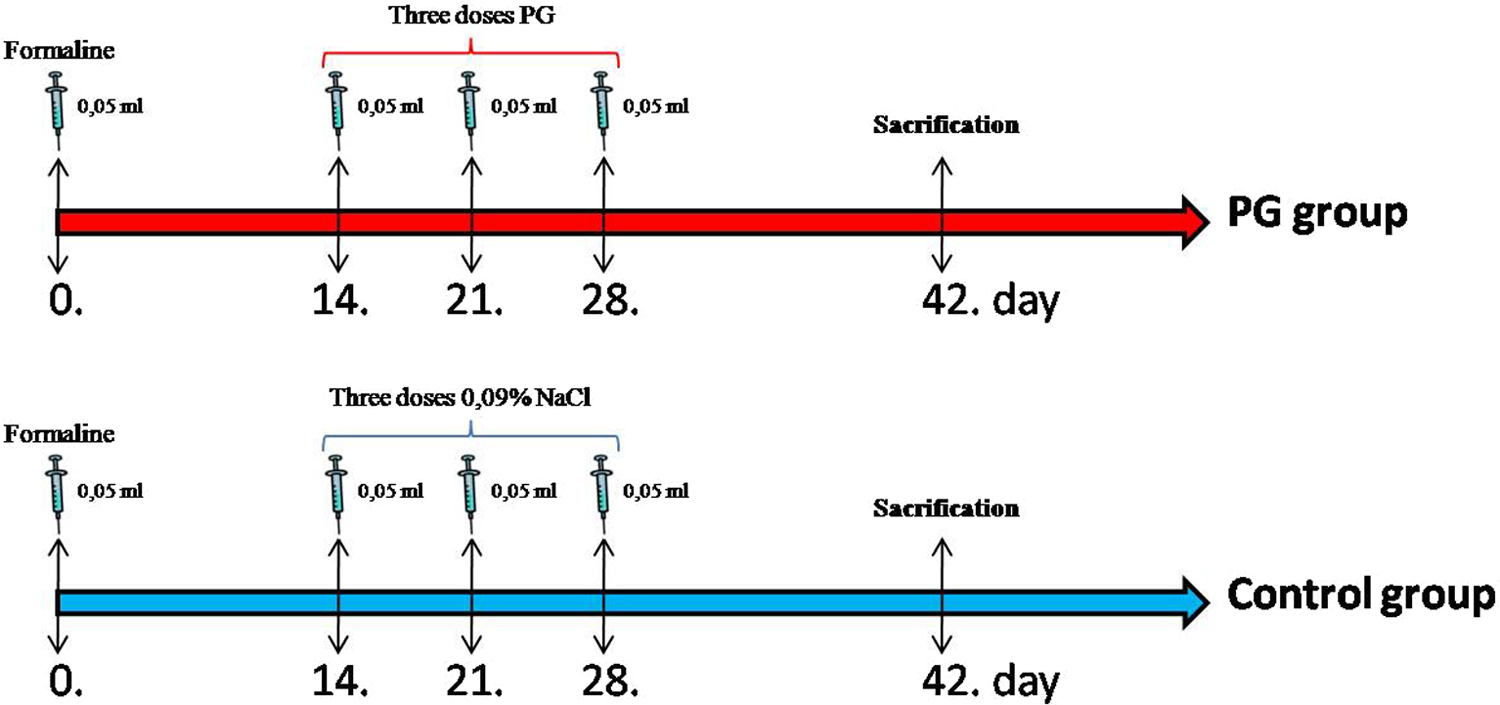
Protocols of administration. After 2 weeks of formaline injections, PG or saline was administered on the day as indicated by arrows. Two weeks after the final administration, the rats were sacrificed by ether asphyxiation.
Induction of OA
Osteoarthritis was induced by a single intra-articular injection of 200 μL of 1% formaline through the patellar ligament of the right knee joint with the leg flexed at an angle of 90°. 11 Osteoarthritis was formed after a 2-week period. The general health of the rats was monitored daily.
Preparation of PRP
The PRP was prepared from 5 male Wistar rats. Whole blood from the 5 rats was drawn through cardiac puncture into ethylene diamine tetraacetic acid-coated tubes. The blood was subjected to centrifugation for 10 minutes at 5600 rpm, and the supernatant was transferred to another tube. The supernatant was subjected to centrifugation for 10 minutes at 2400 rpm to yield platelet poor plasma (PPP) and PRP. The top layer, which consisted of the PPP, was aspirated and put into a new tube. The remaining layer was the PRP. 12
The PRP was separated into 3 parts; 1 unit was used for the first injection and the other 2 units were stored at −60°C. Injections were administered every 7 days; for the second and third treatments, the samples were thawed at room temperature (24-26°C) for 60 minutes just before application.
Histopathological Evaluation
The knee joint surface was separated into 8 equal areas (4 tibial areas and 4 femoral areas) for a histological analysis of the level of degeneration in the cartilage zones. The degeneration level was rated as follows: the absence of degeneration was scored as 0 points (Figure 2A), cartilage splinting in zone I (Figure 2B) was 1 point, cartilage splinting in intermediate zone II (Figure 2C) was 2 points, cartilage splitting involving zone III (Figure 2D) was 3 points, and the complete loss of the cartilage layer was 4 points. 13 The degeneration severity observed in each location in each group was summed to obtain the degeneration score. For the 8 locations, the degeneration was considered as mild, moderate, and severe when the score was 1 to 24 points, 25 to 58 points, and 49 to 72 points, respectively. 14
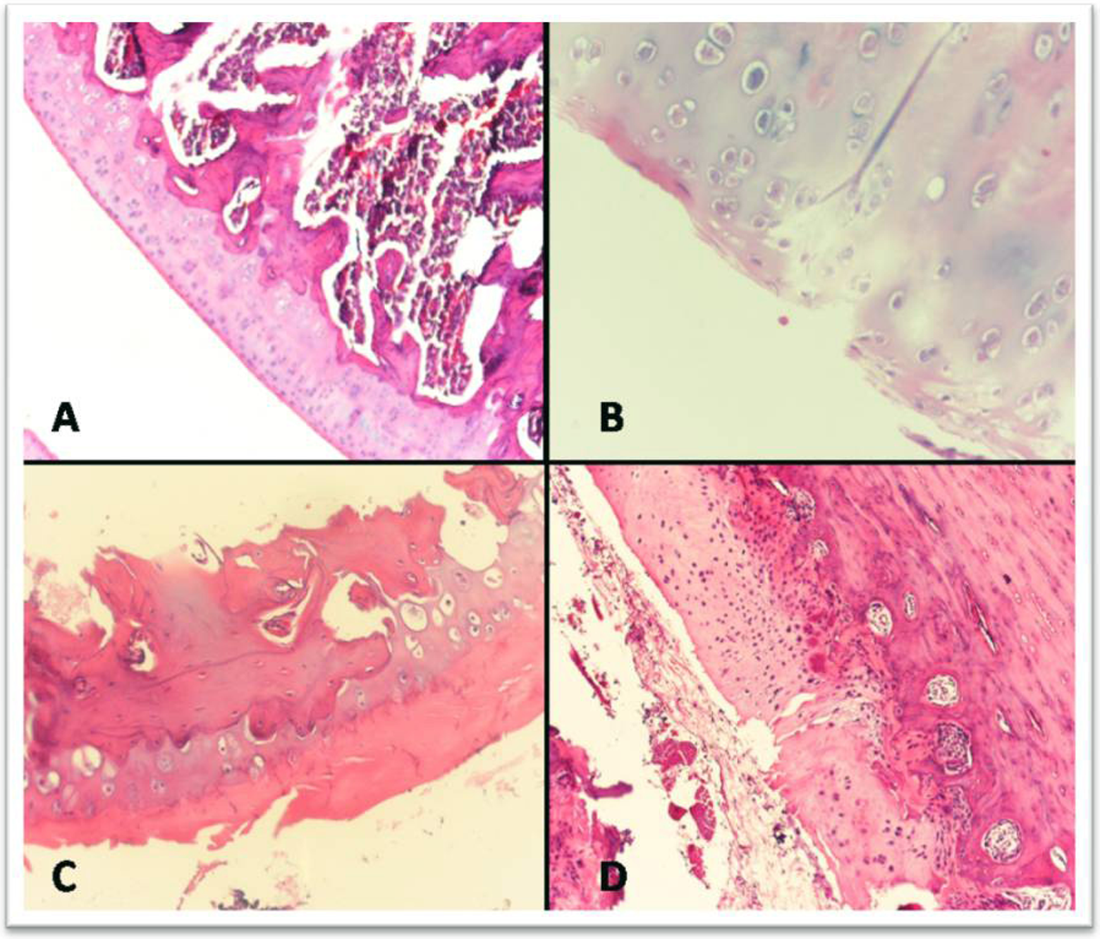
Degeneration level. A, Absence of degeneration; B, cartilage splinting in zone I; C, cartilage splinting in intermediate zone II; and D, cartilage splitting involving zone III.
Statistical Analysis
All data analyses were carried out using the SPSS (Statistical Package of Social Sciences) for Windows software program version 15.0, and the data were analyzed statistically using the Mann-Whitney U test. A P value of less than .05 was considered statistically significant. The degeneration scores are expressed as median (range) values.
Results
Platelet Gel Group
The absence of degeneration was observed in 3 rats, mild degeneration was observed in 1 rat, moderate degeneration was observed in 4 rats, and severe degeneration was observed in 2 rats.
Control Group
The absence of degeneration was observed in 2 rats, mild degeneration was observed in 2 rats, moderate degeneration was observed in 3 rats, and severe degeneration was observed in 3 rats.
We did not observe a complete loss of cartilage layer in either group.
The degeneration scores were found to be 28.5 (61) in the study group and 36 (58) in the control group. No statistically significant differences were found between the groups regarding cartilage healing (Z = −0.643; P > .05; Table 1).
Degeneration Scores and Degeneration Levels of Articular Cartilage in Rat Knee Joints.
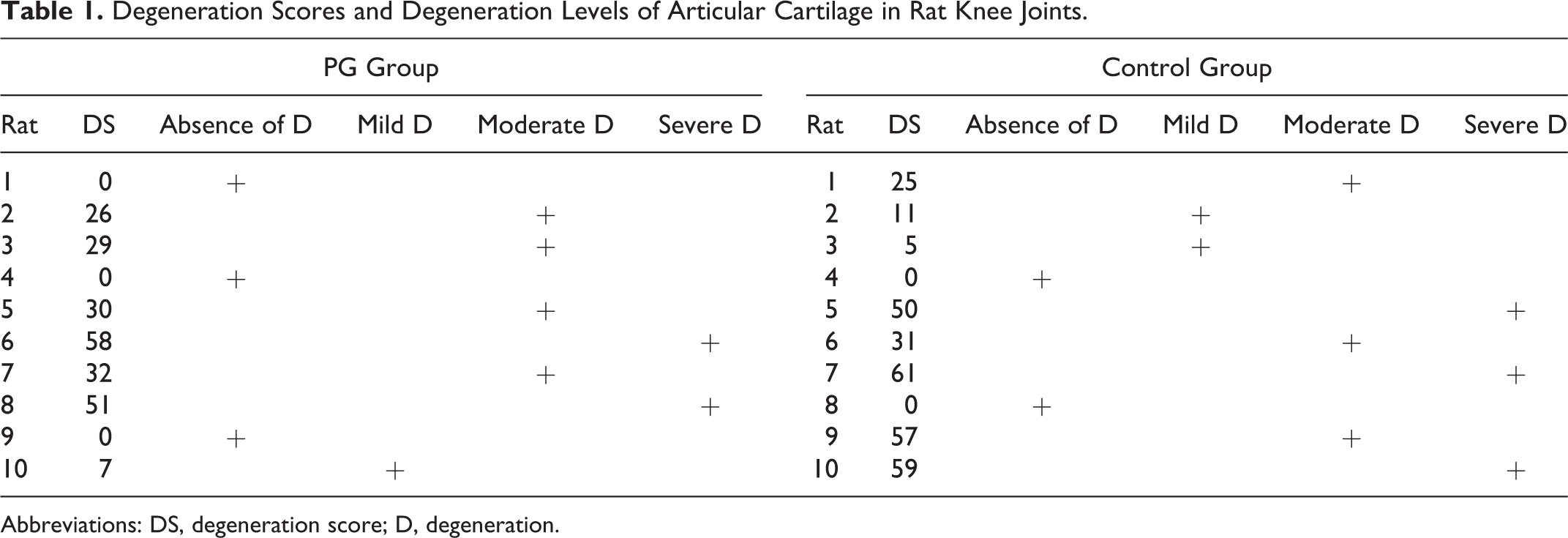
Abbreviations: DS, degeneration score; D, degeneration.
Discussion
Osteoarthritis is the most common form of arthritis that causes significant pain and functional disability. It is a degenerative joint disease that is typically described by the deformation and eventual loss of joint cartilage. 15 Osteoarthritis is the result of mechanical and biological events that destabilize the normal degradation and synthesis processes of articular cartilage chondrocytes, extracellular matrix, and subchondral bone. 16 Unfortunately, the treatment of chondral lesions is challenging due to an intrinsically low cure potential. Current OA treatments are usually targeted to provide a symptomatic relief of pain and may only have restricted effects on joint degeneration. 17
Platelet gel has been used over the past 10 years in an increasing number of fields, particularly in novel orthopedic treatments. 18,19 Platelet gel is formed through the combination of PRP, thrombin, and calcium chloride. Thrombocyte alpha granules are also known to be activated when PRP is combined with thrombin and calcium chloride. 20 We therefore used calcium and thrombin to activate PRP. Activated alpha granules release GFs. 21,22 These GFs include transforming growth factor β, platelet-derived growth factor, epidermal growth factor, insulin-like growth factor, and vascular endothelial growth factor. 23 –25 These factors stimulate mitosis, chemotaxis, angiogenesis, and bone growth. 23
We reviewed the scientific literature in English to find and evaluate studies in which patients with OA were treated with intra-articular PRP injections. In a study carried out by Filardo et al in which 91 patients were evaluated in a previous 12-month follow-up study, 90 were still available for a 2-year follow-up (24 patients presented a bilateral lesion, in a total of 114 knees treated). The results showed a reduction in pain and an improvement in knee function and quality of life with short-term efficacy. 26 Also, Sampson et al treated 14 symptomatic patients with 3 autologous PRP intra-articular injections and were evaluated prospectively at enrollment. Their study demonstrated significant and almost linear improvements in Knee Injury and Osteoarthritis Outcome Scores, including pain and relief from symptoms 12 months after the treatment. 27 In a prospective study by Kon et al, patients with degenerative cartilage lesions in the knee joint were treated with intra-articular PRP injections. The 115 knee joints were clinically evaluated before and at the end of treatment and during the follow-up using appropriate questionnaires. They reported that intra-articular PRP injections reduced pain and improved knee function and quality of life in younger patients with a low degree of articular degeneration. 28 In these studies, joint pain and knee function were evaluated but histopathological assessments were not made. Published data regarding the histopathological assessment following intra-articular PG injection on OA in the English scientific literature is limited.
This work represents the experimental study to examine the effects of intra-articular PG injections on OA in joint. This study was aimed to provide a contribution to the literature. In our study, we researched the efficacy of PG on the healing of the articular cartilage in OA-induced rat knee joints. Through a comparative histopathological assessment of PG and control groups, we found no significant differences in cartilage repair.
Conclusions
In the present study, we were unable to find any beneficial or harmful effects of PG on joint cartilage healing in OA. This study examines the effects of PG on joint cartilage healing in OA and needs to be supported by further animal experiments using higher doses of PG.
Footnotes
Declaration of Conflicting Interests
The authors declared no potential conflicts of interest with respect to the research, authorship, and/or publication of this article.
Funding
The authors received no financial support for the research, authorship, and/or publication of this article.