
Abstract
The aim of this study was to assess the values of mean platelet volume (MPV) in regular smokers and the effect of smoking cessation on MPV. The study group consisted of 116 regular smokers (57 females and 59 males; mean age 46.3 ± 12.7 years) and the control group was composed of 90 healthy volunteers (49 females and 41 males; mean age 47.7 ± 8.3 years). Platelet indices were assessed in regular smokers and control participants. Platelet indices were measured at 3 months after smoking cessation in these 101 participants. The MPV values were significantly higher in smokers than those of controls (8.8 ± 0.9 vs 8.0±0.8 fL, respectively; P < .001). The MPV values decreased significantly at 3 months when compared with the baseline values (8.9 ± 1.0 vs 7.9 ± 0.7 fL, respectively; P < .001). We have found that serum MPV values were significantly higher in regular smokers than in controls. Serum MPV values decreased significantly at 3 months after smoking cessation.
Introduction
Chronic smoking is a major modifiable risk factor for the cardiovascular diseases. 1 it not only accelerates atherosclerosis but also causes endothelial dysfunction and hemostatic disorders.2,3 Previous studies have demonstrated that chronic smoking cause platelet activation.3–9 Mean platelet volume (MPV) is a potential marker of platelet reactivity.10,11 In comparison to smaller ones, larger platelets have more granules, aggregate more rapidly with collagen, have higher thromboxane A2 level, and express more glycoprotein Ib and IIb/IIIa receptors.12–14 Some studies have reported that smoking has no effect on MPV.6,15 On the other hand, Kario et al have found increased MPV values in smokers. 16
Both the adverse effects of cigarette smoking and the benefits of smoking cessation on cardiovascular health occur rapidly. The risk of myocardial infarction falls by half within a year of cessation. 17 Although the risk of coronary artery disease after smoking cessation drops by approximately 50% one year after cessation, it approaches that of a person who has never smoked within 3 to 4 years. 18 It has been shown that smoking cessation improves platelet function.2,19 To the best of our knowledge, there is no data about the effect of smoking cessation on MPV in smokers as a separate study. In this study, we aimed to assess MPV values in regular smokers and the effect of smoking cessation on MPV values.
Patients and Methods
The study group consisted of 116 regular smokers (57 females and 59 males; mean age 46.3 ± 12.7 years). Smokers were selected from persons who admitted to smoking cessation outpatient clinic. An age-, gender- and body mass index–matched control group was composed of 90 healthy volunteers (49 females and 41 males; mean age 47.7 ± 8.3 years). In the present study, participants who smoke at least 8 cigarettes per day at least 3 years were considered as smokers. Nonsmokers (control group) were defined as those who had never smoked and were not exposed to environmental tobacco smoke. Biochemical measurements and platelet indices were assessed in116 regular smokers and 90 control participants. From 116 regular smokers, 101 participants successfully discontinued smoking. Platelet indices including MPV, platelet count and platelet distribution width (PDW), and white blood cell (WBC) count were measured at 3 months after smoking cessation in these 101 participants, and these values were compared with baseline platelet indices values of these 101 participants. Blood samples were drawn, at least 120 minutes after the last cigarette at admission to avoid the acute effects of smoking on platelet function.
Hypertension was considered to be present if the systolic pressure was >140 mm Hg and/or diastolic pressure was >90 mm Hg or if the individual was taking antihypertensive medications. Diabetes mellitus was defined as a fasting blood glucose level >126 mg/dL or current use of a diet or medication to lower blood glucose. Exclusion criteria were history of heart disease, hypertension, diabetes mellitus, obesity, history of renal or liver disease, malignancy, current use of anticoagulant or antiplatelet drugs, or psychiatric disorders. The study was approved by the institutional ethics committee, and all patients gave their informed consent.
Blood Sampling
Blood sampling was performed at admission and at 3 months after smoking cessation. Blood samples were drawn from the antecubital vein by careful venipuncture in a 21 G sterile syringe without stasis between 08.00 and 10.00
Statistical Analysis
Data were analyzed with the SPSS software version 10.0 for Windows. Continuous variables from the study groups were reported as mean ± standard deviation and categorical variables as percentages. To compare continuous variables, the Student t test or Mann-Whitney U test was used where appropriate. Categorical variables were compared with the chi-square test. Paired samples t test was used to compare platelet indices between before and after smoking cessation. The correlations between MPV and other clinical and laboratory parameters were performed with Pearson correlation analysis. Statistical significance was defined as P < .05.
Results
Clinical and laboratory findings of the study and control groups were summarized in Table 1. There were no statistically significant differences between the 2 groups with respect to age, gender, body mass index, systolic and diastolic blood pressures, and levels of glucose, creatinine, total cholesterol, triglyceride, low-density lipoprotein (LDL) cholesterol, high-density lipoprotein (HDL) cholesterol, hemoglobin, and PDW. The average number of cigarettes smoked per day was 18.9 ± 8.2 (8-40 cigarettes/d) for smokers. Average smoking period was 17.9 ± 8.1 years (3-35 years).
Comparison of the Clinical and Laboratory Characteristics of the Smokers and Controls.

Abbreviations: M/F, male to female; BMI, body mass index; SBP, systolic blood pressure; DBP, diastolic blood pressure; LDL-cholesterol, low-density lipoprotein cholesterol; HDL-cholesterol, high-density lipoprotein cholesterol; WBC, white blood cell; MPV, mean platelet volume; PDW, platelet distribution width.
a P value is for comparison between control and study population.
The MPV values were significantly higher in smokers than those of controls (8.8 ± 0.9 vs 8.0 ± 0.8 fL, respectively; P < .001). Platelet count was significantly lower in smokers than that of controls (253.2 ± 60.3 vs 305.9 ± 76.6 ×10 9 /L [normal range: 156-373 × 109/L], respectively; P < .001). The WBC count was significantly higher in smokers than that of controls (7.7 ± 1.9 vs 7.2 ± 1.2 ×10⁹/L, respectively; P = .02). The correlation analysis in 116 smokers indicated that MPV was positively correlated with age (P < .001, r = .42), cigarettes smoked per day (P < .001, r = .81), smoking period (P < .001, r = .68), and negatively correlated with platelet count (P < .001, r = −.35).
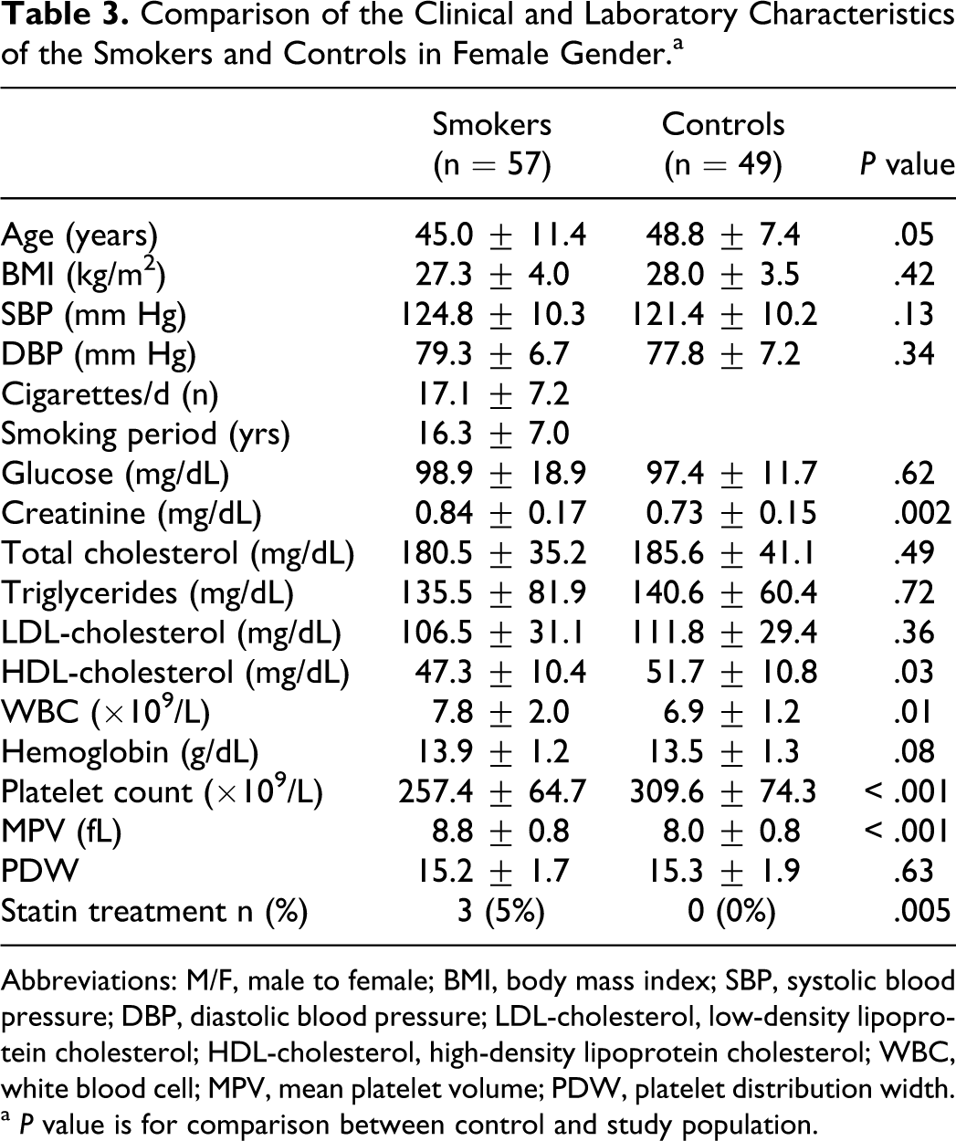
We also compared the smokers and the control groups separately according to the gender. Clinical and laboratory findings of the study and control groups in male and female gender are summarized in Tables 2 3. The MPV values were significantly higher in smokers than those of controls (8.9 ± 1.1 vs 8.0 ± 0.8 fL, respectively; P < .001) and the platelet count was significantly lower in smokers than that of controls (249.1 ± 55.9 vs 301.5 ± 80.0 × 10⁹/L, respectively; P < .001) of male gender. The MPV values were significantly higher in smokers than those of controls (8.8 ± 0.8 vs. 8.0±0.8 fL respectively; P < 0.001) and the platelet count was significantly lower in smokers than that of controls (257.4 ± 64.7 vs 309.6 ± 74.3 × 10⁹/L, respectively; P < .001) of female gender.
Comparison of the Clinical and Laboratory Characteristics of the Smokers and Controls in Male Gender.
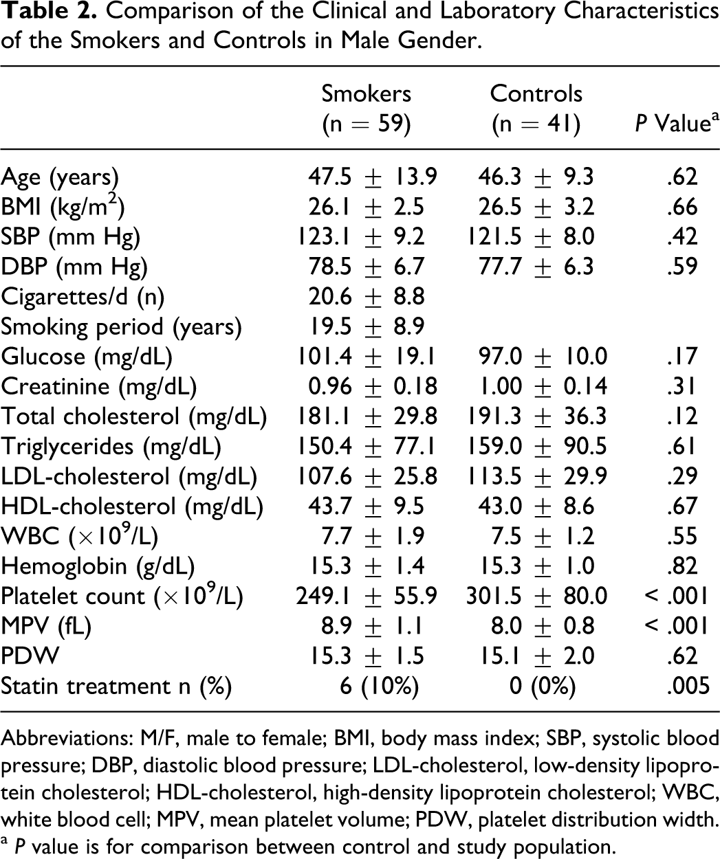
Abbreviations: M/F, male to female; BMI, body mass index; SBP, systolic blood pressure; DBP, diastolic blood pressure; LDL-cholesterol, low-density lipoprotein cholesterol; HDL-cholesterol, high-density lipoprotein cholesterol; WBC, white blood cell; MPV, mean platelet volume; PDW, platelet distribution width.
a P value is for comparison between control and study population.
Comparison of the Clinical and Laboratory Characteristics of the Smokers and Controls in Female Gender.a
Abbreviations: M/F, male to female; BMI, body mass index; SBP, systolic blood pressure; DBP, diastolic blood pressure; LDL-cholesterol, low-density lipoprotein cholesterol; HDL-cholesterol, high-density lipoprotein cholesterol; WBC, white blood cell; MPV, mean platelet volume; PDW, platelet distribution width.
aP value is for comparison between control and study population.
The values of platelet indices before and after 3 months of smoking cessation in participants who succeeded to quit smoking are given in Table 4. The MPV decreased significantly at 3 months when compared with the baseline values (8.9 ± 1.0 vs 7.9 ± 0.7 fL, respectively; P < .001). Platelet count increased significantly at 3 month when compared with the baseline values (252.9 ± 60.0 vs 302.1 ± 79.5 × 10⁹/L, respectively; P < .001). However, WBC and PDW values did not change at 3 months of smoking cessation.
Platelet Indices Before and After 3 Months of Smoking Cessation.
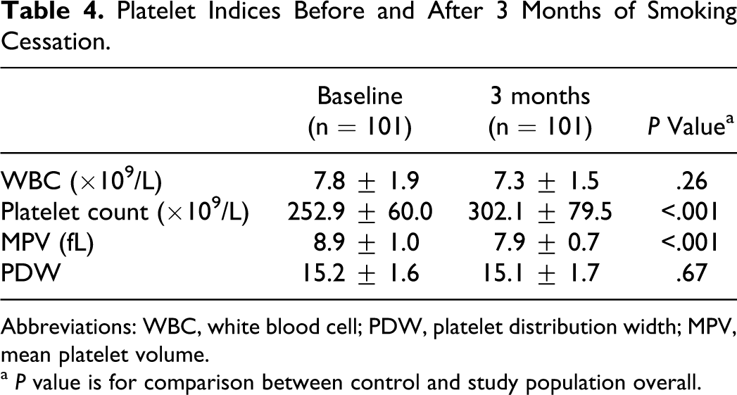
Abbreviations: WBC, white blood cell; PDW, platelet distribution width; MPV, mean platelet volume.
a P value is for comparison between control and study population overall.
Discussion
In this study, we have found that MPV values were significantly higher in regular smokers than those of controls. The MPV was positively correlated with age, cigarettes smoked per day, and smoking period and negatively correlated with platelet count. The MPV values decreased significantly at 3 months after smoking cessation.
The previous reports have shown that chronic smoking causes platelet activation.3–9 It has also been shown that smoking cessation improves platelet function.2,19 The MPV is a simple and easy method of assessing platelet function; and to the best of our knowledge, there is 1 data as a separate study, about the effect of smoking cessation on MPV values in smokers. Kario et al studied the effects of cigarette smoking and atherosclerotic risk factors on MPV. 16 They found that MPV was increased in elderly patients with atherosclerotic risk factors. They also reported a statistically significant decrease in MPV values in 8 smoking participants in the atherosclerotic group who successfully discontinued smoking. They suggested that smoking may increase platelet consumption in atherosclerotic vessels and that subsequently megakaryocytes are activated to produce more active larger platelets.
Butkiewicz et al studied the effect of smoking on thrombocytopoiesis, platelet activation, and some morphological parameters including MPV in 60 healthy women (mean age 30 ± 10 years) and 65 healthy men (mean age 33 ± 9 years). 6 They found that, in neither of the sexes smoking had an effect on MPV. Recently, Arslan et al investigated the effects of smoking on MPV in young healthy male population (smokers 56 and nonsmokers 46, medium age: 22). 15 They found no significant difference in MPV between the smoking and nonsmoking young aged healthy male participants.
Our sample volume was greater than previous studies, and our study participants were composed of middle-aged participants. As a difference from previous studies,6,15 we have found increased MPV in regular smokers in middle-aged participants. Our results confirm the results of the study performed by Kario et al in which the MPV was assessed in elderly patients with atherosclerotic risk factors.
In these studies, only Kario et al reported the effect of smoking cessation on MPV in only 8 participants who successfully discontinued smoking. They reported a statistically significant decrease in MPV values after smoking cessation in 8 smoking participants. We also found a significant decrease in MPV values after 3 months of smoking cessation in 101 participants who successfully discontinued smoking. Our results also confirm the results of the study performed by Kario et al from this aspect.
Smoking cessation is associated with a reduction in the risk of cardiovascular disease. 18 The risk of myocardial infarction falls by half within a year of cessation. Smoking cessation improves platelet function and this obviously has an important role in reduction of the cardiac events.2,19 There are really few studies on the effect of smoking cessation on platelet function.
Activated platelets release a wide variety of substances, 2 of which, platelet factor 4 and β-thromboglobulin, are commonly used as markers of platelet activation in vivo. Caponnetto et al studied changes in blood levels of surrogate markers of endothelial cell activation/injury (von Willebrand factor [vWF] and soluble thrombomodulin), clotting activation (
Morita et al investigated whether and how soon smoking cessation ameliorates impaired platelet-derived nitric oxide bioactivity and augmented platelet aggregability by improving the imbalance of the intraplatelet redox state. 19 It is well known that platelet aggregability is more augmented in chronic smokers than in nonsmokers. 4 They reported that only 2 weeks of smoking cessation can ameliorate the enhanced platelet aggregability and intraplatelet redox imbalance in long-term smokers, possibly by decreasing oxidative stress.
In this study, we have also found that the regular smokers had lower platelet count than that of controls. The MPV has been shown to inversely correlate with the total platelet count, which could even suggest the consumption of small platelets and a compensatory production of larger reticulated platelets. 20
We do not know the exact cause of the decrease in MPV values after smoking cessation. Platelet activation is one of the major factors by which tobacco smoke mediates the pathogenesis of cardiovascular diseases, which may be related to endothelial dysfunction and/or direct effects of oxidant chemicals. 21 Smoking-related endothelial dysfunction results in reduced release of nitric oxide that normally inhibits platelet activation. 22
As a result, restoration of endothelial function and/or decrease in oxidative stress may possibly improve platelet function within months after smoking cessation.
The present study has some study limitations. Smoking status was monitored from self-reports given by participants. Abstinence from smoking was not reviewed objectively by measuring the concentrations of carbon monoxide in expired breath.
In conclusion, we have found that the MPV values were significantly higher in smokers than those of controls. The MPV was positively correlated with age, cigarettes smoked per day, and smoking period and negatively correlated with platelet count. The MPV values decreased significantly at 3 months after smoking cessation. Our results may contribute to the understanding of the pathophysiological link of smoking cessation to beneficial cardiovascular effects. Our results may strengthen the motivation for smokers to quit smoking.
Footnotes
Declaration of Conflicting Interests
The author(s) declared no potential conflicts of interest with respect to the research, authorship, and/or publication of this article.
Funding
The author(s) received no financial support for the research, authorship, and/or publication of this article.