
Abstract
Apolipoprotein A-1 (Apo A-I) Milano, a naturally occurring Arg173 to Cys mutant of Apo A-1, has been shown to reduce atherosclerosis in animal models and in a small phase 2 human trial. We have shown the superior atheroprotective effects of Apo A-I Milano (Apo A-IM) gene compared to wild-type Apo A-I gene using transplantation of retrovirally transduced bone marrow in Apo A-I/Apo E null mice. In this study, we compared the effect of dietary lipid lowering versus lipid lowering plus Apo A-IM gene transfer using recombinant adeno-associated virus (rAAV) 8 as vectors on atherosclerosis regression in Apo A-I/Apo E null mice. All mice were fed a high-cholesterol diet from age of 6 weeks until week 20, and at 20 weeks, 10 mice were euthanized to determine the extent of atherosclerosis. After 20 weeks, an additional 20 mice were placed on either a low-cholesterol diet plus empty rAAV (n = 10) to serve as controls or low-cholesterol diet plus 1 single intravenous injection of 1.2 × 10¹² vector genomes of adeno-associated virus (AAV) 8 vectors expressing Apo A-IM (n = 10). At the 40 week time point, intravenous AAV8 Apo A-IM recipients showed a significant regression of atherosclerosis in the whole aorta (P < .01), aortic sinuses (P < .05), and brachiocephalic arteries (P < .05) compared to 20-week-old mice, whereas low-cholesterol diet plus empty vector control group showed no significant regression in lesion size. Immunostaining showed that compared to the 20-week-old mice, there was a significantly reduced macrophage content in the brachiocephalic (P < .05) and aortic sinus plaques (P < .05) of AAV8 Apo A-IM recipients. These data show that although dietary-mediated cholesterol lowering halts progression of atherosclerosis, it does not induce regression, whereas combination of low-cholesterol diet and AAV8 mediated Apo A-I Milano gene therapy induces rapid and significant regression of atherosclerosis in mice. These data provide support for the potential feasibility of this approach for atherosclerosis regression.
Introduction
Apolipoprotein A-I (Apo A-I) is the major structural protein component of high-density lipoprotein (HDL), and apolipoprotein A-I Milano (Apo A-IM) is a rare naturally occurring Arg173 to Cys173 mutant of apo A-I carried by a small number of inhabitants of Limone sul Garda. 1,2 This mutation appears to confer greater resistance to atherosclerosis in the carriers despite being associated with markedly lower HDL and Apo A-I levels. 2 –4 We have previously shown superior atheroprotective effects of Apo A-IM gene compared to the wild-type Apo A-I gene in Apo A-I/Apo E null mice using transplantation of gene-transduced bone marrow. 5
We have also recently demonstrated superior atheroprotective effects of recombinant adeno-associated virus 8 (rAAV8)-mediated gene transfer compared to rAAV2-mediated gene transfer. 6 The precise reasons for these superior atheroprotective effects are unclear but may include greater cholesterol efflux-promoting capacity and/or antioxidant/anti-inflammatory actions. 1,7 Some laboratories, using different models, different vectors or route of administration, and short duration of treatment, have shown comparable atheroprotective effects of Apo A-IM and wild-type Apo A-I. 8,9
A series of experimental studies from our laboratory and later from other laboratories have established rapid atheroprotective effects of intravenous infusion of recombinant Apo A-IM in animal models, and these results were also supported by a small phase 2 human trial. 10 –15 These findings suggest that therapy based on Apo A-IM could have potential as an effective antiatherogenic strategy. 16 Although changes in plaque composition and reduced progression of lesions with Apo A-IM gene transfer have been demonstrated, the effects of such therapy on regression of preexisting lesions especially in the context of lipid lowering have not been reported. 6,17 In this study, we evaluated the effects of lowering dietary cholesterol alone or cholesterol lowering plus rAAV8-mediated Apo A-IM gene transfer on preexisting atherosclerosis in Apo A-I/Apo E null mice. We found that adeno-associated virus (AAV) 8-mediated Apo A-IM gene therapy combined with dietary cholesterol lowering induces significant regression of atherosclerosis compared to lowering cholesterol alone over a 20-week period.
Materials and Methods
Production of rAAV
The AAV8 vectors encoding Apo A-IM were constructed based on vectors previously constructed and utilized in our laboratory for the purpose of Apo A-IM expression. 17 Briefly, the AAV packaging plasmid p5E18-VD287 (AAV rep2 and cap8) was mixed with adenovirus helper plasmid XX6-80, and plasmid encoding Apo A-IM to be used for transfection of NautCell packaging cell line (Microbix Biosystems Inc., Canada). The expression of human Apo A-IM gene is driven by a cytomegalovirus immediate-early promoter/enhancer that includes the enhanced green fluorescent protein marker gene. The empty plasmid for AAV8 was used as a vector control. Plasmids were transfected into NautCells using Effectene Transfection Reagent (Qiagen, Valencia, CA). The transfected cells were harvested, the cell extracts were prepared, and the rAAV were purified by discontinuous iodixanol density gradient centrifugation. The viral preparations were concentrated and desalted by centrifugation through the Amicon ultra-15 centrifugal filter devices (100,000 nominal molecular weight limit, Millipore, Billerica, MA).
The viral genome titers were determined by dot blot hybridization using RNA Detector Northern Blotting Kit (KPL Inc., Gaithersburg, MD) according to the manufacturer’s instructions. The rAAV2/8 titer used for this experiment was 4 × 10¹² genome copies/mL for empty vector and Apo A-IM–containing vector. Viral transducing units (Tu) were determined by transduction of the 293 cells in the presence of rAAV vectors followed by fluorescence-activated cell sorting analysis.
Animal Procedures and Study Design
Apolipoprotein A-I/Apo E double knockout (KO) mice of either gender were used for this study, of which half were males and half were females. The animals were placed on a high-fat diet (HFD) containing 0.15% cholesterol and providing 42% calories as fat (TD.88137; Harlan, Tekklad Laboratories, Madison, WI) from 6 to 20 weeks of age. After reaching 20 weeks of age, mice were randomly divided into 3 groups (N = 10/per group): group 1 was euthanized at 20 weeks of age, and groups 2 and 3 were placed on normal chow and received through the tail vein an intravenous injection of 1.2 × 10¹² genome copies in 300 µL phosphate-buffered saline of either the empty rAAV8 vector (group 2) or rAAV8–Apo A-IM (group 3). The mice were euthanized 20 weeks after injection at 40 weeks of age. Blood and tissues were harvested. Blood samples were periodically collected from the retro-orbital plexus at indicated time points (before and after injection). The use of experimental animals was in accordance with the guidelines of the Cedars-Sinai Medical Center (CSHS) Institutional Animal Care and Use Committee.
Biodistribution of Human Apo A-IM
The plasma level of human Apo A-IM was measured by enzyme-linked immunosorbent assay (ELISA) using antihuman Apo A-I monoclonal antibody (Calbiochem, San Diego, CA) as described previously. 17 For detecting human Apo A-IM gene expression in organs, total RNA was extracted from different tissues of mice in both groups 2 and 3 (N = 3/per group) with TRIzol (Invitrogen, Carlsbad, CA) and then analyzed by quantitative polymerase chain reaction (qPCR; with human Apo A-I–specific primer sequences [223 base pair]: sense, 5′-AAG GAC CTG GCC ACT GTG TA-3′; antisense, 5′-TCT CCT CCT GCC ACT TCT TC-3′. Glyceraldehyde 3-phosphate dehydrogenase was used as an internal control.
Assessment of Atherosclerosis
Atherosclerosis was assessed in the whole aorta, brachiocephalic artery, and the aortic sinuses. The extent of the atherosclerotic lesions in the total aorta was quantified after Oil red O staining, as described previously. 6,12 Briefly, the vascular tree was dissected intact, and then the luminal surface was exposed. After tissue fixation, the adventitial tissue was removed and aorta was stained with Oil red O for lipids. The aortas were placed on a glass slide, and images were captured using a Spot digital camera (Scientific Instrument Company, Sunnyvale, California). The extent of the atherosclerotic lesions was quantified using Image-Pro Plus (version 4; Media Cybernetics, Silver Spring, Maryland). Lesions were reported as percentage of the total aortic area consisting of thoracic aorta and abdominal aorta ending at the iliac bifurcation (N = 9-10/per group).
For brachiocephalic artery sections, the aortic arch was fixed in 4% paraformaldehyde overnight, then embedded in optimal cutting temperature (OCT), and 5-µm serial sections were used for staining and lesion assessment. For aortic sinus lesion assessment, 5-µm serial sections of OCT-embedded aortic sinus were stained with Oil red O and hematoxylin. The quantitative analysis of lesion area and lipid content of lesion for brachiocephalic artery and aortic sinus was measured by Image-Pro Plus using manual tracing of the lesion, which was identified by a combination of lipid staining and histological morphology (mean of 6-8 sections per mouse, 6-7 mice per group).
To determine macrophage content, serial sections from aortic arch and sinus were stained with antimurine monocyte/macrophage (MOMA-2; Serotec, UK) antibody at 1:100 dilutions and counterstained with hematoxylin, and the positive areas in brachiocephalic artery and aortic sinus lesion reflecting macrophage immunoreactivity were analyzed by Image-Pro Plus.
Plasma Cholesterol Measurement
Fasting blood samples were collected from all groups of mice by retro-orbital venous plexus puncture using heparinized capillary tubes under isoflurane anesthesia (Medeva Pharmaceuticals, Rochester, New York). Total cholesterol was measured using a cholesterol reagent enzymatic kit (Cholesterol E; Wako Pure Chemical Industries, Ltd, Richmond, Virginia).
Phenotype of Circulating Monocyte/Macrophages
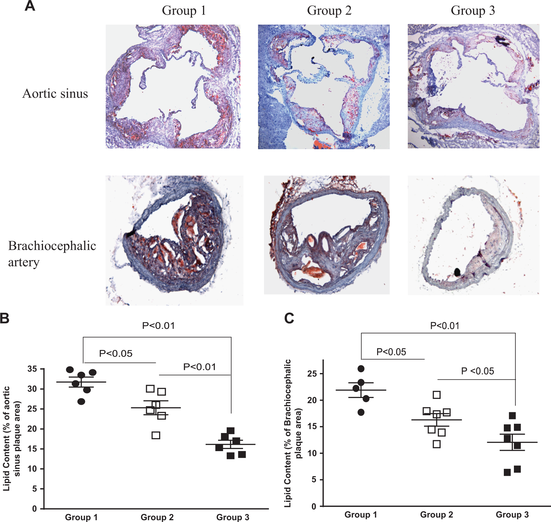
Peripheral blood from groups 2 and 3 was used for monocyte/macrophage M1/M2 marker analysis. Briefly, RNA from blood (after red blood cell lysing) was extracted by TRIzol, and qPCR was performed using primers listed in Table 1.
List of Primers.
Abbreviations: Arg-1 indicates arginase 1; F, forward; iNOS, inducible nitric oxide synthase; IL-10, interleukin 10; R, reverse; TNF-α, tumor necrosis factor α.
Statistical Analysis
Values are presented as mean ± standard error of the mean (SEM). Statistical significance was evaluated with the unpaired Student t test for comparisons between 2 mean values. Comparisons between 3 groups were performed with 1-way analysis of variance followed by a Tukey test for pairwise comparisons. A P value <.05 was considerate significant.
Results
Circulating Levels of Transgene
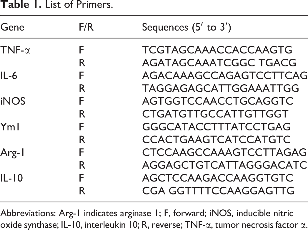
To investigate the efficacy of Apo A-IM gene therapy in atherosclerosis, we first determined the tissue expression of transgene, followed by the biological activity of Apo A-IM in regression of atherosclerosis. To determine the level of transgene expression, plasma samples were collected from each group of mice at various times post-injection, and the level of human Apo A-IM was measured by ELISA (Figure 1). Apolipoprotein A-I Milano was detected in the plasma at all time points in group 3 mice following rAAV8 Apo A-IM injection and remained detectable at the time of euthanasia at 40 weeks of age. As expected, there was no detectable Apo A-IM in the plasma from group 1 (data not shown) and group 2 mice.
Kinetics of Apo A-IM expression after gene transfer. Plasma levels of Apo A-IM at various time points following IV injection of rAAV8 vector control (group 2) or rAAV8–Apo A-IM injection (group 3; data = mean ± SEM, N = 12) are shown. Apolipoprotein A-I Milano was detectable at all time points after injection in group 3 mice but not in group 2 mice. Apo A-IM indicates apolipoprotein A-I Milano; rAAV8, recombinant adeno-associated virus 8; SEM, standard error of the mean.
Organ Distribution of Transgene Expression
To determine the presence of Apo A-IM gene in various organs, we collected the tissues, and the level of transgene expression was quantified by qPCR using primers specific to human Apo A-IM. The Apo A-IM transgene expression was detected in the brain, heart, liver, lung, spleen, and kidney. The level of gene expression in the heart tissue was found to be higher compared with other organs in all 3 mice groups (Figure 2). As expected, no Apo A-IM transgene was detected in group 2 mice (data not shown).

Tissue distribution of Apo A-IM in the double KO mice injected with Apo A-IM. The mouse groups were injected with AAV8 encoding Apo A-IM or empty control vectors, and the mice were euthanized at 24 weeks. The indicated organs were collected, and total RNAs were analyzed by qPCR. The qPCR data are normalized against GAPDH. Real-time PCR (qPCR) shows Apo A-IM transgene expression in the brain, heart, liver, lung, spleen, and kidney, with heart tissue showing higher expression compared with other organs. Data represent mean ± SEM, N = 3 mice per group. AAV8 indicates adeno-associated virus 8; Apo A-IM, apolipoprotein A-I Milano; GAPDH, glyceraldehyde 3-phosphate dehydrogenase; KO, knockout; PCR, polymerase chain reaction; qPCR, quantitative polymerase chain reaction; SEM, standard error of the mean.
Effect on Atherosclerosis
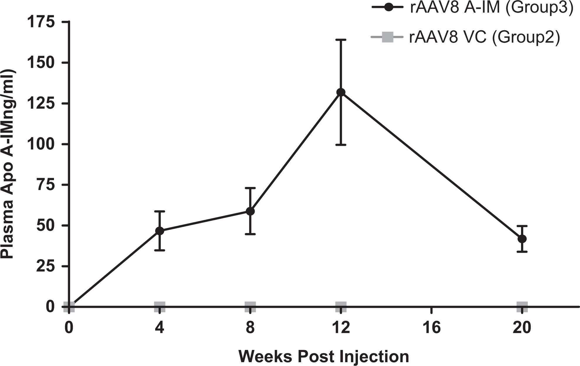
Each group was comprised male and female mice, of which half were males and half were females. We were unable to detect a significant difference in the degree to which each gender developed atherosclerosis. To determine the biological efficacy of Apo A-IM gene therapy, we evaluated atherosclerosis in the mice that were on HFD for 14 weeks. One group of mice was killed to evaluate the lesion extent at baseline (group 1). One remaining group received vector control (group 2), and the other group was injected with the AAV8-expressing Apo A-IM (group 3). The injected groups were fed a normal chow diet for additional 20 weeks, at which time, they were killed and the lesions were evaluated by staining the aortas with Oil red O. Quantitative analysis showed that the extent of atherosclerosis in group 2 mice was similar to that of group 1 mice, indicating stabilization of atherosclerosis progression with a switch to normal chow diet without evidence of regression. However, in group 3 mice, the aortic atherosclerosis was reduced by 41% compared to group 1 (7.5% ± 0.66% vs 12.75% ± 1.55%; P < .01) and by 45% compared to group 2 (7.5% ± 0.66% vs 13.6% ± 1.56%; P < .01), indicating regression of atherosclerosis (Figure 3A). Group 3 mice also showed a significant reduction in the size of brachiocephalic artery lesion compared to group 1 (10.81 ± 0.74 vs 15.74 ± 1.26; 31% reduction; P < .01) and group 2 (10.81 ± 0.74 vs 14.44 ± 0.82; 25% reduction; P < .05; Figure 4A). Group 3 mice also showed a reduction in aortic sinus plaque size compared to group 1 mice (40.41 ± 1.6 vs 61.13 ± 6.8; 34% reduction; P < .05) as well as group 2 mice (40.41 ± 1.6 vs 53.77 ± 4.0; 25% reduction; P < .05; Figure 4B). These findings are overall consistent with significant and robust regression of atherosclerosis induced by a combination of a cholesterol-lowering diet and AAV8-mediated Apo A-IM gene transfer compared to a cholesterol-lowering diet alone (Figure 3B).

Regression of aortic atherosclerotic plaque by Apo A-IM gene transfer. A, Extent of whole aortic atherosclerosis is shown (data = mean ± SEM, N = 8-10). Significant regression of atherosclerosis is observed in group 3 mice compared to group 1 and group 2 mice. B, Representative examples of Oil red O-stained whole aorta are shown from groups 1, 2, and 3. The regression of aortic atherosclerosis in group 3 compared to groups 1 and 2 is depicted. Apo A-IM indicates apolipoprotein A-I Milano; SEM, standard error of the mean.
Regression of atherosclerotic lesions in the brachiocephalic artery and aortic sinus after Apo A-IM gene transfer. A, Extent of brachiocephalic artery atherosclerosis is shown (data = mean ± SEM, N = 6/group). Significant regression of atherosclerosis is observed in group 3 mice compared to group 1 and group 2 mice. B, Size of aortic sinus plaque is shown (data = mean ± SEM, N = 6/group). Significant regression of atherosclerosis is observed in group 3 mice compared to group 1. Apo A-IM indicates apolipoprotein A-I Milano; SEM, standard error of the mean.
Effect of Apo A-IM Gene Therapy on Plaque Phenotype
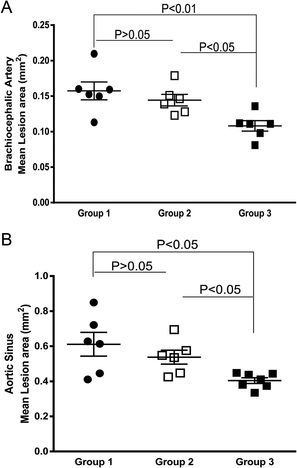
In addition to plaque burden, we investigated the effect of Apo A-IM gene therapy on the phenotype of plaques by measuring lipid content and macrophage content of the lesions (Figure 5A). The lipid content in aortic sinus (Figure 5B) and brachiocephalic artery (Figure 5C) plaques was measured. In group 1 (mice killed at the age of 20 weeks after being fed HFD for 14 weeks), lipid formed 30% or 20% of total aortic sinus and brachiocephalic artery areas, respectively (Figure 5B, C). The lipid content was significantly lower in group 3 compared to groups 1 and 2. Aortic sinus plaque lipid content was significantly lower in group 3 compared to group 2 (16.15% ± 1.0% vs 25.29% ± 1.7%; P < .01). Similarly, the lipid content in the brachiocephalic artery was lower in group 3 compared to group 2 (12.0 ± 1.5 vs 16.3 ± 1.1, P < .05).
Lipid content of the atherosclerotic lesions after Apo A-IM gene transfer. A, Representative examples of aortic sinus and brachiocephalic artery plaques are shown depicting evidence of regression of lesion size and lipid content in group 3 mice compared to group 1 and group 2 mice. B, The lipid content in aortic sinus plaques is shown (data = mean ± SEM, N = 6/group). Marked reduction in lipid content is observed in group 3 compared to group 1 and group 2 mice. A small but significant reduction in lipid content was also noted in group 2 compared to group 1 mice. C, The lipid content in brachiocephalic artery plaques is shown (data = mean ± SEM, N = 6-7/group). Marked reduction in lipid content is observed in group 3 compared to group 1. A significant reduction in lipid content was also noted in group 2 compared to group 1 mice. Apo A-IM indicates apolipoprotein A-I Milano; SEM, standard error of the mean.
In addition to lipid content, we studied the inflammatory phenotype of the plaques by investigating macrophage content of the lesions using an anti–MOMA-2 antibody (Figure 6A). In group 1, macrophage content of the aortic sinus lesions (Figure 6B) and brachiocephalic artery lesions (Figure 6C) represented 22% and 12% of the lesion areas, respectively. The macrophage immunoreactivity in aortic sinus (Figure 6B) and brachiocephalic artery (Figure 6C) plaques was significantly lower in group 3 mice compared to group 2 mice (aortic sinus lesion: 10.10 ± 1.1 vs 14.82 ± 1.7; a 32% reduction, P < .05; brachiocephalic artery lesion: 3.8% ± 0.7% vs 7.5% ± 1.5%; a 49% reduction, P < .05).

Analysis of macrophage content of the lesions after Apo A-IM gene transfer. A, Representative examples of aortic sinus and brachiocephalic artery plaques are shown depicting evidence of regression of lesion size as well as reduction in macrophage immunoreactivity in group 3 mice compared to group 1 and group 2 mice. The top panels show lower magnification of sinus lesion, and the lower panels show the higher magnification of the lesions. B, Analysis of macrophage immunoreactivity in aortic sinus plaques is shown (data = mean ± SEM; N = 6-7/group). Marked reduction in macrophage content is noted in group 3 compared to group 1, and group 2 also showed a significant reduction in macrophage content compared to group 1. C, Macrophage immunoreactivity in brachiocephalic artery plaques is shown (data = mean ± SEM; 6-7/group). Marked reduction in macrophage content is noted in group 3 compared to group 1, and group 3 also showed a significant reduction in macrophage content compared to group 2. Apo A-IM indicates apolipoprotein A-I Milano; SEM, standard error of the mean.
Plasma Cholesterol
To determine whether the expression of Apo A-IM by the AAV vectors affects plasma cholesterol level, we measured cholesterol level in plasma collected from mice at various time points throughout the course of the study (Figure 7). The mean plasma cholesterol level at 6 weeks of age before HFD feeding was 267.3 ± 19 mg/dL. After HFD feeding for 14 weeks, the average plasma cholesterol level increased to 1433 ± 135.8 mg/dL. Four weeks following a switch to normal chow diet, the mean plasma cholesterol level was down to 425.9.3 ± 36.32 mg/dL, which was further reduced to 383.2 ± 26.77 mg/dL at the time of euthanasia. There was no significant difference in the plasma cholesterol level between group 2 and group 3 mice.

Analysis of plasma cholesterol level after the Apo A-IM gene transfer. The plasma cholesterol levels at different time points (N = 9-10/group) are shown. No significant differences are shown between groups 2 and 3 at various time points. Apo A-IM indicates apolipoprotein A-I Milano.
Apolipoprotein A-I Milano Gene Therapy Influences Macrophage Polarization
We have previously shown that following intravenous injection of rAAV8 encoding Apo A-IM in Apo A-I/Apo E double KO mice, Apo A-IM can be detected in peripheral blood mononuclear cells (PBMCs). 6 To determine whether Apo A-IM expression in PBMC can influence macrophage polarization, PBMCs were collected at the time of euthanasia from group 2 and group 3 mice (N = 4/per group), and the expression of M1 and M2 macrophage markers (inducible nitric oxide synthase [iNOS], tumor necrosis factor [TNF] α, interleukin (IL) 6, Ym1, arginase 1, and IL-10) was determined by qPCR. Glyceraldehyde 3-phosphate dehydrogenase was used as an internal control. Quantitative polymerase chain reaction analysis revealed that there is a trend toward reduced expression of M1 marker (iNOS, TNFα, and IL-6) in group 2 mice compared to group 3 mice (Figure 8).
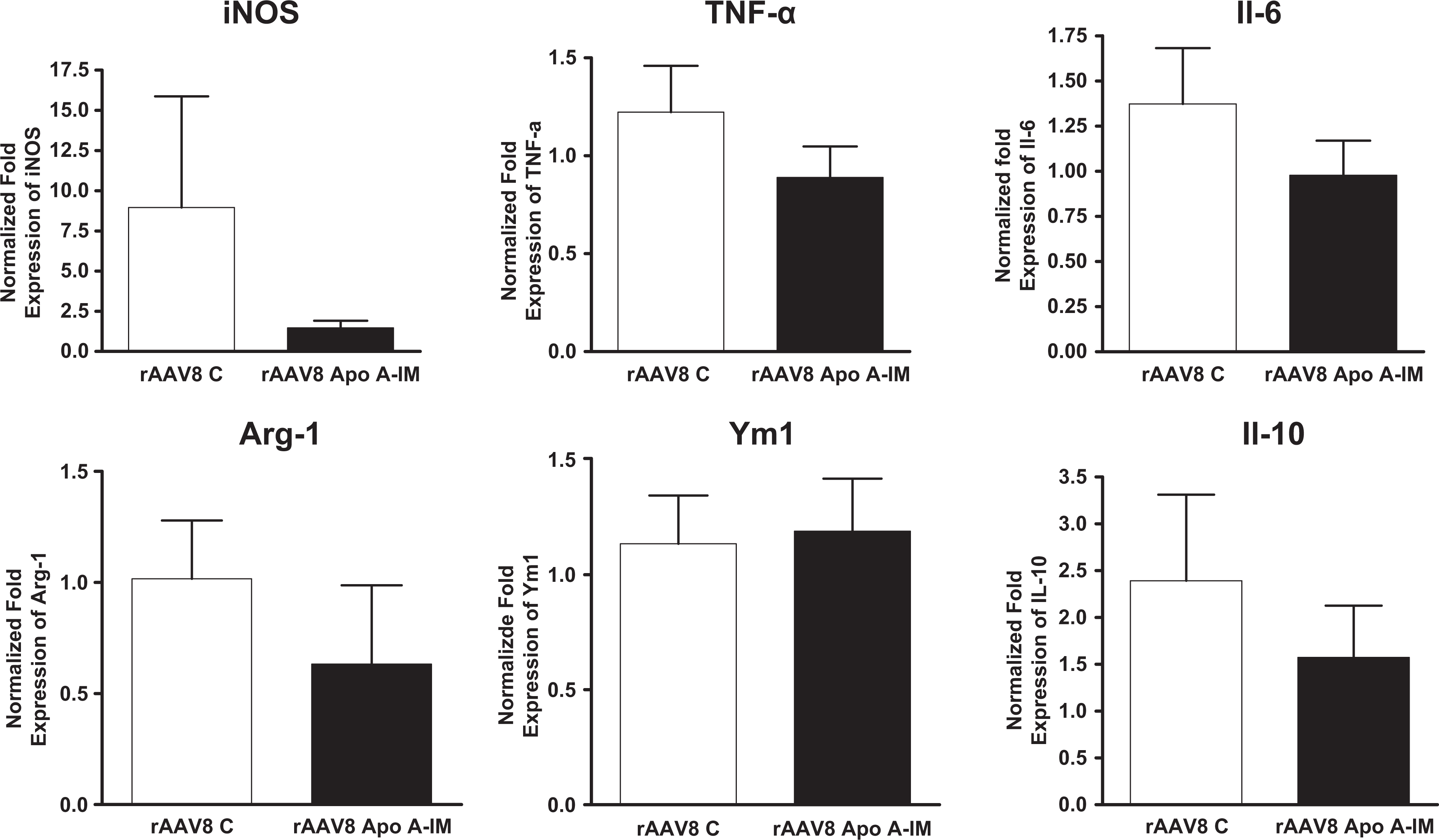
Analysis of macrophage polarization in response to Apo A-IM gene transfer. Quantitative polymerase chain reaction depicting messenger RNA expression for various macrophage markers (M1 and M2) in peripheral blood mononuclear cells (data = mean ± SEM, N = 4/group). Results show reduced expression of M1 markers (iNOS, TNF-α, and IL-6) in group 3 compared to group 2. Apo A-IM indicates apolipoprotein A-I Milano; iNOS, inducible nitric oxide synthase; IL-6, interleukin 6; SEM, standard error of the mean; TNF-α, tumor necrosis factor α.
Discussion
Apolipoprotein A-IM gene transfer is a potentially attractive approach for the amelioration, stabilization, and regression of atherosclerosis because of its pleiotropic biological actions. These include promotion of reverse cholesterol transport, anti-inflammatory and immune modulating effects, antioxidant actions, and proven efficacy in animal models. 16 Gene transfer is a potentially attractive approach to harness the atheroprotective effects of Apo A-I and Apo A-IM, avoiding the need for the large-scale manufacture of the recombinant protein required for intravenous injection.
In this study, we demonstrate that cholesterol-lowering diet alone stabilizes plaques by halting progression without inducing regression, whereas Apo A-IM gene transfer plus a cholesterol-lowering diet produces a significant regression of atherosclerosis in the whole aorta, aortic sinuses, and brachiocephalic arteries in a hyperlipidemic mouse model without significant differences in circulating cholesterol levels between the groups. We further showed that AAV8 encoding Apo A-IM gene also reduces plaque lipid content and plaque macrophage immunoreactivity consistent with a more stable plaque phenotype. We selected AAV vectors because they have several advantages as outlined earlier and are currently being evaluated in several gene therapy trials in humans. In this study, we used Apo A-I−/−Apo E−/− double KO recipient mice in order to eliminate the interplay between endogenous Apo A-I activity and exogenous Apo A-IM activity; therefore, the biological activity of Apo A-IM that we observed would be exclusively related to the activity of the delivered gene. In addition, using the double KO mice allowed us to investigate tissue distribution of the virally produced Apo A-IM without any interference from endogenous Apo A-I.
In summary, we have demonstrated that dietary lipid lowering fails to regress atherosclerosis, whereas the combination of dietary lipid lowering and AAV8-mediated Apo A-IM gene transfer using intravenous injection produces robust regression of atherosclerosis and favorably modifies plaque composition in Apo A-I/Apo E null mice. Further investigation of this strategy is warranted as a novel athero-regressing intervention.
Footnotes
Acknowledgments
The authors gratefully acknowledge the support of The Heart Foundation, The Eisner Foundation, the Spielberg Foundation, Corday Foundation, and the Skirball Foundation.
Author Contributions
L. Wang, B. G. Sharifi, and P. K. Shah contributed to conception, contributed to acquisition, drafted the manuscript, critically revised the manuscript, gave final approval, and agrees to be accountable for all aspects of work ensuring integrity and accuracy. F. Tian and M. Yang contributed to design and acquisition. A. Arias contributed to acquisition.
Declaration of Conflicting Interests
The author(s) declared no potential conflicts of interest with respect to the research, authorship, and/or publication of this article.
Funding
The author(s) disclosed receipt of the following financial support for the research, authorship, and/or publication of this article: The evolution of this approach was initially supported by program project grant from NHLBI Grant 1POHI60898.