
Abstract
Antibody-coated stents to capture circulating endothelial progenitor cells (EPCs) for re-endothelialization appear to be a novel therapeutic option for the treatment of atherosclerotic disease. Hydroxybutyl chitosan (HBC), a linear polysaccharide made from shrimps and other crustacean shells, is biocompatible, nontoxic, and hydrophilic, making it ideal for biomedical applications. In this study, HBC was explored for the immobilization of anti-CD133 antibodies. We demonstrated that CD133 antibodies mediated by HBC were successfully coated on cobalt–chromium alloy discs and metal stents. The coating was homogeneous and smooth as shown by electronic microscopy analysis. Balloon expansion of coated stents did not cause cracking or peeling. The HBC discs promoted CD133+ EPCs and human umbilical vein endothelial cell growth in vitro. The CD133 antibody-coated but not bare discs bound CD133+ EPCs in vitro. Implantation of CD133 antibody-coated stents significantly inhibited intimal hyperplasia and reduced restenosis compared with implantation of bare stents in a porcine model of atherosclerosis. These findings suggest HBC is a valuable anchoring agent that can be applied for bioactive coating of stents and that CD133 antibody-coated stents might be a potential therapeutic alternative for the treatment of atherosclerotic disease.
Keywords
Introduction
Coronary stent implantation is an effective revascularization strategy for the treatment of coronary artery disease.1 –4 Although the incidence of restenosis, the reduction in luminal gain caused by intimal hyperplasia after stent implantation,5 –8 was dramatically decreased by use of drug-eluting stents (DESs), late catch-up restenosis resulting from inflammation-related neoatherosclerosis remains a significant problem.9 –15 Stent thrombosis is an uncommon but serious complication that often presents as death and almost always results in a myocardial infarction. The incidence of stent thrombosis appears to occur with similar frequency in patients with bare metal stents (BMSs) or DESs as long as patients are treated with dual antiplatelet therapy (DAPT) for the recommended duration for the particular stent. In the absence of DAPT, the period of high risk for stent thrombosis is longer with DES than with BMS due to a delay in endothelial coverage.12,14
Maintaining the endothelial integrity is key to the prevention of thrombosis, inhibition of vascular smooth muscle cell (VSMC) proliferation, and the control of inflammatory reaction after vessel injury. 16 Since the discovery of endothelial progenitor cells (EPCs),17,18 an active area of study, which focuses on antibody (Ab) coating of intravascular devices to capture circulating EPCs to promote re-endothelialization, has emerged.19 –21 Although a consensus on specific markers that identify an EPC has not been reached, both CD34 and CD133 have been consistently used as surrogate markers for putative EPCs. 22 Coronary stents coated with antihuman CD34 Ab for capturing EPCs have been developed and widely studied.19,21,23,24 In contrast, few studies have explored CD133 Ab to coat stents for capturing EPC.
Chitosan, a linear polysaccharide made from shrimps and other crustacean shells, has been explored for biomedical applications because of its biocompatible and nontoxic properties. 25 However, the insolubility of chitosan under neutral conditions limits its use. Hydroxybutyl chitosan (HBC), a derivative of chitosan, is water soluble under neutral conditions and thermosensitive, which is more suitable for application on biomedical devices. 26 In the present study, HBC was explored to functionalize cobalt–chromium alloy (CCA) discs and metal stents for immobilization of anti-CD133 Ab. Bioactivity of coated discs was assessed in vitro. The CD133 Ab stent was analyzed for its ability to inhibit neointimal hyperplasia and reduce restenosis in a porcine model of atherosclerosis.
Materials and Methods
Hydroxybutyl Chitosan-Mediated Coating
Hydroxybutyl chitosan was prepared as described previously. 26 Cobalt–chromium alloy discs (10 mm in diameter) and metal stents (both obtained from MicroPort Medical, Shanghai, China) were cleaned in an ultrasonic bath of acetone, followed by both an isopropyl alcohol and ddH2O rinse. The metal substrates were then incubated in HBC solution (5 mg/mL in ddH2O) at room temperature for 30 to 60 minutes. Immobilization of CD133 Ab was accomplished by incubation of HBC processed materials in CD133 Ab solution (100 µg/mL, prepared in phosphate buffered saline [PBS], Clone CD133/2 (293C3), Miltenyi Biotech, Bergisch Gladbach, Germany) at room temperature for 60 minutes. After 3 washes with PBS, Ab-coated discs and stents were sterilized by γ-irradiation and stored at 4°C.
Cell Proliferation Assay
The CD133+ EPCs were isolated and cultured as described previously. 27 Human umbilical vein endothelial cells (ATCC, Manassas, Virginia) were maintained in endothelial basal medium 2 containing 10% fetal bovine serum and several growth factors (vascular endothelial growth factor, basic fibroblast growth factor, and insulin-like growth factor 1). Bare- or HBC-coated discs were placed in a 24-well plate (1 disc/well); 5 × 10⁴ CD133+ EPCs or HUVECs in 0.5 mL culture medium were added to each disc/well. After 6-hour culture, discs with cells attached were transferred to another 24-well plate with each well filled with 0.5 mL fresh medium. Human umbilical vein endothelial cell proliferation was assayed after culture for 24 hours, while CD133+ EPC proliferation was detected at 24-, 48- and 72-hour time points after culture using the CCK-8 Kit (TransGen Biotech, Beijing, China) according to the manufacturer’s instructions. Briefly, the medium was removed, and cells were washed twice with PBS. Fresh medium of 500 µL (without phenol red) containing 10% CCK-8 reagent was added to each well and incubated at 37°C for 3 hours. Afterward, 200 µL of supernatant from each well was transferred to a 96-well plate for optical density (OD) measurement at 450 nm using a microplate reader.
In Vitro Cell Binding Assay
The CD133+ EPCs were labeled by Dil-Ac-LDL (10 µg/mL) as described previously. 27 Bare- or CD133 Ab-coated discs were blocked by incubating with PBS containing 2% bovine serum albumin (BSA) at room temperature for 1 hour. The 1 × 10⁵ CD133+ cells in 0.5 mL PBS was added to each disc/well of a 24-well plate and incubated at room temperature for 1 hour. After 4 to 6 washes with PBS to remove unbound cells, attached cells were fixed with 2% paraformaldehyde in PBS for 10 minutes and visualized under fluorescence microscopy.
Analysis of CD133 Ab-Coated Discs and Stents
CD133 Ab coated on the surface of discs and stents was analyzed using a fluorescein isothiocyanate (FITC)-conjugated antimouse secondary Ab. The coated material was blocked in PBS containing 2% BSA followed by immersion in the secondary Ab solution (1:1000 diluted in PBS) and incubated at room temperature for 1 hour. After 4 to 6 washes with PBS, the coated discs and stents were visualized under fluorescence microscopy. Bare discs and stents were used as controls. To determine whether balloon expansion affects the structural integrity of the stent coating, coated stents were mounted onto standard compliant angioplasty balloons and the balloons expanded at a pressure of 10 atm. The postexpansion stent was examined by scanning electron microscopy (SEM).
Porcine Model of Neointimal Hyperplasia and Stent Implantation
All animal protocols were approved by the Animal Care and Use Committee of Fudan University and conformed to the Guide for the Care and Use of Laboratory Animals published by the US National Institutes of Health (NIH Publication No. 85-23, revised in 1996). Domestic male pigs (˜20 kg) were obtained from the Shanghai Animal Administration Center (Shanghai, China) and fed a diet containing 1.25% cholesterol, 15% lard, 0.5% cholic acid, and 0.2% methylthiouracil. After 3 months, balloon overstretch injury to the left anterior descending and/or left circumflex coronary arteries was performed with standard coronary angioplasty balloons as described previously. 28 Briefly, pigs were anesthetized with intramuscular injection of ketamine/xylazine (20/2 mg/kg). An angioplasty balloon was advanced to the left coronary artery via right femoral arterial access. The balloon was inflated to 10 atm for 30 seconds with a 1-minute rest period followed by inflation at the same site for a total of three 30-second inflations. Bare metal stents (n = 20) or CD133 Ab-coated stents (n = 20) were implanted (stent-to-artery ratio was about 1.1-1.2:1) by random assignment. Aspirin and clopidogrel were administered daily to each animal and the end point study was performed 12 weeks poststent implantation.
Scanning Electron Microscopy
To evaluate cellular coverage of stents, SEM was performed as described previously.27,29 Briefly, the stented segments at weeks 1 and 2 postimplantation were explanted and prepared for SEM by fixation in 10% buffered formalin/PBS for 30 seconds and further fixed in 2% paraformaldehyde (PFA) with 2.5% glutaraldehyde (BDH Inc, Shanghai, China) in 0.1 mmol/L sodium cacodylate buffer (Sigma, Shanghai, China) overnight. Postfixation will be completed with 1% osmium tetroxide (Sigma) in 0.1 mmol/L cacodylate buffer followed by serial dehydration with ethanol and subsequent critical point drying. Microscopy analysis was then performed at the SEM facility, Qingdao University, following established protocols.
Quantitative Coronary Angiography
Quantitative coronary angiography (QCA) was performed from multiple orthogonal views using standard techniques. The angiographic images were evaluated using Centricity Cardiology CA1000 Cardiac Review 1.0 (SpaII) QCA software (GE Healthcare, Beijing, China) in a blinded manner. Absolute area stenosis and the percent diameter stenosis were recorded for each treated coronary artery segment.
Histopathological Analysis
Immediately after QCA, animals were killed with intracoronary injection of potassium chloride. The coronaries were pressure fixed with 10% formalin at a pressure of 80 to 100 mm Hg for 30 minutes. The stented arterial segments were then excised and immersed in 10% formalin overnight. After dehydration with ethanol solutions of increasing concentrations, the arteries were embedded in methacrylate. A total of 3 cross-sections (proximal, mid, and distal within the stented segment) were obtained from each vessel on a rotary microtome (cut thickness 3-6 µm) and stained with hematoxylin and eosin and van Gieson methods. Neointimal areas were calculated in a blinded manner.
Statistical Analysis
Data were presented as mean ± standard error of the mean. Continuous variables were compared by 1-way analysis of variance in Origin 7.0 (Microcal) and P < .05 was considered statistically significant.
Results
CD133+ EPCs and HUVECs Grown on Coated Discs Had a Higher Proliferation Rate
A cell proliferation assay was performed to assess the effect of HBC-coated discs on CD133+ EPC and HUVEC growth. Using the CCK-8 cell proliferation assay kit, we showed that OD450 for HUVECs cultured for 24 hours on HBC-coated discs was 0.60 ± 0.24, which was significantly higher than 0.37 ± 0.07 of cells seeded on the control discs (n = 6, P < .05; Figure 1). In contrast, we did not observe significant difference in cell growth at 24-hour and 48-hour time points for CD133+ EPCs seeded on bare and HBC discs. However, 72 hours after culture, CD133+ EPCs on the HBC surface had a significantly higher proliferation rate than those on bare discs (n = 6, P < .05; Figure 1).

Human umbilical vein endothelial cells (HUVECs) and CD133+ endothelial progenitor cells (EPCs) grown on hydroxybutyl chitosan (HBC)-coated discs had a higher proliferation rate. Cells were seeded on coated and bare discs. The HUVECs were cultured for 24 hours; CD133+ EPCs were cultured for different time points as indicated. Cell proliferation was comparatively assessed by the CCK-8 Kit as described in Materials and Methods section. The results showed that OD450 value was significantly higher for HUVECs grown on coated discs for 24 hours compared with those on bare discs. The CD133+ EPCs grown on HBC discs for 72 hours demonstrated a significantly higher proliferation rate than those on bare discs. *P < .05, compared with bare disc controls.
Analysis of CD133 Ab Coated on the Surface of CCA Discs and Stents
CD133 Ab coated on the surface of discs and stents was detected using an FITC-conjugated antimouse secondary Ab. As shown in Figure 2, a green fluorescence layer was seen on the surface (left top and bottom panels for a disc and a stent, respectively), suggesting a reasonable CD133 Ab coating. We noticed a less uniformed signal on the stent surface due to difficulties in focusing on stent structure. Scanning electron microscopy confirmed the integrity of the coating, which remained continuous and smooth with no cracking or peeling, after balloon expansion (Figure 2, right panel).

CD133 antibody coating analysis and stability test. CD133 antibody coated on discs and stents was determined using a fluorescein isothiocyanate (FITC)-conjugated antimouse secondary antibody. After incubation with the second antibody, a layer of green fluorescence was seen on surfaces of both discs (left top panel) and stents (left bottom panel) under fluorescence microscopy, suggesting a successful CD133 antibody coating. The balloon expansion test revealed that after dilation, the coating remained smooth and intact and no cracking or peeling was observed as examined by scanning electron microscopy (SEM; right panel).
CD133 Ab-Coated Surfaces Bound CD133+ EPCs In Vitro
In vitro cell binding assay showed that CD133 Ab-coated discs bound CD133+ EPCs (Figure 3, left panel). The EPCs were labeled with Dil-Ac-LDL and appeared red. In contrast, bare discs did not bind CD133+ EPCs (Figure 3, right panel).
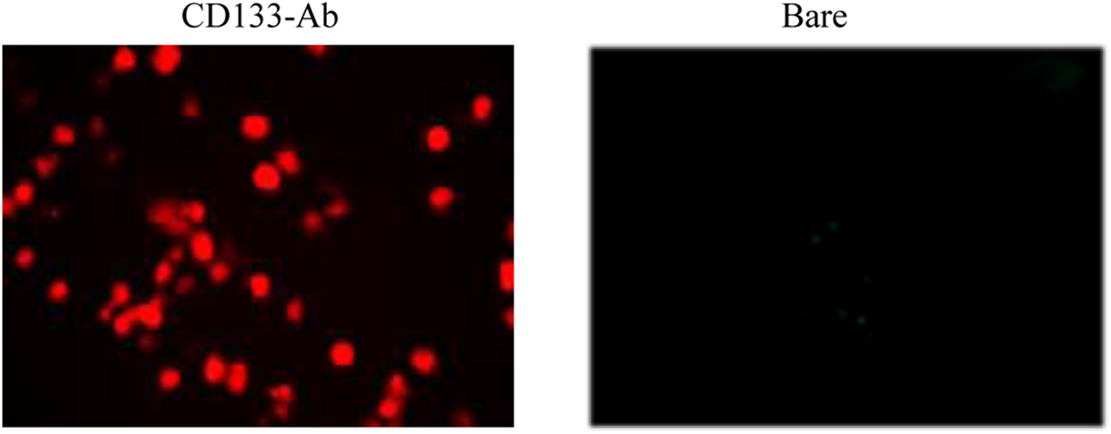
CD133 antibody-coated discs bound CD133+ endothelial progenitor cells (EPCs) in vitro: CD133 antibody-coated and bare discs were incubated with Dil-Ac-LDL-labeled CD133+ EPCs (appeared red). A representative image taken under fluorescence microscopy showed coated discs bound EPCs (left panel) but bare discs did not (right panel).
CD133 Ab-Coated Stents Enhanced Re-Endothelialization
Most CD133 Ab-coated stents from atherosclerotic coronaries 1 week postimplantation were seen covered by a thin intimal layer as analyzed by SEM. An image inserted in the right panel of Figure 4 showed typical intima formed on CD133 Ab stents. However, complete cellular coverage on bare stents was rarely seen (inserted in the left panel, Figure 4). Further analysis revealed that the intima on CD133 Ab-coated stents was smooth (right panel, Figure 4) compared with fibrous tissues and excrescences often seen on the bare stent (left panel, Figure 4). Both bare and CD133 Ab stents were similarly covered by intima examined at week 2 after implantation (data not shown).
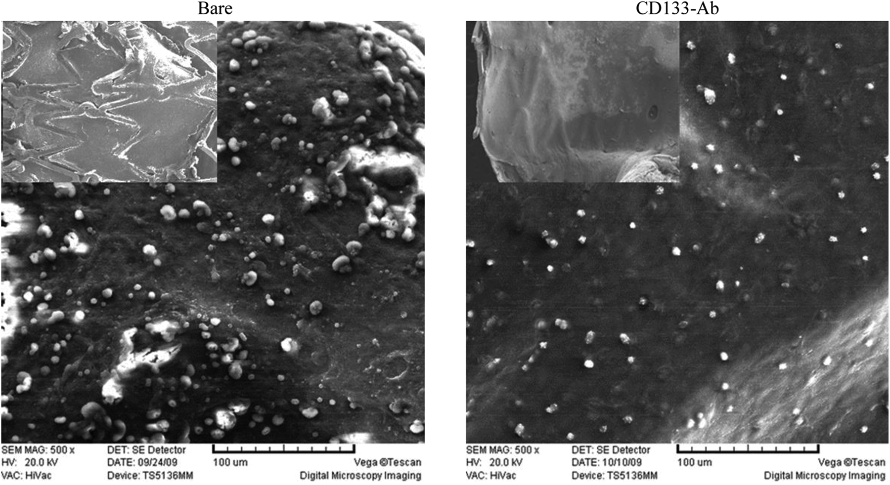
CD133 antibody (Ab) stents enhance cellular coverage. A thin layer of intima was discovered on almost all CD133 Ab stents after 1 week implantation in atherosclerotic coronaries (inserted in the right panel). In contrast, cellular coverage were rarely seen on bare stents (inserted in the left panel). Further analysis revealed a smooth and integrated cellular coverage on CD133 Ab stents (right panel) compared with fibrous tissues and excrescences often seen on the bare stent (left panel).
CD133 Ab-Coated Stents Inhibited Neointimal Hyperplasia and Reduced Restenosis
In a porcine model of atherosclerosis, 12 weeks after implantation of CD133 Ab-coated stents, the arteries had a decreased absolute area stenosis and a reduced percent diameter stenosis as measured by QCA, compared to those implanted with BMSs (Figure 5). The area stenosis in the CD133 Ab stent group was 0.89 ± 0.31 mm2 (n = 10), which was significantly lower than 3.48 ± 0.77 mm2 (n = 10) of bare stent group (P < .01). The percent diameter stenosis was 11.77% ± 1.35% and 50.73% ± 6.80% for CD133 Ab stented- and bare stented-arteries, respectively (P < .01). Histopathological data demonstrated that the neointimal area was 1.01 ± 0.27 mm2 for animals with CD133 Ab stents, which was significantly lower than 2.79 ± 1.62 mm2 of those with bare stents (n = 8, P < .05).

Implantation of CD133 antibody (Ab) stents inhibited intimal hyperplasia and reduced restenosis in a porcine model of atherosclerosis. Representative coronary angiograms showed an increase in stenosis in the bare-stented region of control artery (arrows) compared with the CD133 Ab stented artery (left top panel). The quantitative coronary angiography (QCA) data indicated both absolute area stenosis and percent diameter stenosis were significantly higher in the bare-stented arteries (right top plots). Representative photomicrographs of hematoxylin and eosin (HE)-stained arterial cross-sections were shown in the left bottom panel. Statistical analysis revealed that neointimal area was significantly higher in the control artery compared with the CD133 Ab-stented artery (right bottom plot). *P < .05, **P < .01, compared with bare-stented controls. I indicates intima; L, lumen.
Discussion
Although effective in the treatment of coronary artery disease, coronary stent implantation also causes injury to the arterial wall, which can lead to an inflammatory reaction, stent thrombosis, VSMC proliferation, and eventually vessel renarrowing.5 –8 Complete re-endothelialization of the stented segment is crucial for the prevention of these adverse events, as functional endothelial covering provides a variety of vasoactive, antiinflammatory, antithrombotic, and antiproliferative substances.16,30,31 It is well documented that EPCs participate in endothelial repair. 32 Recently, Ab coating of stents to capture circulating EPCs to promote natural endothelialization has drawn great attention.19 –21 In the present study, we immobilized CD133 Ab on CCA discs and metallic stents using a modified chitosan, namely, HBC, and tested the Ab bioactivity.
The HBC has a number of advantages over its parent polysaccharide. Particularly, it is water soluble under neutral conditions and thermosensitive. 26 Studies have shown that HBC promotes HUVEC proliferation and possesses appreciable histocompatibility,26,33 making it a promising agent for biomedical applications. We explored HBC for the functionalization of CCA discs and metal stents. As HBC gelation happens at 25.2°C when its concentration reaches 25 mg/mL, 26 a significantly lower concentration of HBC was used in this study. The HBC-coated discs were assessed in vitro. Using a cell proliferation assay, we showed that HUVECs grown on HBC-coated discs had a higher proliferation rate compared with those seeded on bare discs, which is in agreement with a previous finding by Wei et al. 33 More relevantly, CD133+ EPCs grown on HBC discs had a significantly higher proliferation rate, although delayed 2 days compared with HUVECs, suggesting HBC-mediated anti-CD133 Ab coating is conducive to CD133+ EPC growth.
Immobilization of CD133 Ab was achieved by incubation of HBC functionalized CCA discs and stents in CD133 Ab solution. Using an FITC-conjugated antimouse secondary Ab, we showed a homogenous coating of CD133 Ab on the surface of both discs and stents. After balloon expansion of the coated stents, there was no cracking or peeling of the coated film. An in vitro cell binding assay revealed that the CD133 Ab-coated surface captured CD133+ EPCs. After this validation, we implanted CD133 Ab stents in a porcine model of coronary artery injury and compared their effect on the reduction in restenosis with bare stents. It is important that the lesion levels between the 2 groups of animals receiving different stents are comparable when implantation of stents takes place. It has been well documented that the degree of lesion progression is similar among pigs postangioplasty injury if the same procedure is done. 28 We applied the same angioplasty procedure for all animals as described previously. The results showed that explanted CD133 Ab stents had significantly lower levels of neointimal area, absolute area stenosis, and percent diameter stenosis compared with control pigs with bare stents (Figure 5).
The first CD133 Ab-coated stent was reported by Sedaghat et al who covalently immobilized CD133 Ab on metallic stents. 29 In vitro assessment confirmed that the Ab-coated devices were able to bind CD133+ EPCs. However, in a porcine coronary artery model, implantation of CD133 Ab stents did not enhance re-endothelialization or reduce restenosis. In a previous study, also using a covalent linking method, we coated stents with CD133 Ab and tested the stents in a porcine model. At 4 weeks poststent implantation, we did not observe significant reduction in restenosis between animals with CD133 Ab stents and those with bare stents. 27 These data contrast what we observed in the present study. The difference might be explained by the different coating methods that were employed. The HBC may be superior to covalent bond in the immobilization and bioactivity maintenance of CD133 Ab. In addition, HBC was conducive to CD133+ EPC growth as shown in this study.
We hypothesized that CD133 Ab stents reduce in-stent restenosis by capturing CD133+ EPCs for improved endothelialization. Using SEM, we compared cellular coverage on explanted stents and indeed found at week 1 postimplantation, there was a thin layer of intima formed on CD133 Ab stents while most bare stents were still exposed (Figure 4). In a previous study, we examined endothelium function by measuring the coronary vasomotor reactivity in response to acetylcholine and found, although not significant, there was a trend that the segments distal to the bare stents were more strongly constricted to the acetylcholine infusion compared to the distal part of coronaries implanted with CD133 Ab-coated stents. 27
Conclusion
In conclusion, HBC is successfully employed to functionalize CCA discs and metallic stents. The HBC processed surface is conducive to CD133+ EPC and HUVEC growth. The HBC can mediate CD133 Ab coating, and the coated Ab binds CD133+ EPCs in vitro. Implantation of CD133 Ab stents significantly reduced restenosis in a porcine model of atherosclerosis. These findings suggest that HBC is a valuable agent for bioactive coating of stents and that CD133 Ab stents might be a potential therapeutic option for the treatment of coronary artery disease.
Footnotes
Acknowledgments
The authors would like to thank Drs Xiguang Chen and Xiaojie Cheng from the College of Marine Life Science, Ocean University of China, Qingdao and Drs Junbo Ge and Li Shen from Shanghai Institute of Cardiovascular Diseases, Zhongshan Hospital, Fudan University, Shanghai, for their help in completing this project.
Author contributions
Jian Li contributed to acquisition, analysis, and interpretation and drafted the manuscript. Qiuwang Zhang contributed to acquisition, analysis, and interpretation and drafted the manuscript. Dan Li contributed to acquisition, analysis, and interpretation. Yi An contributed to conception and design, analysis, and interpretation; critically revised the manuscript; gave final approval; and agreed to be accountable for all aspects of work ensuring integrity accuracy. Michael B. J. Kutryk contributed to design, analysis, and interpretation; critically revised the manuscript; and gave final approval.
Declaration of Conflicting Interests
The author(s) declared no potential conflicts of interest with respect to the research, authorship, and/or publication of this article.
Funding
The author(s) disclosed receipt of the following financial support for the research, authorship, and/or publication of this article: This work was supported by the National Natural Science Foundation of China (81071246 and 81370323); Shandong Provincial Natural Science Foundation, China (ZR2010HM081); and China Postdoctoral Science Foundation (2012M511462).