




Abstract
Infections with Mycobacterium bovis can lead to clinical tuberculosis in many mammals. Here, we describe M. bovis infections in 4 of 7 cats from 1 household in Germany. These cats had respiratory disorders at intervals of several months. Two of 4 euthanized animals (cats 3 and 4) were submitted for postmortem examination; lungs failed to collapse and were firm in both cats. In cat 4, a severely enlarged pulmonary lymph node was found, as well as small white granulomas in the spleen, liver, and kidney. Bacterial cultivation identified M. bovis spoligotype SB0120 as the causative pathogen in cats 3 and 4. Molecular genetic fine typing revealed that this genotype had not been reported previously in animals or humans in Germany. Serum from cat 4 shortly before euthanasia, and from cats 5–7, were tested for TB-specific antibodies by ELISA. Cat 4 was strongly positive. In contrast, cats 5–7 were negative and remained negative 4 mo later. Based on our case series, tuberculosis should be considered as a differential diagnosis in pet animals, even in countries that are officially free of the disease. A lack of awareness about tuberculosis could increase the risk of zoonotic infections with M. bovis—for both owners and other animals living in affected households.
Tuberculosis (
Feline tuberculosis is a well-known disease. Case descriptions exist from as early as the 1950s in Switzerland 52 and the 1980s in New Zealand. 14 The number of feline TB cases has decreased in concert with the decreasing numbers of bovine TB cases. Hence, awareness of the disease by veterinarians and owners has declined. In cats, 2 main groups of MTC infections share some of their clinical signs but differ in lesions, causative pathogen, and mean age of affected cats.21,32 Cases of the first group, with cutaneous nodules and lymphadenopathy, are infected by M. microti. 39 The reservoir host for M. microti is the wild field vole 10 and most infections occur via scratches in vole-hunting cats. The second group of feline MTC infections is caused by M. bovis or M. caprae. These systemic infections originate in the respiratory or digestive tracts and occur predominantly in regions where TB is endemic in cattle.4,15,21
Rarely, MTC infections are reported from officially tuberculosis-free (
All members of the MTC are considered zoonotic pathogens. 16 Therefore, an infectious risk for humans exists, especially with pet animals, which often live in close contact with their owners.5,13,27,34,37 Here, we describe an outbreak of feline TB in an urban household in Germany that included 7 cats. Six rescue animals had been transferred from Turkey to Germany by a private pet agency; 4 of these 6 cats had respiratory disorders.
Material and methods
Animals and history
Seven cats were kept in an urban household on the outskirts of a large German city. Six of these cats were imported from Turkey by a private pet agency (

Feline tuberculosis in a German household. Timeline for the available information on breed, sex, birthdate, transfer to Germany, duration of clinical signs, blood sampling, and death of the cats. ESC = European Shorthair cat; GER = Germany; NFC = Norwegian Forest cat; NK = not known; TUR = Turkey; * = year of birth.
All 7 animals had access to their owner’s house and garden, but did not leave the property. There were no livestock farms nearby or accessible to the cats. In Germany, the animals were fed a commercial diet (dry and wet food) and had no access to unpasteurized raw milk or raw meat.
The examinations performed and results were part of the diagnostic approach and follow-up by the official authorities (local veterinary office and public health department in North Rhine-Westphalia). Therefore, no ethical approval was required.
Postmortem examination
The postmortem examinations of cats 3 and 4 were conducted by pathologists at the Chemical and Veterinary Investigations Office Rhein-Ruhr-Wupper (CVUA-RRW; Krefeld, Germany) following internal standardized operating procedures. Samples of various organs were taken for histologic analysis. Additionally, samples of the lung of cat 3 and tracheobronchial lymph node of cat 4 were examined via bacterial culture and molecular testing.
For histologic examination of tissue sections, H&E, Grocott (fungi), Azan (connective tissue), and Ziehl–Neelsen or Kinyon (acid-fast bacteria) stains were applied according to standard protocols. Immunohistochemistry (IHC) was performed as an ancillary test with a polyclonal antibody specific for M. bovis (BYT-ORB100411; Biozol). A negative control using a polyclonal antibody against an unrelated antigen as the primary antibody was included for each section. Bovine skeletal muscle tissue was spiked with M. bovis to serve as the positive control.
Microbiologic and molecular genetic detection of mycobacteria
Cultivation of tissue samples
Tissue samples from the lung of cats 3 and 4 and tracheobronchial lymph nodes of cat 4 were homogenized and decontaminated with N-acetyl-L-cysteine-NaOH (final concentration 1%, 25 min, shaking). After 2 washing steps, 1 part of the suspension was used for DNA extraction and the other part was cultured on Stonebrink agar slants (supplemented with PACT [polymyxin, amphotericin, carbenicillin, trimethoprim]; Becton Dickinson), Herrold egg-yolk agar, 59 Middlebrook 7H11 agar slants supplemented with vancomycin and amphotericin, and Middlebrook 7H10 agar slants (all in-house), as well as in MGIT/PANTA/OADC broth (Becton Dickinson). Cultures were incubated at 37 ± 2°C for up to 12 wk and examined weekly for suspicious colonies.
Real-time PCR from tissue samples
DNA was extracted from homogenized tissues (DNeasy blood and tissue kit; Qiagen) and tested for MTC-specific DNA according to the German official manual of diagnostic procedures for epizootics. 19 In brief, 2 real-time PCR (rtPCR) assays targeting the MTC-specific IS1081 and a putative helicase gene, respectively (each in combination with the amplification of a fragment of the host’s β-actin gene), were used (QuantiTect multiplex PCR NoROX kit; Qiagen).
Molecular characterization of the isolates
End-point PCR assays for species identification
Species identification of the MTC isolates was done using end-point PCR assays targeting regions of difference (
Spoligotyping
Spoligotyping was performed using a DNA microarray format (ArrayStrip platform; Alere Technologies) according to the manufacturer’s instructions 46 and using the mbovis.org database to identify the profile. 49
Mycobacterial interspersed repetitive unit-variable number of tandem repeats (MIRU-VNTR)
MIRU-VNTR typing of the M. bovis strains was carried out using an in-house technique, 7 according to published protocols 53 with slight modifications.20,48 Overall, we tested 26 VNTR loci: VNTR154 (MIRU-2), VNTR424 (Mtub04), VNTR577 (ETR-C), VNTR580 (MIRU-4, ETR-D), VNTR802 (MIRU-40), VNTR960 (MIRU-10), VNTR1644 (MIRU-16), VNTR1955 (Mtub21), VNTR2059 (MIRU-20), VNTR2163b (QUB-11b), VNTR2165 (ETR-A), VNTR2347 (Mtub29), VNTR2401 (Mtub30), VNTR2461 (ETR-B), VNTR2531 (MIRU-23), VNTR2687 (MIRU-24), VNTR2996 (MIRU-26), VNTR3007 (MIRU-27, QUB-5), VNTR3171 (Mtub34), VNTR3192 (MIRU-31, ETR-E), VNTR3690 (Mtub39), VNTR4052 (QUB-26), VNTR4156 (QUB-4156), VNTR4348 (MIRU-39), VNTR2163a (QUB-11a), and VNTR3232 (QUB-3232).
Whole-genome sequencing
For DNA preparation, isolates were grown on Coletsos agar (Artelt-Enclit) for at least 6 wk. The DNA was extracted using the cetyltrimethylammonium bromide method 55 with additional RNase A digestion and washing steps. DNA was sent to a commercial sequencing service (Eurofins Genomics) to generate whole-genome sequencing (WGS) data using NovaSeq technology (Illumina). The raw sequencing data of both isolates were deposited in the European Nucleotide Archive (ENA) under project PRJEB104949.
Bioinformatic analysis of WGS data
The raw sequence data were uploaded to the TB profiler pipeline (https://tbdr.lshtm.ac.uk/) to determine the spoligotype, affiliation to known genetic lineages, as well as prediction of anti-tuberculosis drug resistances (accession date 2024 Aug 16).12,40 Core genome SNP calling was conducted (Snippy v.4.6.0, https://github.com/tseemann/snippy) with M. bovis strain AF2122/97 (GCF_000195835.3) as reference. Based on the core genome single-nucleotide polymorphism (
Serology
One serum sample was available from cat 4 ~2 d before euthanasia. Additionally, sera from cats 5–7 were collected in parallel to cat 4 as well as 4 mo later. The sera were analyzed (Ingezim DR ELISA, 1:25 diluted; Gold Standard Diagnostics) according to the manufacturer’s instructions. The cutoff was determined with the kit internal positive and negative controls.
Results
Case descriptions
Cats 1 and 2
The cat owners reported that cats 1 and 2, both imported from Turkey, fell ill in May 2022 with severe respiratory signs similar to those of cats 3 and 4. Cats 1 and 2 were euthanized by local veterinarians given their deteriorating general condition. No further examinations were carried out on these cats.
Cat 3
Cat 3 was a 3-y-old, female European Shorthair cat that was initially in very good nutritional condition. Starting in November 2022, the cat was dyspneic, had a reduced appetite, and lost weight. The local veterinarian performed a radiographic examination, which revealed compacted lung tissue with condensation of the lung parenchyma (

Tuberculosis in cat 3 in a German household.
Hematology and serum biochemistry values outside their RIs for cat 3.

The body condition declined rapidly over the following weeks, and, in mid-January 2023, the cat was euthanized for animal welfare reasons. The dominant alterations at autopsy were marked anemia and consolidated lungs. The tracheobronchial lymph nodes were not detectable. Liver and spleen were slightly enlarged. The liver, spleen, and kidneys were hyperemic. The right cardiac ventricle was slightly dilated.
Histologically, granulomas in the lung were surrounded by diffuse mixed-cell infiltrates; alveolar histiocytosis was moderate to severe (
MTC-specific DNA was detected by rtPCR testing of the lung tissue. Subsequently, M. bovis was identified by culture.
Cat 4
Cat 4 was an ~4-y-old, female European Shorthair cat. The radiographic examination performed in November 2022 revealed consolidation of the left cranial lung lobe (

Tuberculosis in cat 4 in a German household.
At autopsy, the cat was in good nutritional condition. The lungs were firm, nodular, and diffusely infiltrated by solid white material. The tracheobronchial lymph nodes were markedly enlarged and had white spots (

Tuberculosis in cat 4 in a German household.
Histologically, numerous coalescing granulomas (
In the tracheobronchial lymph nodes, most of the lymphoid tissue was replaced by confluent granulomas with extensive areas of caseous necrosis (
For direct rtPCR analysis of DNA extracts of the tissue and culture, lung, lymph node, liver, spleen, and kidney tissues were pooled and further processed. In the pooled sample, DNA specific for MTC bacteria was detected, and M. bovis was identified by culture.
Epidemiologic investigations
Genomic analysis
Using the classical spoligotyping method, both isolates were identified as M. bovis spoligotype SB0120 (IT482). Finer genetic typing was performed using PCR-based MIRU-VNTR analysis of 26 different loci. The MIRU-VNTR-pattern found (1-2-6-3-2-2-3-3-2-2-6-3-4-3-4-2-3-3-3-3-2-4-1-2-10-5) was identical for the isolates from both cats and had not been reported previously in M. bovis isolates from animals in Germany. Analysis of the genome sequences with TB profiler confirmed the spoligotype SB0120 (IT482), additionally revealed lineage La1.4, and detected no resistance determinants against commonly used anti-tuberculosis drugs, aside from the characteristic M. bovis pyrazinamide resistance.
The isolates from both cats were identical in single-nucleotide polymorphism (SNP) analysis of the WGS data, with only a single cgSNP different. To test if the current isolates were genetically close to previously isolated German M. bovis spoligotype SB0120 strains, other WGS data,
28
which also comprised animal isolates, were included in the analysis. The data of 2 strains from that study
28
were excluded after preliminary analysis because of quality issues. In total, 3,756 core genome SNPs were called. In the midpoint-rooted maximum likelihood tree (
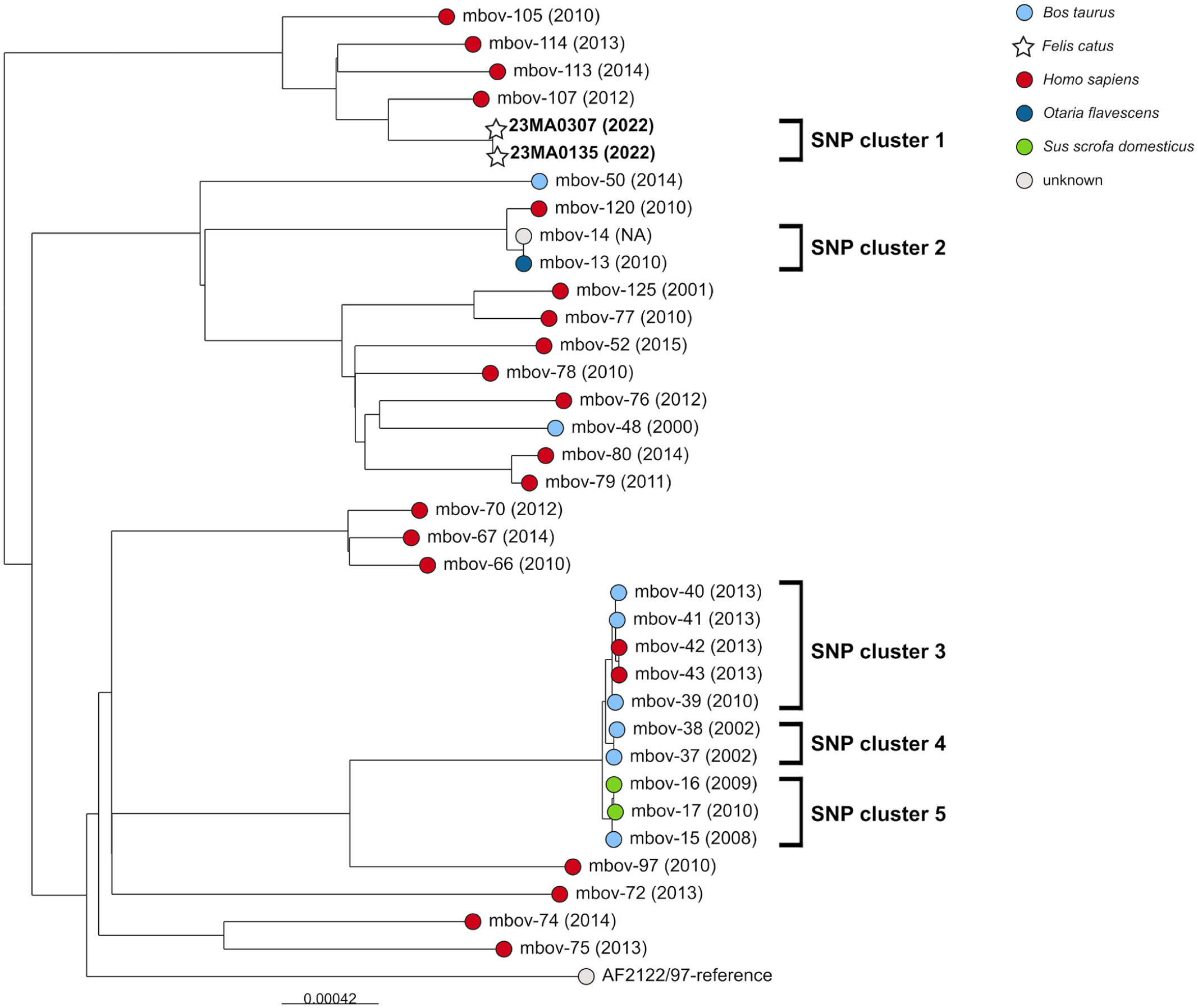
Feline tuberculosis in a German household. Midpoint-rooted maximum-likelihood tree based on cgSNP alignment of German Mycobacterium bovis spoligotype SB0120 isolates (data from Kohl, Kranzer, 28 and our study). The leaves are colored according to the host. Scale bar = the nucleotide changes per base position.
Serology
Cat 4 serum was strongly ELISA-positive (value >4 in the original 1:25 dilution). Performing log10 dilution steps, cat 4 still tested positive at a 1:2,500 dilution. In contrast, cats 5–7 were negative in ELISA testing, which remained the case 4 mo later when the animals were re-tested.
Zoonotic impact
Both owners as well as the local veterinarians were asked by the local health agency to undergo an interferon-gamma release assay (IGRA). Only the 2 owners accepted the offer to test, and 1 of them was positive (data not shown). Radiography of the positive owner did not reveal any suspicious lung changes; therefore, latent TB was diagnosed. The patient was still in a TB monitoring program at the last contact made in December 2023.
Discussion
M. bovis has the widest known host range of all MTC members. In humans, ~150,000 new infections and 12,500 deaths caused by M. bovis are estimated each year worldwide.
58
However, the most important hosts for this pathogen are cattle. Bovine tuberculosis (
Contrary to cattle, the tools for antemortem diagnosis of TB in pet animals are limited. In cattle, the diagnosis of bTB is based on the measurement of cell-mediated immunity using intradermal tuberculin skin tests with bovine purified protein derivates (
In the early stages of infection, clinical signs of TB may be inapparent. As disease progresses, nonspecific signs (e.g., reduced body condition, cachexia, lymphadenopathy, or respiratory disorders) can occur. 35 Depending on the affected organ system, cats may be blind 51 or lame. 31 Results of analysis of blood measurands (CBC, biochemistry) are mostly nonspecific. Sometimes, a shift in the albumin:globulin ratio caused by decreased albumin and increased globulin concentrations is apparent in animals with mycobacteriosis.26,47 We did note this change in cat 3. Radiography or computed tomography may be informative in detecting abnormalities not discernible on physical examination (e.g., disseminated lymphadenopathies or consolidated lung parenchyma). 24 In our cases, radiographic findings included severely consolidated lung tissue in cat 3 and consolidation of the left cranial lung field in cat 4.
Although many publications have described one or more enlarged lymph nodes as prominent pathology findings,1,13,15,21,44 we found an enlarged tracheobronchial lymph node only in cat 4. Furthermore, although both animals had firm, consolidated lungs at autopsy, pathognomonic granulomas were only macroscopically visible in the lung, tracheobronchial lymph nodes, and liver of cat 4. Thus, for accurate diagnosis, inclusion of microbiologic identification of the causative agent as a part of the postmortem examination should be considered the gold standard.
In cat 3, lesions were detected only at the primary site of infection. This is characteristic of the post-primary phase of infection, resulting in chronic organ TB. In cat 4, lung, bronchial lymph node, and multiple other organs were affected. Necrotic material and inflammatory cells from granulomas were present within airways, indicating a post-primary phase infection. Dissemination to other organs is indicative of late generalization, which occurs by lympho-hematogenous spread. These lesions are, as seen in cat 4, poorly mineralized. They may occur in anergic animals.
Differences in the morphology of the granulomas between the 2 cats detected by histologic examinations may be the result of differences in the duration of infection (thus, stage of TB) or differences in the individual immune response. In agreement with other authors, multinucleate giant cells of the Langhans type were not seen in granulomas in either cat.13,22,32,33 As also commonly described for M. bovis, bacteria were rarely detected in lesions. Acid-fast staining and IHC results were congruent, which indicates that this lack of detection was not based on changes in the mycobacterial cell wall resulting in different staining properties. Mycobacteria were detected only in the tracheobronchial lymph node of cat 4, which was the organ with the most extensive necrosis. IHC allowed easier detection and revealed larger numbers of bacteria compared with Ziehl–Neelsen staining. Detection by microscopy, as well as by cultivation and molecular genetic analysis, were further complicated by the lack of even distribution of M. bovis within the necrotic tissue.
Genetic characterization of both strains revealed a common spoligotype, SB0120, which is frequently detected in cattle and humans in several European and African countries.7,25,28,29,45 Strains with this spoligotype have also been isolated from cattle and humans in Turkey.23,54 Comparing the 4 MIRU-VNTR profiles among 9 M. bovis isolates from cattle, 23 we found at least 5 differing loci in 24 loci comparable to the strains from our study. Unfortunately, WGS data were not available from M. bovis spoligotype SB0120 isolates from Turkey. Therefore, we compared the data with published German M. bovis spoligotype SB0120 isolates of human or animal origin. Both of our feline M. bovis spoligotype SB0120 isolates clearly separated from the German isolates, with a minimum difference of 120 SNPs. For M. tuberculosis, the mutation rate was estimated between 0.5 and 1 SNPs/y, 56 highlighting the large genetic distance between these human and animal German isolates and the feline ones from our study. Nevertheless, regarding the high diversity of M. bovis spoligotype SB0120 in Germany, the source of the cats’ infection cannot be determined unambiguously, based on the available genomic data.
Considering the long incubation time typical for TB, it is likely that the animals may have been infected before they were brought to Germany. In OTF countries, the awareness for TB is low, especially when animal species other than cattle are affected. In 2025, several colleagues published feline TB case studies (e.g., M. bovis infections in Argentina 5 and Brazil, 15 M. africanum infection in Italy, 1 M. bovis infections in the Netherlands 13 ), reflecting an increased alertness for this disease in pet animals. Although no mandatory national control measures exist for TB in cats, the detection of a positive TB test in owners shows that the zoonotic risk should not be underestimated.
Conclusion for practice
Importing animals from certain regions and countries carries the risk of introducing diseases that occur only rarely or not at all in Germany.
TB should be included as a differential diagnosis in cats with respiratory disorders or cutaneous lesions, especially if the animals originate from countries with higher TB prevalence.
For diagnosis in animals with no obvious tissue lesions, the analysis of multiple organs might increase the sensitivity of detection, given that the distribution of the pathogen in tissues is not homogeneous.
Intense testing is needed to estimate the zoonotic risk for humans in close contact with infected animals.
Our case series highlights the problem of inadequate TB test procedures in pet animals. First, antemortem, when even limited test options are not fully available or the available tests are not utilized; and second, postmortem, when owners—for financial, ethical, or emotional reasons—do not have their animals autopsied after death.
Footnotes
Acknowledgements
For excellent technical assistance in the microbiological and molecular genetic analyses, we thank Boris Coonen, Lea van den Ham, and Marion Lütz (CVUA-RRW, Krefeld); for the genetic characterization of the isolates, Uta Brommer, Gesine Kauth, and Tyrell Pötzsch (FLI, Jena); and for immunohistochemistry, Lisa Wolf and Kevin Hause (FLI, Jena). We thank Marcus Pfau (FLI, Jena) for support in the graphical presentation.
Data availability
The raw sequencing data of both isolates are publicly available in the European Nucleotide Archive (ENA) under project PRJEB104949.
Declaration of conflicting interests
The authors declared no potential conflicts of interest with respect to the research, authorship, and/or publication of this article.
Funding
The authors received no financial support for the research, authorship, and/or publication of this article.
