
Abstract
Astylus atromaculatus Blanchard is a native beetle of South America that feeds on pollen. During the summer of 2022–2023 in Argentina and Uruguay, an explosive infestation of these insects occurred in pastures in which ruminants were grazing. This was believed to be associated with a severe drought, which had significantly reduced the flowering of crops. Three farms in Uruguay and one in Argentina were visited to examine the flocks and perform autopsies. Affected sheep had watery diarrhea, anorexia, depression, and ruminal atony. The average morbidity, mortality, and case fatality rates were 7.5%, 4.3%, and 68%, respectively. The main gross findings in all animals were in the jejunum; the serosa had multifocal hemorrhages, and the mucosa was necrotic and covered by a pseudomembrane. Microscopically, the mucosa had partial-to-complete necrosis of the lamina propria, as well as loss of villus and crypt epithelium with neutrophilic infiltration. Overlying the necrotic mucosa was a pseudomembrane of fibrin, cell debris, desquamated epithelial cells, degenerate neutrophils, and bacteria. Many specimens of A. atromaculatus were in all paddocks in which sheep grazed, as well as in the ruminal content of the autopsied animals.
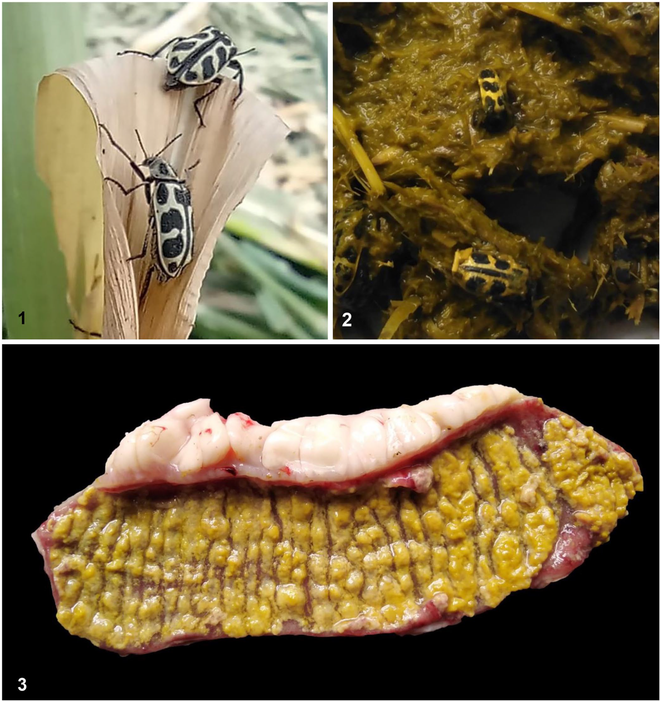
The pollen beetle, Astylus atromaculatus Blanchard, also known by the Spanish names “Astilo moteado” or “7 de oro,” is an insect native to the southern region of South America, which was introduced into South Africa in the 1960s.1,4 The adult insect is 7–9 mm long and is yellow with black spots on the prothorax and elytra. 1 Adults emerge and are active during the summer when they can be observed in clusters mainly on flowers of maize, soybean, sunflower, and sorghum, where they feed on pollen. 1
During the summer of 2022–2023 in the southern hemisphere, the central region of Argentina, the whole of Uruguay and Rio Grande do Sul state (southern Brazil) suffered a severe, unprecedented drought (Table 1). Throughout this period, unusually large numbers of A. atromaculatus were reported in the areas affected by the drought on several crops and pastures, including those used for livestock grazing (https://www.engormix.com/equinos/articulos/mortandad-bovinos-equinos-ovinos-t52061.htm, Spanish; http://www.inia.uy/Paginas/siete-de-oro.aspx, Spanish; Fig. 1).
Historical summer rainfall compared with 2022–2023 summer rainfall in Uruguay, Argentina, and southern Brazil.
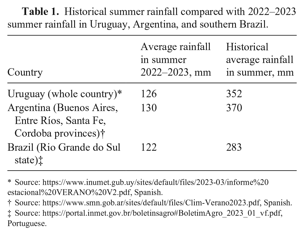
Source: https://www.inumet.gub.uy/sites/default/files/2023-03/informe%20estacional%20VERANO%20V2.pdf, Spanish.
Source: https://www.smn.gob.ar/sites/default/files/Clim-Verano2023.pdf, Spanish.
Source: https://portal.inmet.gov.br/boletinsagro#BoletimAgro_2023_01_vf.pdf, Portuguese.
Astylus atromaculatus intoxication in sheep.
A. atromaculatus is a common insect in South America. However, no toxicity caused in animals by this beetle in the American continent has been described until recently, when several outbreaks of intoxication were reported in cattle. 2 In South Africa, cases of diarrhea and mortality in cattle in the 1970s were believed to be caused by ingestion of A. atromaculatus. 4 The toxic principle of the insect was not determined. We describe here 4 outbreaks of spontaneous intoxication of sheep by A. atromaculatus in Uruguay and Argentina.
The outbreaks occurred in January and February 2023 on 3 farms in Soriano county, southwestern Uruguay (outbreaks 1–3) and on 1 farm in Santa Fe province, Argentina (outbreak 4; Table 2). Affected farms in Uruguay and Argentina were visited to collect epidemiologic information, to examine the pastures in which affected animals had been grazing, and to perform clinical examinations and field autopsies.
Epidemiologic data of 4 outbreaks of spontaneous intoxication of sheep by Astylus atromaculatus.
The pastures on which the affected sheep had been grazing at the time of onset of clinical signs consisted of alfalfa (Medicago sativa; outbreaks 1, 3, 4) and lotus (syn. bird’s-foot trefoil, Lotus corniculatus; outbreak 2). In outbreaks 1 and 4, ~90% of the plants were in the flowering stage, and the flowers were covered by large numbers of A. atromaculatus. In outbreaks 2 and 3, very few trefoil and alfalfa plants, respectively, had flowers. However, these pastures had been invaded by many thistle plants (Cirsium vulgare), most of which were blooming and had flowers heavily colonized by A. atromaculatus.
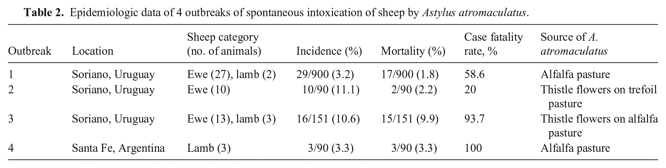
Most of the affected sheep were ewes, although a few lambs were also affected (Table 2). Morbidity was 3.2–11.1% (x̄ = 7.5%), mortality was 1.8–9.9% (x̄ = 4.3%), and case fatality rate was 20–100% (x̄ = 68%). Clinical signs in all animals included watery diarrhea, depression, anorexia, and ruminal atony. In addition, a few animals were bloated. Two autopsies were performed in each of outbreaks 1–3, and one in outbreak 4. Lesions were found mostly in the gastrointestinal tract and were similar in the 7 autopsied animals (Table 3). Briefly, the rumen was slightly distended, and there were large numbers of whole bodies or parts of A. atromaculatus beetles in the ruminal content (Fig. 2). The intestinal content was very watery and yellow or red. The mucosa of the small intestine was markedly congested, had multifocal areas of necrosis and hemorrhage, and was covered by a fibrinous pseudomembrane (Fig. 3). The serosa of the small intestine had petechiae. In addition, there was diffuse reddening of the mucosa of the rumen and abomasum in 2 animals, and of the cecal mucosa in another. No other significant gross abnormalities were observed in any of the animals examined.
Clinical signs and gross findings in 4 outbreaks of spontaneous intoxication of sheep by Astylus atromaculatus.
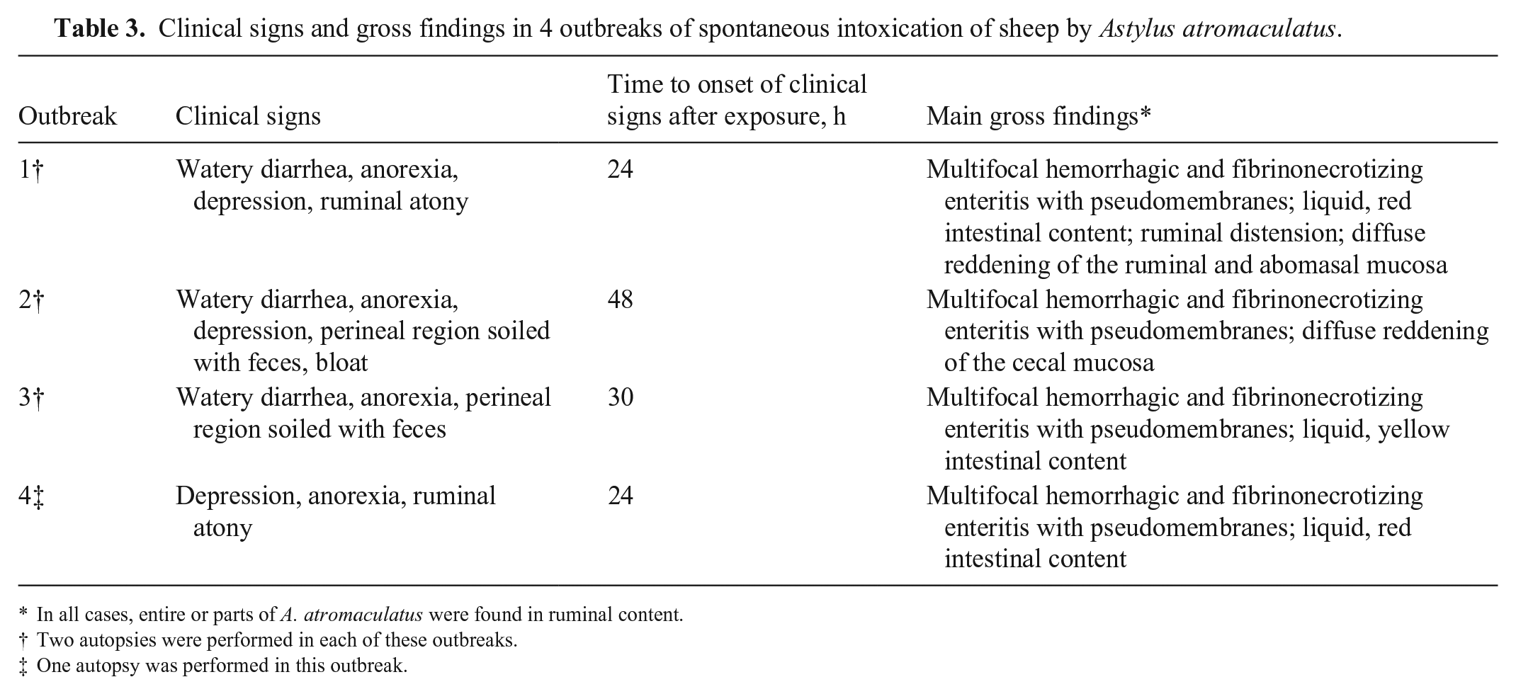
In all cases, entire or parts of A. atromaculatus were found in ruminal content.
Two autopsies were performed in each of these outbreaks.
One autopsy was performed in this outbreak.
A diagnostic workup following División de Laboratorios Veterinarios (DILAVE), Instituto de Innovación para la Producción Agropecuaria y el Desarrollo Sostenible (IPADS), and/or California Animal Health and Food Safety Laboratory (CAHFS) standard operating procedures was performed. Briefly, aerobic bacterial cultures of the liver, lung, spleen, mesenteric lymph node, and small intestinal content, and Salmonella enrichment culture of small intestinal content and liver, were performed. Samples of rumen, abomasum, small intestine, cecum, colon, rectum, lung, liver, lymph nodes, heart, kidney, urinary bladder, and brain were fixed by immersion in 10% neutral-buffered formalin and processed routinely for the production of 4-µm thick, H&E-stained sections for histologic examination. Selected ruminal and intestinal sections were processed by immunohistochemistry (IHC) for bovine viral diarrhea virus (BVDV), bovine alphaherpesvirus 1 (BoAHV1), bovine coronavirus (BoCV), bovine rotavirus (BoRV), Listeria spp., Clostridium septicum, C. chauvoei, C. novyi, and Paeniclostridium sordellii. Feces from all autopsied animals were processed by a McMaster test for parasitologic examination. Beetles collected from the ruminal content and pastures on which affected sheep had been grazing were submitted to the Laboratorio de Entomología, Facultad de Agronomía, Universidad de la República, Uruguay and Instituto Nacional de Tecnología Agropecuaría, Argentina, for identification.
Histologically, the most significant lesion was in the jejunum of the 7 animals examined. The mucosa had multifocal areas of superficial necrosis, eosinophilic debris free in the lumen, and an edematous submucosa (Fig. 4). In these areas, loss of villus and crypt epithelium with distended crypts was observed (Fig. 5). The mucosal necrosis was partial-to-complete with hypereosinophilic areas intermingled with cell debris, fibrin, red blood cells, viable and degenerate neutrophils, and fewer lymphocytes and plasma cells. A pseudomembrane of fibrin, cell debris, desquamated epithelial cells, degenerate neutrophils, and mixed bacteria, overlaid the necrotic mucosa multifocally. Many crypts were dilated and had necrotic or attenuated epithelium, and necrotic cells mixed with mucus, fibrin, and cell debris in the lumen. The submucosa was expanded by large amounts of fibrin and edema, and lymphatic vessels were dilated (Fig. 6). Mucosal and submucosal blood vessels were congested or thrombotic. One sheep had severe fibrinoid vasculitis of arterioles of the submucosa. Four animals had mild, multifocal congestion of the mucosa and submucosa of the abomasum. Two animals had moderate, multifocal congestion of the cecal mucosa and submucosa, with mild, diffuse infiltration of the lamina propria by lymphocytes, plasma cells, and eosinophils. One sheep had mild congestion of the ruminal mucosa and submucosa. A few gram-positive rods were observed in the lumen and within the pseudomembrane covering the intestinal mucosa of one sheep. Most of these rods stained positively for Clostridium perfringens by IHC. All other IHCs for viral and bacterial pathogens were negative in all cases.
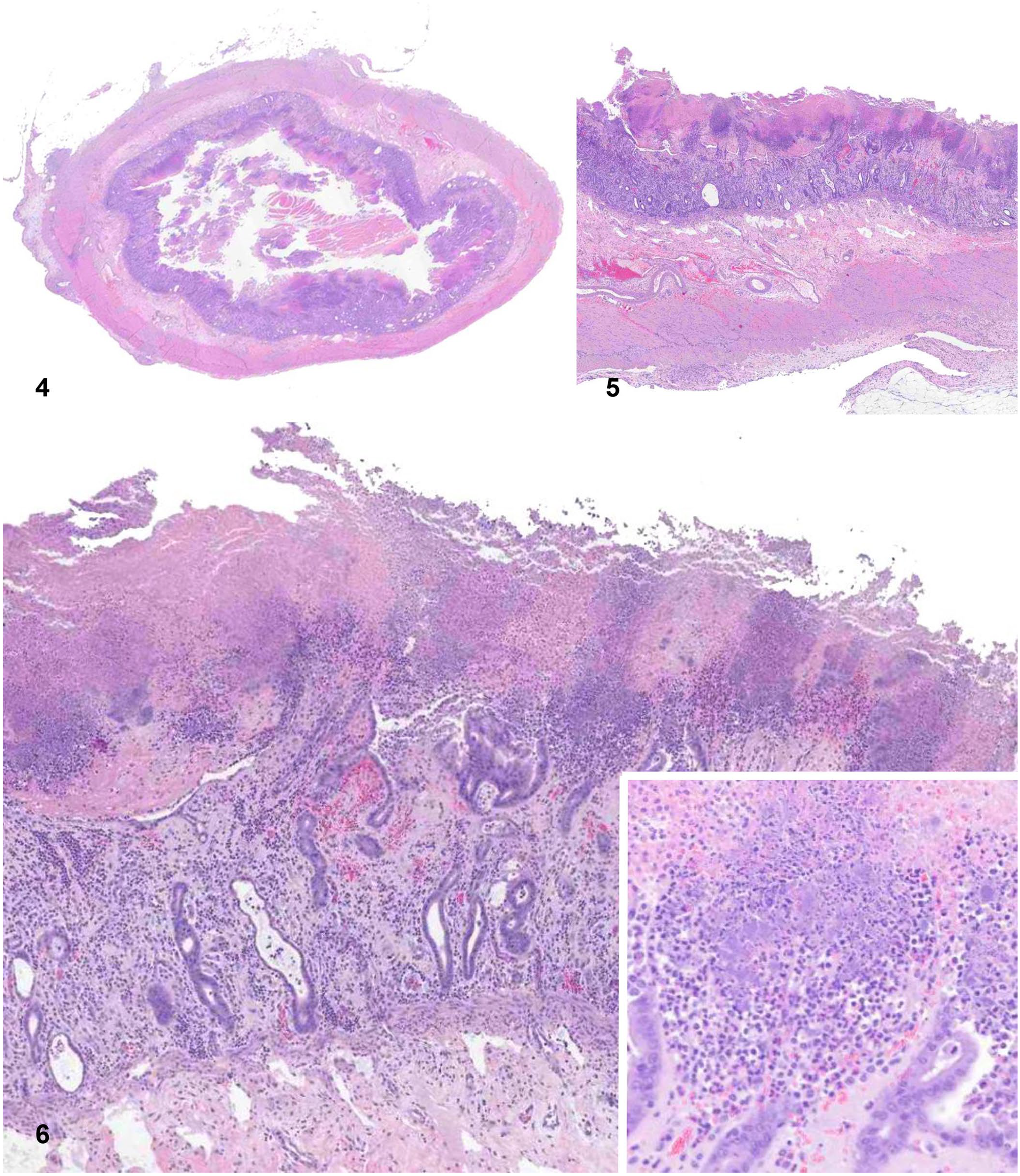
Main histologic lesions in the jejunum of a sheep spontaneously intoxicated by Astylus atromaculatus.
Beetles collected from pastures and the ruminal content of the 7 sheep autopsied were identified as A. atromaculatus Blanchard. No pathogenic bacteria were isolated from any of the animals investigated, and the McMaster test revealed low egg counts per gram of feces in all affected animals.
Based on the presence of large numbers of A. atromaculatus in the pastures on which the affected sheep had been grazing and in the ruminal content of the dead animals, the clinical signs, gross and microscopic findings, and ruling out other common causes of intestinal disease, a presumptive diagnosis of intoxication by A. atromaculatus was established. Intoxication by these beetles was previously reproduced experimentally in sheep in South Africa. 4 In addition, the disease was recently experimentally reproduced in sheep (García y Santos and Corro, unpublished) and cattle 3 by oral dosing with A. atromaculatus specimens collected in the affected zone of Uruguay in February 2023. Several outbreaks of severe spontaneous intestinal disease associated with ingestion of A. atromaculatus were recently described in cattle in Uruguay and Argentina 2 in the same area in which the ovine cases of our report occurred. The lesions in our cases were very similar to those in the bovine cases. 2
In an extensive search of Google, PubMed, CAB Direct, Web of Science, and Scopus, using search terms “sheep” and “Astylus atromaculatus,” we retrieved no cases of spontaneous intoxication by A. atromaculatus, suggesting that this condition has not been reported previously in sheep. We speculate that the severe drought that occurred during the summer of 2023 in Uruguay and the central region of Argentina (Table 1) predisposed to explosive multiplication of this beetle and increased the likelihood that ruminants would ingest toxic amounts of the insects. Also, the drought was responsible for a delayed or reduced flowering of crop species that the insect typically pollinates, and as a result, they shifted to less-common host plants, such as alfalfa or trefoil, which are grazed by ruminants. The toxic principle of A. atromaculatus remains unknown. In a 2024 study of natural intoxication of cattle, extensive toxicologic investigation was performed on samples of A. atromaculatus collected from the same areas that the animals in our study had been grazing. Although the toxic principle of the beetle could not be determined, several toxic substances, including cantharidin and batrachotoxin, were ruled out. 2
Before the recently reported natural intoxication of cattle by A. atromaculatus 2 there was only one report of suspected spontaneous intoxication of cattle by this beetle. 4 However, to demonstrate the toxicity of A. atromaculatus in the latter study, 4 sheep were orally dosed with this beetle; it was toxic at 3.0 g/kg BW after 3 daily oral doses and 5.0 g/kg BW after a single oral dose. The lesions in those sheep were very similar to those described in the sheep of our study. No cantharidin was identified in extracts of the A. atromaculatus specimens used in that study. 4
In the 4 outbreaks reported here, most of the affected sheep were adults with an estimated weight of 50–60 kg. Based on the results of the aforementioned report, 4 250–300 g of beetles at a single oral dose and 150–180 g of beetles daily for 3 d should be enough to cause intoxication. Based on a beetle weighing 27.3 mg in recent experimental trials in calves, 3 the single oral dose of 250–300 g would be ~10,990 insects in a day, or ~6,500 beetles per day for 3 d. Although the number of beetles might seem large, adult beetles gather in clusters over flowers, attracted by the secretion of an aggregation pheromone, 1 which would have facilitated the consumption of large numbers of insects by the sheep in our study and perhaps explain why only a few animals per flock were affected.
Differential diagnosis in the outbreaks described here included intoxication by Baccharis coridifolia, gastrointestinal helminthosis, coccidiosis, yersiniosis, and several viral infections. 6 Intoxication by B. coridifolia was ruled out based on the absence of the plant in the areas in which the sheep were grazing and by the lack of ruminal lesions; also, the intestinal lesions in the animals of our study were much more severe than those previously described in cases of B. coridifolia intoxication.5,6 Helminthosis was ruled out based on the absence of parasites in the gastrointestinal content, the negative McMaster tests, and the severity of the intestinal lesions. Yersiniosis was ruled out based on the negative results of the bacterial cultures and the lack of large bacterial colonies in histology. Clostridial disease, listeriosis, and viral infections (BVDV, BoAHV1, BoCV, BoRV) were ruled out based on the gross and microscopic lesions and the negative results of IHC for these agents. In one sheep, C. perfringens was present in the lumen of the jejunum; this was considered an incidental finding, given that C. perfringens is present in the intestine of most clinically normal ruminants and no lesions consistent with clostridial enterotoxemia were observed.
Footnotes
Acknowledgements
We thank Alberto Scarpa (Uruguay) and Eugenia Ferrero Regis (Argentina) for the submission of samples, and Horacio Silva (Facultad de Agronomía, Universidad de la República, Uruguay) for beetle identification.
Declaration of conflicting interests
The authors declared no potential conflicts of interests with respect to concerning the research, authorship, and/or publication of this article.
Funding
The authors received no financial support for the research, authorship, and/or publication of this article.