
Abstract
Distinct solitary dermal nodules, either covered by an alopecic, or sometimes ulcerated, epidermis, were noticed on the head of a stillborn Holstein calf. The head was submitted for autopsy, and the nodules were found to consist of homogeneous, diffuse pale-yellow, soft-tissue masses with distinct margins that elevated the epidermis above the adjacent skin. Histologically, the dermal nodules were well-delineated on the deep margin approaching the cutaneous muscle and consisted of perivascular neoplastic infiltrates of round cells that in some places coalesced into sheets that extended into the dermis and subcutis. Neoplastic cells separated adnexa and collagen. Immunohistochemistry revealed intense tumor cell expression of vimentin, Iba1, E-cadherin, and CD204; expression of CD18 was faint. The masses were diagnosed as Langerhans cell histiocytosis. Congenital cutaneous Langerhans cell histiocytosis has not been reported previously in cattle, to our knowledge, and should be included in the differential diagnosis of congenital nodular skin lesions.
A stillborn, purebred, Holstein calf was born at 278 d gestation. The herd veterinarian was consulted regarding multiple skin lesions on the calf’s head and submitted the head of the calf to the University of Copenhagen (Frederiksberg, Denmark) for diagnosis.
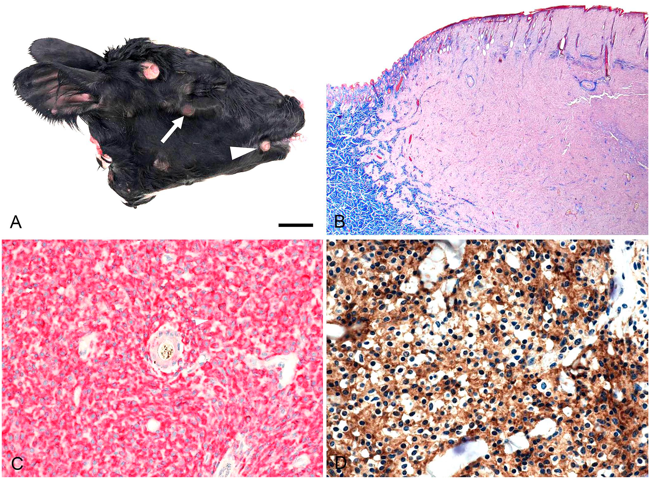
Distinct, solitary, round-to-oval nodules of up to 24-mm diameter were disseminated in the skin of the head, including the pinnae. Some nodules were raised 1–2 mm above the surrounding skin and were covered by intact alopecic epidermis. Other nodules were more prominent with a yellow, centrally depressed ulcer and raised alopecic margins (Fig. 1A). The cut surface of the soft tissue masses was homogeneous, diffuse pale-yellow, with distinct margins. Lymph nodes of the head were grossly normal. Specimens were collected from representative lesions and from submandibular and parotid lymph nodes. Tissue was fixed in 10% neutral-buffered formalin, processed routinely, and stained with H&E and Masson trichrome stains.
Cutaneous Langerhans cell histiocytosis in a stillborn Holstein calf.
Histologically, dermal nodules elevated the epidermis above the adjacent skin and were covered by intact epidermis. The base of the mass was well-delineated toward the cutaneous muscle and was separated only by a thin border of connective tissue. Peripherally, coalescing nests of neoplastic cells infiltrated along blood vessels into the dermis and subcutis. Within the neoplastic masses, neoplastic cells infiltrated between adnexa and collagen bundles and distorted the dermal and subcutaneous architecture (Fig. 1B). Necrosis and hemorrhage were not observed. In the pinnae, invasion of the auricular cartilage was not observed. The epidermal surface of ulcerated lesions was necrotic and covered by cellular debris.
Neoplastic cells were round, 9–12-µm diameter, with oval, occasionally round, rarely reniform, 4–6-µm nuclei. Chromatin was diffusely distributed, often dense (perhaps influenced by moderate autolysis), with occasional clumping. The cytoplasm was eosinophilic. Mitotic figures were infrequent (mitotic count: 3 per 2.37 mm2).
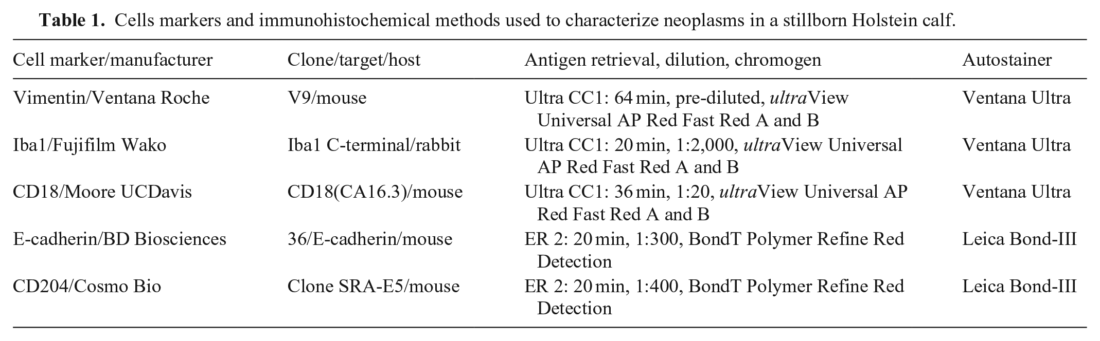
Immunohistochemical (IHC) staining of tissue sections at the Nebraska Veterinary Diagnostic Laboratory (Lincoln, NE, USA) revealed intense cytoplasmic expression of vimentin and Iba1 in tumor cells (Fig. 1C); expression of CD18 was faint. Additional IHC staining performed at the Colorado State University–Veterinary Diagnostic Laboratory (Fort Collins, CO, USA) revealed strong cytoplasmic expression of E-cadherin (Fig. 1D) and CD204 (Table 1). The lymph nodes examined were histologically normal, and no neoplastic cell infiltrates were observed in H&E- or IHC-stained sections.
Cells markers and immunohistochemical methods used to characterize neoplasms in a stillborn Holstein calf.
The neoplasm shared gross features of canine cutaneous Langerhans cell histiocytosis (LCH) being localized on the head and pinnae, with a distinct dome-shaped nodular alopecic appearance and with crater-like ulceration. 4 Histologically, the cell size, shape, cytoplasmic volume, and distribution were consistent with a Langerhans cell origin. The nuclear morphology was not well preserved in H&E sections due to moderate autolysis. IHC staining was necessary to establish the diagnosis and the Langerhans cell origin.
Histiocytic proliferative disease includes neoplasia and inflammatory histiocytic proliferative disorders. In animals, this group of histiocytic diseases is most common in dogs, and several entities have been characterized. 4 Reports of histiocytic proliferative diseases in cattle are sparse and include cutaneous histiocytoma, 7 intestinal benign fibrous histiocytoma, 5 malignant fibrous histiocytoma,3,6 and malignant histiocytosis,1,8 all of which occurred in adult cattle. However, most reports are > 20-y-old and lack thorough IHC characterization; updated information on histiocytic proliferative disease in cattle is limited.
As reviewed in 2021, 2 congenital tumors in cattle have been reported only rarely, and congenital cutaneous LCH has not been reported previously, to our knowledge. We retrieved no cases of congenital histiocytosis in cattle in a search of Google, PubMed, CAB Direct, Web of Science, and Scopus, using the search terms “histiocytosis, cattle, bovine,” suggesting that this condition has not been reported in cattle previously. Our case adds cutaneous LCH to the list of recognized congenital tumors.
The role of the cutaneous LCH in the calf’s stillbirth remains unsolved as only the head was available for autopsy. However, canine cutaneous LCHs rarely involve internal organs; the stillbirth may be independent of the cutaneous LCH.
Footnotes
Acknowledgements
We thank Dr. Marianne R. Schmidt, Tinglev Animal Hospital, Denmark for submitting the case.
Declaration of conflicting interests
The authors declared no potential conflicts of interest with respect to the research, authorship, and/or publication of this article.
Funding
The authors received no financial support for the research, authorship, and/or publication of this article.