
Abstract
Bovine abortion is a critical problem in the cattle industry. Identifying causes of abortion is key to establishing appropriate herd management and prevention strategies. We used pathology examinations, detection of etiologic agents, and serology to determine the cause of bovine abortions in Korea. We analyzed 360 abortion and stillbirth cases submitted to the Animal and Plant Quarantine Agency from December 2014 to January 2020. The putative cause of abortion was identified in 140 of 360 (38.9%) cases; 124 of the 140 (88.6%) cases were attributed to infections. The most common etiologic agents detected were bovine viral diarrhea virus (65 of 360; 18.1%), Coxiella burnetii (19 of 360; 5.3%), Leptospira spp. (13 of 360; 3.6%), Listeria monocytogenes (9 of 360; 2.5%), and Neospora caninum (8 of 360; 2.2%). Minor abortifacient pathogens included Brucella abortus (2 of 360; 0.6%), bovine alphaherpesvirus 1 (2 of 360; 0.6%), Akabane virus (2 of 360, 0.6%), and bovine ephemeral fever virus (1 of 360; 0.3%). Non-infectious conditions included congenital anomalies (7 of 360; 1.9%), goiter (7 of 360; 1.9%), and vitamin A deficiency (2 of 360; 0.6%). Our diagnostic rate in cases with placenta submitted (42 of 86; 48.8%) was significantly higher than in cases without placenta (98 of 274; 35.8%), which highlights the value of submitting placentas. Our results confirm the status of the large variety of causative agents associated with abortions in cattle in Korea.
Bovine abortion is a major economic problem in the cattle industry worldwide.12,28 There are diverse causes of abortion, including infectious (viruses, bacteria, protozoa, fungi) and non-infectious (congenital anomalies, metabolic disorders, toxic agents, twinning, dystocia, endocrine problems, nutritional deficiencies, genetic abnormalities) causes.4,23 Certain organisms such as Coxiella burnetii, Leptospira spp., Listeria monocytogenes, Brucella spp., and Chlamydia abortus are zoonotic agents. 30 Most abortifacient pathogens are being reported as sporadic causes in Korea.
Identifying the cause of bovine abortion is essential to prevent economic losses. 12 However, there are several diagnostic challenges, such as autolysis, contamination, and failure to submit crucial samples such as placenta and maternal serum.4,23 An etiologic diagnosis can be established in 23–60% (usually < 50%) of bovine abortion cases submitted to veterinary diagnostic laboratories.1,3,12,18,23,30 No large-scale investigation of the infectious causes of bovine abortion has been reported from Korea since 2002, at which time, Neospora caninum was considered the leading cause. 12 Little information is available regarding newly emerging infectious agents (such as C. burnetii, Leptospira spp., and L. monocytogenes) related to bovine abortion in Korea. No abortigenic infectious diseases have been eradicated from South Korea, and strict surveillance programs have been implemented only for brucellosis. Therefore, to provide basic data to help establish proper herd management in Korea, we investigated, in a 5-y nationwide study, the frequency of causes of bovine abortion.
Materials and methods
Animals
We conducted investigations of aborted fetuses and samples submitted to our laboratory by veterinarians and animal owners. Abortion was defined as expulsion of a non-viable fetus between gestation days 43 and 260; stillbirth was defined as expulsion of the fetus after gestation day 260. 30 Aborted and stillborn bovine fetuses, their maternal sera, and placentas were submitted to the Animal and Plant Quarantine Agency for the differential diagnosis of abortion from December 2014 to January 2020. Based on the submission of the placenta, submitted samples were categorized as follows: group Ⅰ, aborted fetus without placenta; group Ⅱ, aborted fetus with placenta; and group Ⅲ, placenta from aborted dams without fetus. Gestational age was estimated based on the crown-rump length. 13 Abortion was categorized as having occurred during the first, second, or third gestational trimester.
Autopsy and histologic examination
Gross pathology examinations were conducted on all 357 fetuses and 86 placentas. After measuring the crown-rump length of the fetuses, the brain (cerebrum, cerebellum, brainstem), tongue, lungs, heart, liver, spleen, kidneys, skeletal muscle, small and large intestine, endocrine organs, and placenta samples were collected for histologic analysis. The samples were fixed in 10% neutral-buffered formalin and processed routinely using H&E staining.
Bacterial isolation and identification
Samples of lungs, abomasal contents, and placentas were aseptically cultured on 5% sheep blood agar, chocolate agar (Asan Pharm), and MacConkey agar (Becton Dickinson). The plates were incubated aerobically at 37°C and observed daily for up to 7 d for Brucella-like colonies (smooth, small, translucent, glistening, dew drop–like, round, and convex).
L. monocytogenes was isolated as in a previous study. 29 A set of fetal organs (brain, lungs, heart, liver, spleen, kidney, abomasal contents, skeletal muscle, tongue) and placentas (2 g each) were incubated in 20 mL of Listeria enrichment broth (CM0862; Oxoid) at 30°C for 24 h. Subsequently, 0.1 mL of pre-enrichment broth was added to 10 mL of Fraser broth (Becton Dickinson) for a second enrichment culture and incubated at 37°C for 48 h. Finally, the culture was inoculated onto Listeria selective agar (CM0856; Oxoid) and incubated for 48 h at 37°C.
All colonies from the inoculated plates were speciated using matrix-assisted laser desorption/ionization time-of-flight mass spectrometry (MALDI-TOF MS; VITEK MS, bioMérieux). Furthermore, suspected isolates of Brucella spp. and L. monocytogenes were confirmed through Bruce-ladder PCR and amplification of hly, respectively.17,26
DNA/RNA extractions and PCR assays
DNA and RNA from the collected samples (brain, tongue, lungs, heart, liver, spleen, kidneys, skeletal muscle, small and large intestine, endocrine organs, placenta) were extracted separately (Maxwell RSC instrument, Maxwell RSC blood DNA kit, RSC Viral TNA; Promega), according to the manufacturer’s recommendations. PCR assays for N. caninum, Campylobacter fetus, C. abortus, C. burnetii, Leptospira spp., and Yersinia pseudotuberculosis were each performed separately (Mastercycler ep gradient S; Eppendorf) using the extracted DNA from the collected samples, as described previously.5,8,15,25,27
Using the extracted DNA or RNA from the collected samples, we used various PCR kits for viral detection: the LiliF IBR PCR kit (iNtRON Biotechnology) for bovine alphaherpesvirus 1 (BoAHV1; Varicellovirus bovinealpha1); the LiliF BVDV real-time RT-PCR kit (iNtRON Biotechnology), and the VDx single RT-PCR kit (Median Diagnostics) for bovine viral diarrhea virus (BVDV1, Pestivirus bovis; BVDV2, Pestivirus tauri); the VDx Bovine Akabane/Aino MP RT-PCR kit (Median) and VDx Bovine Chuzan/BEF/Ibaraki MP RT-PCR kit (Median) for 5 arboviruses, including Akabane virus (AKAV; Orthobunyavirus akabaneense), Aino virus (AINOV; Orthobunyavirus ainoense), Chuzan virus (CHUV; Palyam virus), bovine ephemeral fever virus (BEFV; Ephemerovirus febris), and Ibaraki virus (IBAV; Sedoreoviridae, Orbivirus), according to the manufacturer’s instructions. All PCR products were directly sequenced by Macrogen (Seoul, South Korea).
Serologic tests
Fetal body fluids collected from thoracic cavities (n = 270) and maternal sera (n = 205) were screened for total anti-Leptospira antibodies using a microscopic agglutination test (MAT) at a dilution of 1:50. 31 The following live cultures of Leptospira spp. were used: L. interrogans serovars Canicola, Grippotyphosa, Hardjo, Copenhageni, and Pomona; and L. borgpetersenii serovars Tarassovi and Sejroe. A titer of ≥ 100 was considered positive. 10
Serologic testing using fetal body fluids and maternal sera against Brucella spp., C. burnetii, C. abortus, N. caninum, BoAHV1, BVDV, and AKAV was performed using commercial ELISA kits (Brucella C-ELISA antibody test kit, Svanova Biotech AB; ID Screen Q fever indirect multi-species kit, ID Screen C. abortus indirect multi-species kit, ID Screen N. caninum indirect kit, IDvet; infectious bovine rhinotracheitis antibody test screening kit, Svanova; BVDV antibody test kit, Idexx; ID Screen Akabane competition kit, IDvet), according to the manufacturers’ instructions.
Diagnostic criteria
The “final diagnosis” of infectious causes of abortion or stillbirth in each case was based on the isolation or detection of the pathogens in the set of fetal tissues and/or placentas, regardless of the detection of gross and/or histologic lesions. The rationale for adopting this approach was the frequent occurrence of advanced postmortem decomposition in fetuses and placentas, which often precluded the identification of lesions. Therefore, in cases in which no lesions were identified, a diagnosis was made by considering the identification of pathogens in the respective cases. Non-infectious conditions with malformation or goiter were diagnosed based on pathology findings.
Vitamin A quantification in serum
In 2 cases of stillbirth, in which a preliminary clinical diagnosis of vitamin A deficiency was suspected based on the occurrence of blindness in some animals in the herd, serum samples from the 2 dams were sent to the EONE laboratory (Incheon, South Korea) for quantification of vitamin A levels. A serum vitamin A level of < 0.7 µmol/L in the dams was consistent with hypovitaminosis A. 11
Statistical analysis
Statistical analyses were performed using Prism v.5.01 (GraphPad). The 2-tailed Fisher exact test was used to assess whether the differences in the proportions of cases with infectious etiologies differed significantly between seasons, breeds, and gestational periods using a 2 × 2 contingency table. Statistical significance was set at p ≤ 0.05.
Results
Case information
We included 360 aborted (n = 299) or stillborn (n = 48) bovine fetuses from 260 different farms nationwide (Table 1). The number of cases per farm varied from a minimum of 1 case from 216 farms to a maximum of 16 cases (3–4 fetuses every year) from 2 farms. The number of cases included in each group was as follows: group Ⅰ, 274 fetuses without placentas (151 with and 123 without sera of the dams); group Ⅱ, 83 fetuses and their respective placentas (51 with and 32 without sera of the dams); and group Ⅲ, 3 placentas and maternal sera from aborted dams without fetuses. Three hundred sixty fetal cases involved 342 native Korean cattle (syn. Hanwoo cattle; Bos taurus coreanae; 342 of 360, 95.0%) from 244 different farms and 18 Holstein cattle (Holstein Friesian, Bos taurus taurus; 18 of 360, 5.0%) from 16 different farms. Among the 205 maternal serum samples, 195 were from native Korean cattle (95.1%) and 10 were from Holstein cattle (4.9%).
Geographic origin, season of occurrence, and gestational trimester of 360 cases of bovine aborted and stillborn fetuses in Korea.
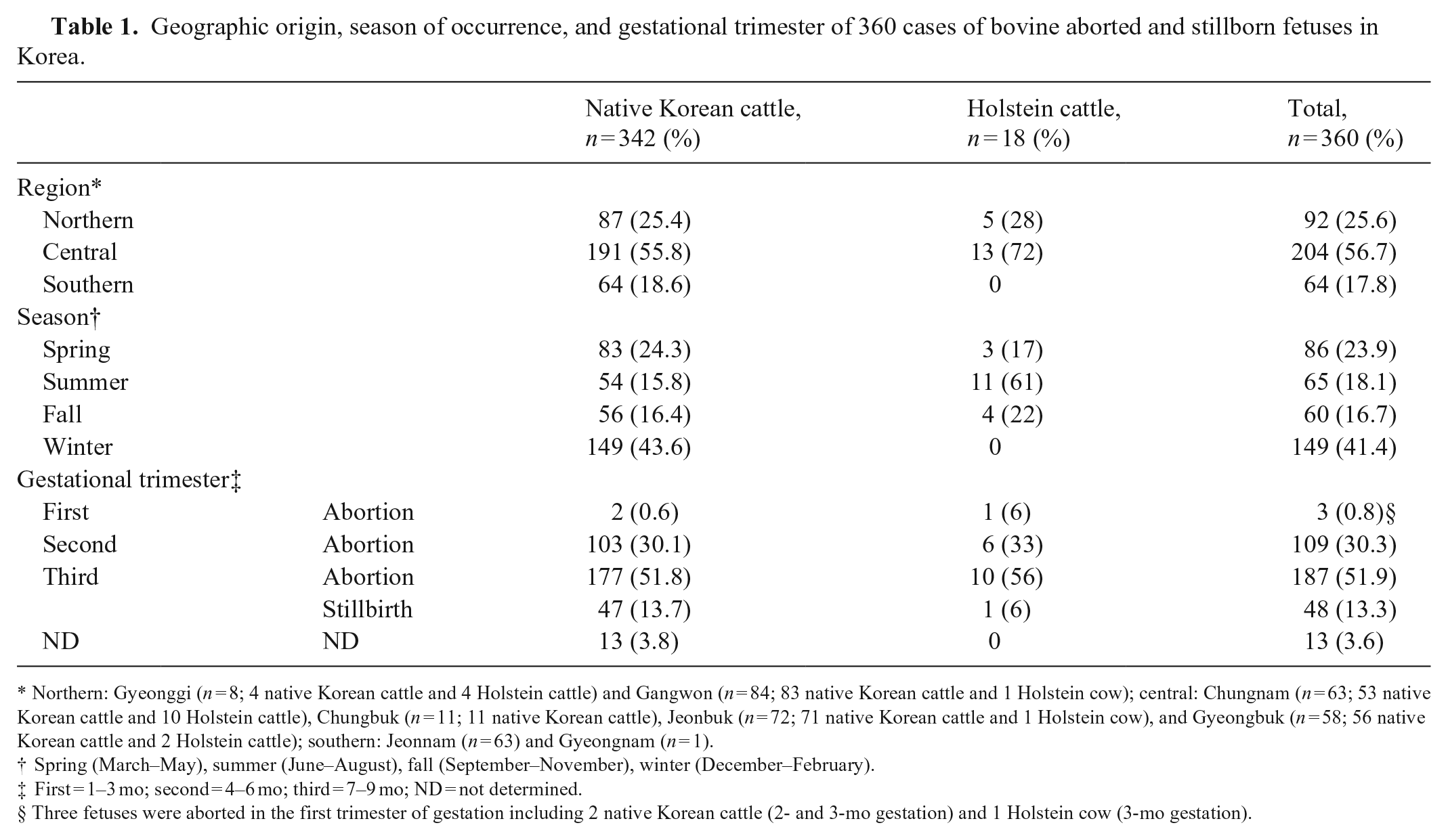
Northern: Gyeonggi (n = 8; 4 native Korean cattle and 4 Holstein cattle) and Gangwon (n = 84; 83 native Korean cattle and 1 Holstein cow); central: Chungnam (n = 63; 53 native Korean cattle and 10 Holstein cattle), Chungbuk (n = 11; 11 native Korean cattle), Jeonbuk (n = 72; 71 native Korean cattle and 1 Holstein cow), and Gyeongbuk (n = 58; 56 native Korean cattle and 2 Holstein cattle); southern: Jeonnam (n = 63) and Gyeongnam (n = 1).
Spring (March–May), summer (June–August), fall (September–November), winter (December–February).
First = 1–3 mo; second = 4–6 mo; third = 7–9 mo; ND = not determined.
Three fetuses were aborted in the first trimester of gestation including 2 native Korean cattle (2- and 3-mo gestation) and 1 Holstein cow (3-mo gestation).
Distribution of agents and conditions detected
Our diagnostic rate for the 360 aborted (112 of 299; 37.5%) and stillborn (22 of 48; 45.8%) bovine fetuses was 140 of 360 (38.9%) at 102 different farms. The diagnostic rate (42 of 86; 48.8%) in groups with placentas submitted, including groups Ⅱ (39 of 83; 47.0%) and Ⅲ (3 of 3; 100%), was significantly higher (p = 0.03) than in group Ⅰ (98 of 274; 35.8%). Of the 140 cases with a final diagnosis, 124 of 140 (88.6%) were from infectious causes, and 16 of 140 (11.4%) were from non-infectious conditions. The proportion of infectious cases in groups Ⅱ (30 of 83; 36.1%) and Ⅲ (3 of 3, 100%) were relatively higher than that of group Ⅰ (91 of 274; 33.2%). According to the frequency outcomes of the etiologic diagnosis (Fig. 1) of the 360 aborted and stillborn cases, the most frequently detected agents in all regions were viruses (60 of 113; 53.1%), followed by bacteria (47 of 113; 41.6%) and protozoa (6 of 113; 5.3%). Non-infectious conditions included malformations (n = 7) and miscellanea (n = 9; Fig. 2). N. caninum was not identified in the southern region of the country (Fig. 1). The detection rates were higher in Holstein cattle (10 of 18; 55.6%) than in native Korean cattle (130 of 142; 38.0%).
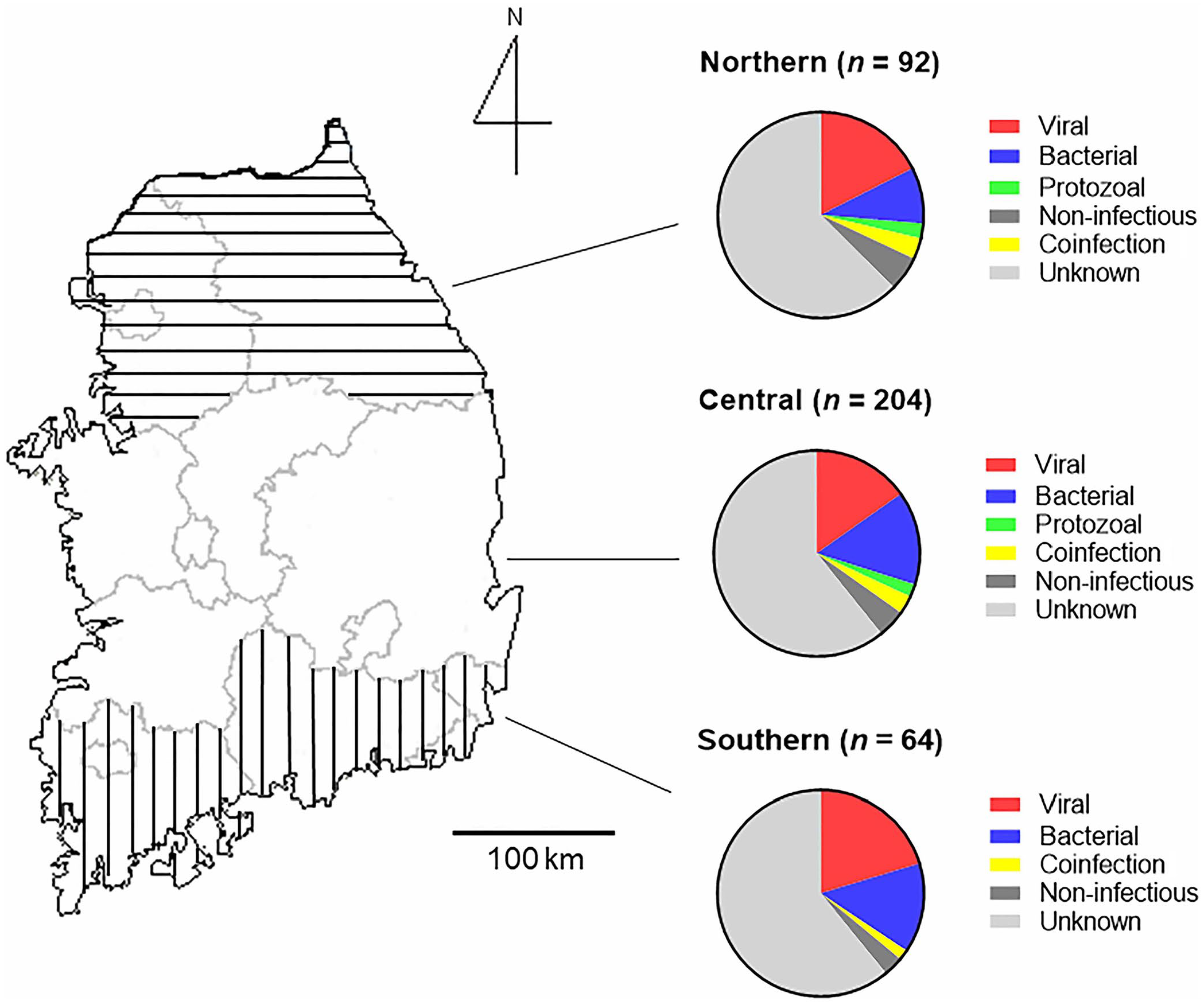
Geographic distribution of cases of bovine abortion in South Korea by etiologic diagnosis.
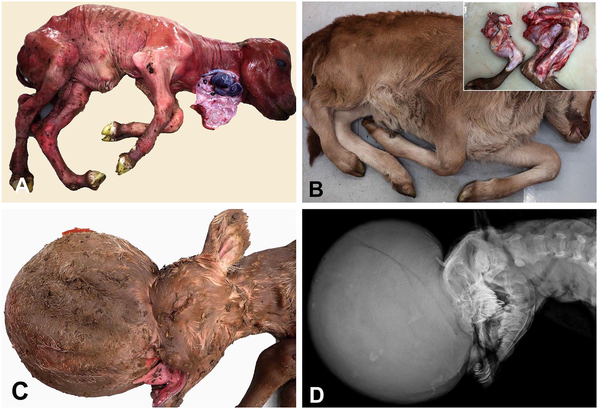
Gross and radiographic findings in cases involving non-infectious conditions associated with bovine abortion in Korea.
Among the infectious agents, a single agent was observed in 113 of 360 (31.4%) cases, followed by 2 pathogens in 10 of 360 (2.8%) and 3 pathogens in 1 of 360 (0.3%) cases (Suppl. Table 1). The most common infectious agent detected was BVDV (65 of 360; 18.1%), followed by C. burnetii (19 of 360; 5.3%), Leptospira spp. (13 of 360; 3.6%), L. monocytogenes (9 of 360; 2.5%), and N. caninum (8 of 360; 2.2%). According to the BVDV subtyping results (n = 47), BVDV1b (n = 21) and BVDV2a (n = 21) were the most frequently detected subtypes, followed by BVDV1a (n = 2), BVDV1n (n = 1), and undetermined (n = 2).
Regarding seasonal outbreaks, the proportion of cases of Leptospira spp. was higher in winter (10 of 149, 6.7%; p = 0.008) than in all other seasons combined (3 of 211; 1.4%). The proportion of cases of L. monocytogenes was higher in spring (6 of 86, 7.0%; p = 0.0026) than in all other seasons combined (3 of 274; 1.1%).
The proportion of cases of Leptospira spp. (11 of 235, 4.7%; p = 0.28) and L. monocytogenes (7 of 235, 3.0%; p = 0.72) in the third trimester were higher than those (2 of 112; 1.8%) in the first and second trimesters. The proportion of cases of N. caninum was significantly higher in Holstein cattle (6 of 18; 33.3%) than in native Korean cattle (2 of 342, 0.6%; p < 0.0001). The proportion of other bacterial abortions with suppurative placentitis or suppurative pneumonia was 15 of 360 (4.2%), including those of Streptococcus spp. (n = 5), Staphylococcus spp. (n = 2), Enterococcus spp. (n = 2), Klebsiella pneumoniae (n = 2), Escherichia coli (n = 1), Trueperella pyogenes (n = 1), Aerococcus spp. (n = 1), and Kocuria spp. (n = 1). There were 2 (0.6%) infectious cases with AKAV, 2 (0.6%) with BoAHV1, and 2 (0.6%) with Brucella abortus. One case of BEFV infection (0.3%) was identified. No infectious abortion cases were identified as caused by C. fetus, C. abortus, Y. pseudotuberculosis, AINOV, CHUV, or IBAV. Among the 16 cases with non-infectious conditions, goiter (7 of 16; 43.8%) with alopecia and thyroid gland hyperplasia was diagnosed most frequently, followed by vitamin A deficiency (2 of 16; 12.5%) and cleft palate (2 of 16; 12.5%).
Gross and histologic findings
Among the 124 cases diagnosed as infectious abortion, 34 of 124 (27.4%) had clear histologic lesions. Suppurative placentitis (n = 14) and suppurative bronchopneumonia (n = 10) were predominant, followed by nonsuppurative hepatitis (n = 4) or myocarditis (n = 3), nonsuppurative encephalitis (n = 2), and suppurative hepatitis (n = 1; Fig. 3). All cases of suppurative inflammation were caused predominantly by bacterial pathogens. Icterus was not present in any of the fetuses with Leptospira spp. infection. Suppurative placentitis (n = 14) was observed in the cases caused by L. monocytogenes (3 of 9; 33.3%), C. burnetii (4 of 19; 21.1%), Leptospira spp. (1 of 13; 7.7%), and other bacterial species (6 of 15; 40.0%), including Streptococcus spp. (n = 2), E. coli (n = 2), Staphylococcus spp. (n = 1), and Kocuria spp. (n = 1). Suppurative bronchopneumonia (n = 10) was observed in 1 case of brucellosis and 9 cases of various bacterial species, including Streptococcus spp., E. coli, Staphylococcus spp., Aerococcus spp., Enterococcus spp., and T. pyogenes. No etiologic agent was identified in 11 cases with histologic lesions, such as suppurative bronchopneumonia (n = 7) and suppurative placentitis (n = 4). In addition, we did not observe fungi (hyphae and yeast) or Sarcocystis cysts in the tissue samples examined in our study.
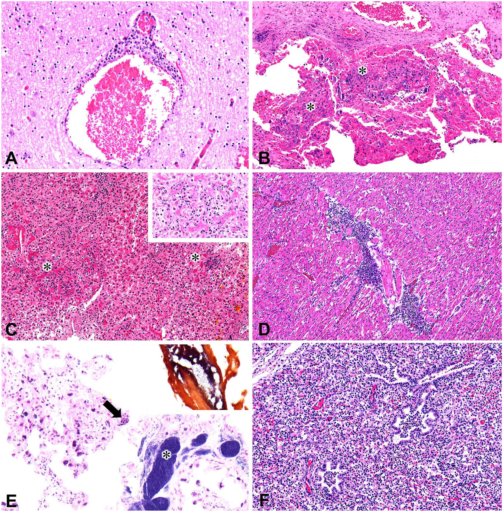
Histopathologic findings in representative infectious cases of bovine abortion.
Considering the main infectious etiologies detected, among the 65 cases with BVDV, 4 cases had nonsuppurative hepatitis, 2 had nonsuppurative myocarditis, and 2 had nonsuppurative encephalitis. In addition, suppurative bronchopneumonia (n = 3) and suppurative placentitis (n = 1) were found in coinfections with BVDV and bacterial species, including Aerococcus spp., K. pneumoniae, and Streptococcus spp.; no lesions were identified in 53 cases. Among the 19 C. burnetii cases, 4 cases had suppurative placentitis; 15 cases had no lesions. Among leptospirosis cases (n = 13), 1 case of suppurative hepatitis and 2 suppurative placentitis cases (one coinfected with L. monocytogenes) were found; 10 cases had no lesions. Among L. monocytogenes cases (n = 9), 3 cases of suppurative placentitis were confirmed, 1 from a L. monocytogenes single infection and the others from coinfection with Leptospira spp. and N. caninum. However, in 6 cases, no lesions were observed. Among neosporosis cases (n = 8), 6 cases had no identifiable lesions and 2 cases had suppurative placentitis (coinfected with L. monocytogenes) and nonsuppurative myocarditis. Of the 2 cases of B. abortus infection, 1 had suppurative bronchopneumonia, whereas no lesion was identified in the other. No lesions were found in the cases of AKAV (n = 2), BoAHV1 (n = 2), and BEFV (n = 1) infection. None of the cases of BVDV, AKAV, and BEFV infection had detectable CNS malformations.
Detection of antibody to infectious agents in body fluid samples of aborted and stillborn fetuses and maternal sera
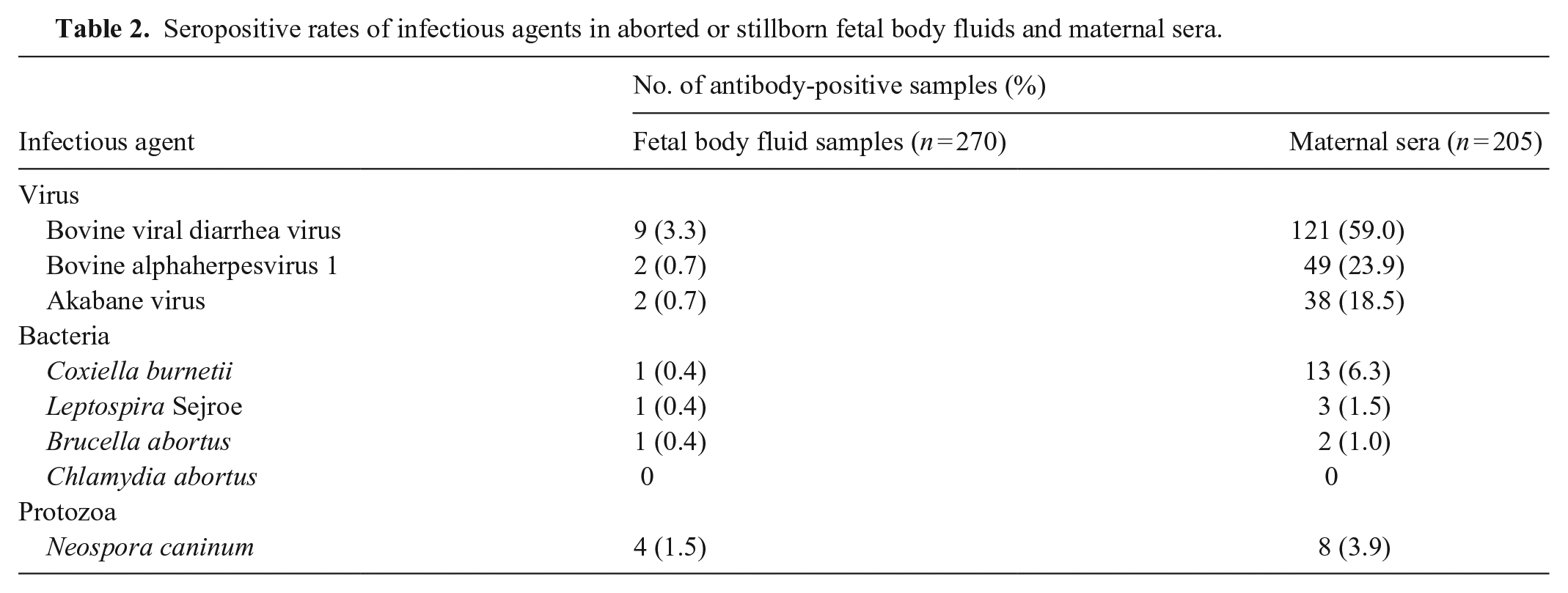
Of the 270 fetal body fluid samples examined, 9 of 270 (3.3%) were positive for BVDV, followed by N. caninum (4 of 270; 1.5%). Antibodies against BoAHV1 (2 of 270; 0.7%), AKAV (2 of 270; 0.7%), C. burnetii (1 of 270; 0.4%), L. Sejroe (1 of 270; 0.4%), and B. abortus (1 of 270; 0.4%) were detected to a lesser extent. Among the 205 maternal sera tested, 121 of 205 (59.0%) were positive for BVDV antibodies, followed by BoAHV1 (49 of 205; 23.9%) and AKAV (38 of 205; 18.5%) antibodies. C. burnetii (13 of 205; 6.3%), N. caninum (8 of 205; 3.9%), L. Sejroe (3 of 205; 1.5%), and B. abortus (2 of 205; 1.0%) specific antibodies were detected in a small number of cases. All fetal body fluid and maternal sera were negative for C. abortus and other Leptospira serovars, except for Sejroe (Table 2).
Seropositive rates of infectious agents in aborted or stillborn fetal body fluids and maternal sera.
Vitamin A quantification in serum
In the 2 dams tested, the vitamin A concentrations were low (0.1 and 0.28 µmol/L, respectively; adult cattle RI: 1.4–1.75 µmol/L).
Discussion
Our diagnostic rate of bovine abortion was 140 of 360 (38.9%) cases, which was within the range reported in most studies.23,30 However, our diagnostic rates might be overestimated compared with those of other studies in which the diagnosis in a given case was based on the detection of the pathogen plus the identification of lesions typically induced by the detected abortifacient pathogens. The diagnostic rates reported in some studies from Korea and California were > 50%.3,12 The disparity in diagnostic rates between our study and a 2000 Korean study is likely the result of the high proportion of N. caninum (21.1%, mostly in Holstein cattle) and AKAV (6.6%) in the previous study. 12
Submission of placental samples is recommended to provide useful diagnostic information. 3 We found that diagnostic rates for cases in which placentas were submitted were significantly higher than those for cases in which placentas were not submitted. In results from Uruguay, the diagnostic rates of cases with placenta (27 of 49; 55.1%) were slightly higher than the cases with only fetuses (27 of 53; 50.9%). 18 According to a report of bovine abortion in Denmark, detection rates of histologic lesions in placenta (78 of 123; 63.4%) were much higher than those of other tissues (20.4–31.1%), including lungs, liver, heart, and brain. 30
In our study, BVDV was the most frequent abortifacient pathogen, detected in 18.1% of cases, which is higher than the frequency reported previously in Korea. 12 The frequencies of BVDV detection in aborted fetuses in other countries, such as Denmark, the United States, Algeria, Australia, New Zealand, and Uruguay, are considerably lower (0.6–2.9%).3,4,18,20,30 However, BVDV has been detected at a high frequency in blood samples of native Korean calves (87 of 143; 60.8%). 7 Furthermore, diverse BVDV subtypes have been reported in Korea.7,21 In a retrospective study conducted in Korea, BVDV was a major enteric and reproductive pathogen, with a higher frequency of detection in native Korean cattle than in Holstein cattle. 16 These results might contribute to the high frequency of BVDV detection in the bovine abortion cases in our study. To prevent economic losses in the Korean cattle industry, continued monitoring of BVDV and appropriate herd management, including detecting and removing persistently infected animals, are necessary and supported by previous studies and our findings. Although the commercial BVDV vaccines used in Korea contain only the inactivated BVDV1a strain, BVDV vaccines destined to prevent BVDV-associated abortions in Korea should contain other BVDV subtypes.
In our study, the proportion of abortions caused by C. burnetii, Leptospira spp., and L. monocytogenes, which all have zoonotic potential, was relatively higher than that reported in other recent studies from Korea.3,12,30 The frequency of abortions caused by C. burnetii infection was higher in Korea (5.3%), which was similar to that reported in Uruguay (6%), than in Denmark, Finland, New Zealand (0%), California (0.1%), and Algeria (1.67%).3,4,18,20,24,30 The gradual increase of C. burnetii diagnostic rates in humans and livestock from 2013 to 2019 observed by the Korea Disease Control and Prevention Agency 14 is reflected in the relatively high frequency of bovine abortion that we found caused by C. burnetii. Therefore, more attention should be paid to C. burnetii, and continuous surveillance will be necessary to improve animal and human health.
N. caninum has been identified as the most frequent causative agent in aborted bovine fetuses subjected to laboratory investigation in other countries (9–29%).3,12,18,30 Our detection rate of bovine abortion caused by N. caninum infection of only 2.2% is considerably lower than the 21.1% reported in a 2000 Korean study. 12 Moreover, 6 of the 8 (75%) cases involved Holstein cattle in our study, which is consistent with findings from the United States where the proportion of abortions caused by N. caninum in Holstein cattle (35.2%) was higher than that in beef cattle (8.4%). 3 The low frequency of neosporosis in our study might be the result of the relatively small number of requested abortion cases in Holstein cattle owing to the small percentage of Holstein dairy farms (6,360 of 102,990 farms; 6.2%) in the domestic cattle industry in Korea. 16 We found no N. caninum abortion cases or seropositive fetal fluid samples in the southern region. Furthermore, only one seropositive maternal serum sample was identified. The detection rate of abortion caused by N. caninum infection in a previous study of native Korean cattle was higher in Gangwon (northern region) and Gyeongbuk (central region), 16 which is consistent with the relatively high frequency of N. caninum (5 of 8; 62.5%) observed in these regions in our study. In addition, there are only 955 (955 of 6,847; 13.9%) commercial dairy farms with a combined total of 66,592 (66,592 of 444,552; 15.0%) cattle in the southern region. This number is much lower than the number of farms and cattle in both the northern (3,045 farms; 199,911 cattle) and central (2,847 farms; 178,049 cattle) regions, as reported in the livestock statistics from the fourth quarter of 2014 by the Korean Statistical Information Service (https://kosis.kr/eng/, https://kosis.kr/statisticsList). These results might affect the low number of N. caninum abortion cases or seropositive fetal fluid samples in the southern region. Investigation of the geographic differences in N. caninum distribution in the Korean cattle population is beyond the scope of our study.
AKAV, which causes epizootic and sporadic abortions in Korea,12,33 was the second most frequent abortifacient pathogen detected (6.6%). Large-scale epidemics of AKAV and BEFV infection occurred in the southern region of Korea (Jeonbuk and Jeonnam) in 2010.12,19,33 In our study, AKAV and BEFV had relatively low detection rates (0.6% and 0.3%, respectively), perhaps as the result of the large-scale provision of AKAV and BEFV vaccines. 33
We found associations between specific pathogens and seasons or gestational periods. In temperate climates, such as that in South Korea, leptospirosis (fall fever) usually occurs in autumn.2,18 Given that leptospiral abortions occur several months after the infection of the dam, 5 it is assumed that this resulted in the high seasonal occurrence of leptospirosis in the winter in our study. 6 Similar to a study in Latvia, 22 the higher listerial abortion rates in spring compared with other seasons demonstrated the seasonality of listerial abortions.
According to a previous study, 20 gestational periods are associated with different pathogens such as BVDV (all gestational periods), N. caninum (the second trimester of gestation), and bacteria causing abortions (the third trimester of gestation), with the exception of some bacteria such as C. fetus subsp. venerealis. Similarly, we found that the most common gestational period of bacterial abortion caused by Leptospira spp. (11 of 235; 4.7%) and L. monocytogenes (7 of 235; 3.0%) was the third trimester. Because there were only 3 cases (C. burnetii, N. caninum, and unknown cases) from the first trimester, comparing gestational associations with pathogens was not possible.
Limited information is available regarding the non-infectious conditions of bovine abortions. We identified non-infectious conditions in 16 of 360 (4.4%). Among the non-infectious conditions, congenital malformations had a low frequency of 7 of 360 (1.9%), consistent with the findings from studies in Finland (21 of 286; 7.3%) and California (11 of 595; 1.8%).9,24 Seven goiter cases from different farms (2 northern regions: Wonju-si and Pyeongchang-gun; 4 central regions: Seosan-si, Jangsu-gun, Imsil-gun, and Wanju-gun; and 1 southern region: Haenam-gun) occurred in indoor housing farms without a history of exposure to goitrogenic substances or iodine deficiency. Of the 2 cases of vitamin A deficiency from the northern and central regions, 1 case involved a pregnant cow with a history of being exclusively fed concentrated growth-stage feed and dried straw without supplements. Management of feedstuffs containing proper diet formulations is required for prevention of abortion or stillbirth caused by vitamin A deficiency.
The seropositive rates for pathogens for which no vaccines are commercially available in South Korea, such as C. burnetii, N. caninum, and B. abortus, are indicative of maternal exposure to the pathogens, which is consistent with the results in our study, except for Leptospira Sejroe. MAT is the standard serologic test used in the diagnosis of leptospirosis. 31 We observed differences in the overall detection rate (13 of 360; 3.6%) and the fetal (1 of 270; 0.4%) and maternal (3 of 205, 1.5%) serologic results for leptospirosis possibly because of limitations of the MAT in detecting chronic infections in individual animals and abortion cases. 31 Further, MAT titers against Leptospira spp. from fetal body fluid can be very low, and some laboratories use a minimum titer of 1:10 to increase the sensitivity of MAT. 31 MAT titers and cutoff values might have affected the seropositive rates in our study. L. Hardjo and Pomona are the most important Leptospira serotypes associated with bovine abortion. 1 However, L. Sejroe was the only serotype detected in our study, consistent with the seropositive rate of bovine leptospirosis reported in Korea from 2005 to 2006. 10 Further studies regarding the monitoring and pathogenesis of L. Sejroe infection in cattle are required.
Commercial vaccines against BVDV, BoAHV1, and AKAV have been used in the cattle industry in Korea. Compared with the seropositivity rate of 49.8% for AKAV in 2012, the proportion of seropositive dams in our study was low (38 of 205; 18.5%). Warnings against diseases with <30% seropositive rates have been issued to prevent epidemics in Korea, including a nationwide caution and encouraging vaccination against these diseases. 33
We used a range of detection methods to determine the causes of bovine abortion; however, our survey has several limitations. First, the collected samples were not fully representative of the current status within Korean cattle farms, given the lack of submission of placentas (274 of 360; 76.1%), breed, and geographic imbalances of the requested samples. Second, maternal information was missing, including the parity of dams or vaccination status. Third, some viruses (bluetongue virus, bovine parainfluenza virus 3, and bovine polyomavirus 1) were not included in in our study. Fourth, histologic lesions were only identified in 34 of 124 (27.4%) cases among cases with etiologic agents. Suppurative bronchopneumonia and suppurative placentitis, which were suggestive of bacterial infection but without etiologic agents, were observed in 11 of 360 (3.1%) cases. Enhanced laboratory techniques, such as culture-independent methods and microaerobic or anaerobic bacterial cultures, are required to increase the detection of specific bacterial agents (e.g., Ureaplasma spp., Mycoplasma spp.). We did not attempt Mycoplasma or Ureaplasma culture. Serology has several limitations when used for diagnosis, including the variable sensitivity of fetal serology (50–84%), incapability of fetal antibody production before the fifth month of gestation, confusion with antibody titers in vaccinated animals, and unclear periods of maternal exposure to infectious agents.3,30,32 Lastly, fetal body fluids were not recommended for use in the commercial ELISA kits according to the manufacturers’ instructions (only plasma and serum). Therefore, serology using fetal body fluids and maternal sera are only components of abortion testing. 3
Supplemental Material
sj-pdf-1-vdi-10.1177_10406387241239919 – Supplemental material for Laboratory investigation of causes of bovine abortion and stillbirth in the Republic of Korea, 2014–2020
Supplemental material, sj-pdf-1-vdi-10.1177_10406387241239919 for Laboratory investigation of causes of bovine abortion and stillbirth in the Republic of Korea, 2014–2020 by Jongho Kim, Jong Wan Kim, Kyoung-Ki Lee, Kyunghyun Lee, Bok-Kyung Ku and Ha-Young Kim in Journal of Veterinary Diagnostic Investigation
Footnotes
Acknowledgements
We thank all of the participating investigators and farm owners who provided fetal samples to the Animal and Plant Quarantine Agency.
Availability of data and materials
The datasets generated or analyzed during this study can be obtained from the corresponding author upon request.
Declaration of conflicting interests
The authors declared no potential conflicts of interest with respect to the research, authorship, and/or publication of this article.
Funding
Our work was supported by a grant from the Animal and Plant Quarantine Agency, Ministry of Agriculture, Food, and Rural Affairs of the Republic of Korea (N-1543069-2015-99-02). The funding body had no role in the design of the study; collection, analysis, and interpretation of data; or writing of the manuscript.
Supplemental material
Supplemental material for this article is available online.
References
Supplementary Material
Please find the following supplemental material available below.
For Open Access articles published under a Creative Commons License, all supplemental material carries the same license as the article it is associated with.
For non-Open Access articles published, all supplemental material carries a non-exclusive license, and permission requests for re-use of supplemental material or any part of supplemental material shall be sent directly to the copyright owner as specified in the copyright notice associated with the article.