
Abstract
On the basis of the scarcity of reports in the veterinary literature, it appears that Propionibacterium spp. are rarely associated with disease or isolated from cattle tissues. Recently, Propionibacterium spp. has been associated with multifocal abscessation in cattle. This report describes a case of necrosuppurative placentitis and abortion in an adult Holstein cow. Numerous colonies of small, pleomorphic, Gram-positive, rod-shaped bacteria were observed within the fibrin lattice associated with placental lesions and within the fetal atelectatic lung. Propionibacterium acnes was isolated in high numbers from the placenta, fetal lung, and stomach contents. To the authors' knowledge, this is the first report of placentitis associated with propionibacteria in a cow.
Members of the genus Propionibacterium are facultatively anaerobic, non–spore-forming, pleomorphic, Gram-positive rods. Propionibacteria have traditionally been grouped according to habitat. Classical propionibacteria (Propionibacterium freudenreichii) are those found in plants and used in food and dairy products, whereas the acnes/cutaneous group (Propionibacterium acnes) are those found on human skin, in the mouth, and in the intestine. Organisms belonging to this second group are responsible for infections in humans and, less commonly, in animals. 10 In humans, propionibacteria have been implicated in inflammatory acne, 14 sporadic abortion (http://www.fertilitysolution.com/PDF/abort.pdf), endophthalmitis, 1 ocular and periocular infections, 9 and periodontal and dental infections 4 and are linked to medical implant-related devices. 15 Isolation associated with animal disease is less common, but P. acnes has been implicated in fibrinous pericarditis in horses. 11 In cattle, Propionibacterium propionicum was isolated from a lesion resembling actinomycosis, 6 whereas Propionibacterium sp. was also associated with extensive granulomatous lesions in cattle involving the head, thorax, abdomen, pelvic area, and skin. 7 Propionibacterium spp. were also isolated from a case of osteomyelitis/arthritis in a dog. 8 Propionibacterium acnes has been tested extensively as an immune stimulant by inducing a nonspecific, cell-mediated response predominantly by activation of macrophages and release of cytokines that elicit a general increase in immune system activity. 18 Propionibacterium acnes is being tested in pigs as a model for inflammatory acne in humans. 20 To the authors' knowledge, this is the first report of a propionibacteria associated with placentitis and abortion in a cow.
A late-term, aborted Holstein fetal calf and placenta were presented to the Department of Pathobiology, University of Tennessee College of Veterinary Medicine (Knoxville, TN), for postmortem examination. The calf was within the chorioallantois, which had dried and adhered to the body. The carcass was in good condition, with little evidence of postmortem decomposition. No significant gross morphologic abnormalities were noted in the calf. The intercotyledonary areas of the placenta that occupied the uterine body were brown, thickened, and not transparent (resembling Moroccan leather), and this additionally involved the placenta originating from 1 uterine horn. Cotyledons were thickened and showed distinct cupping of the edges and brown necrotic debris within the central core. Adventitial placentation, in which additional smaller cotyledons were seen among the preexisting cotyledons, was also noted. Samples of placenta, calf lung, and stomach contents were submitted for aerobic bacterial culture. Multiple fetal tissues (lung, synovium, jejunum, esophagus, skeletal muscle, adrenal, heart, bone marrow, liver, spleen, kidney, thyroid, umbilicus, brain, and stomach) and placenta were fixed in 10% neutral buffered formalin and processed for routine paraffin embedding, sectioning, and staining with hematoxylin and eosin. Brown and Brenn tissue Gram stains were performed on lung and placental sections. Significant microscopic lesions were observed in the fetal calf lungs, which were atelectatic and contained numerous multifocal variably large colonies of pleomorphic Gram-positive rods accompanied by few multifocal necrotic cells identified by karyorrhexis and karyolysis, as well as occasional neutrophils, and squamous cells within the alveoli and larger airways (Fig. 1).
The allantoic surface of the placenta was characterized by a marked regionally extensive ulceration with fibrin deposition and neutrophilic infiltration and accumulation of inflammatory and necrotic cell debris with karyorrhexis and karyolysis, as well as mild congestion, hemorrhage, and edema of the underlying stroma (Fig. 2). The chorionic surface had multifocal to coalescing areas of marked necrosis and loss of villous architecture with necrotic cell debris, as well as neutrophils and fewer lymphocytes, plasma cells, and macrophages. Karyorrhectic debris and bacterial colonies were embedded within the fibrin lattice. The bacteria were small (≤0.5 μm in diameter), irregular, and rod-shaped and demonstrated a Gram-positive staining reaction (Fig. 3).
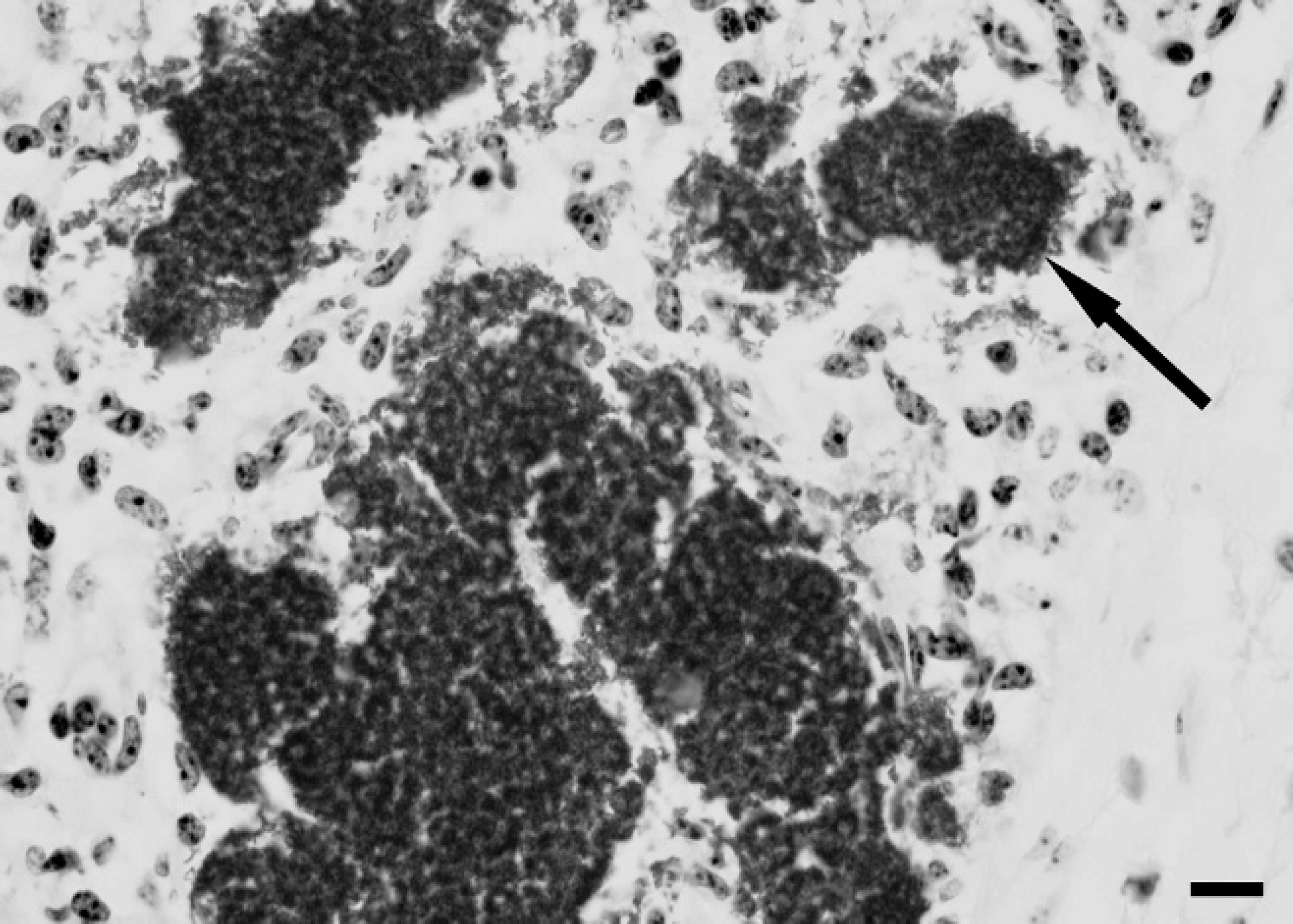
Lung. Atelectatic fetal lung with large aggregates of Gram-positive, pleomorphic bacterial rods within alveoli (arrow). Gram stain. Bar = 25 μm.
Aerobic bacterial culture, Mycoplasma culture, and fungal culture tests were requested on each of the samples submitted. Placenta and calf lung surfaces were seared, and subsurface samples were aseptically removed for culture. Impression smears of calf lung and placental tissue and a smear of stomach contents contained many small, pleomorphic, Gram-positive rods. Lower numbers of Gram-negative rods, Gram-positive cocci, and large Gram-positive rods were also observed in the placental smear. From each of the samples, heavy growth of very small (<1 mm), gray, nonhemolytic colonies was observed on Columbia agar a plates supplemented with 5% sterile defibrinated sheep blood, after 5 days of incubation at 35°Cin 7% CO2. The size of the colonies did not change appreciably on prolonged incubation. This colony morphotype was recovered in pure culture from the stomach contents and lung, whereas the placental culture contained lighter growth of other bacteria, including Escherichia coli, an Enterococcus sp., and a non-fermentative, Gram-negative rod (not further identified). Because the bacterial colonies isolated in greatest numbers contained small, pleomorphic, Gram-positive rods, similar in appearance to those observed in the histopathologic examination and direct impression smears, they were considered to be of clinical significance.
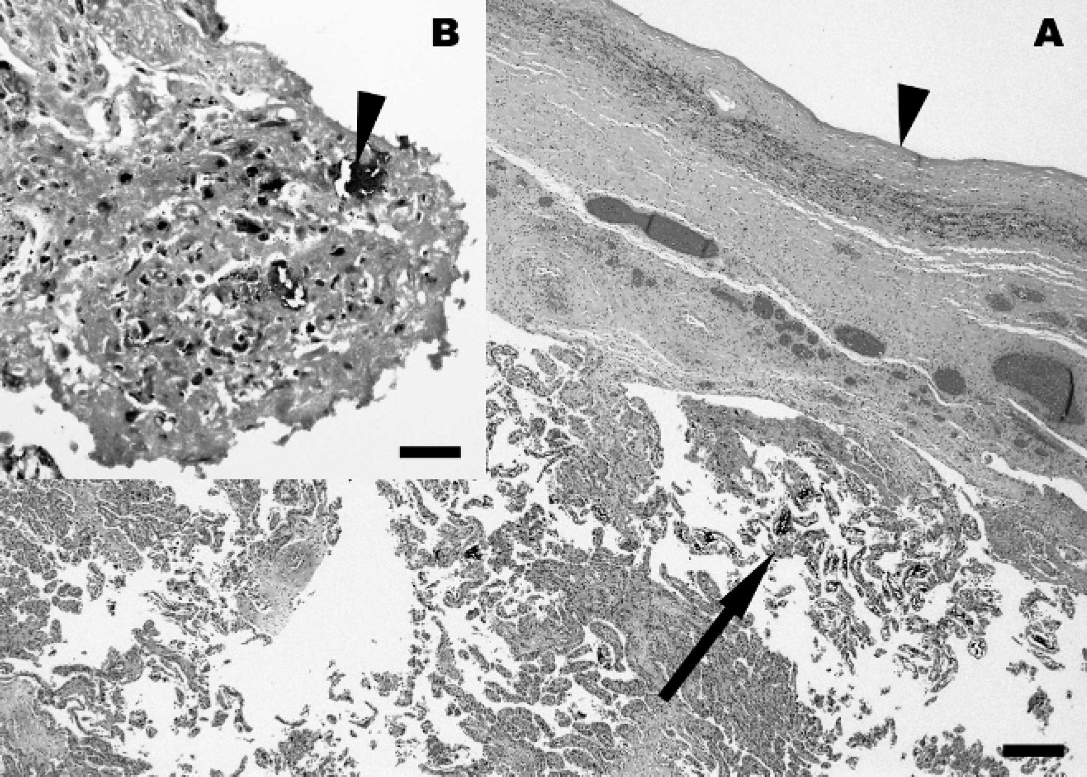
Placenta (chorioallantois).
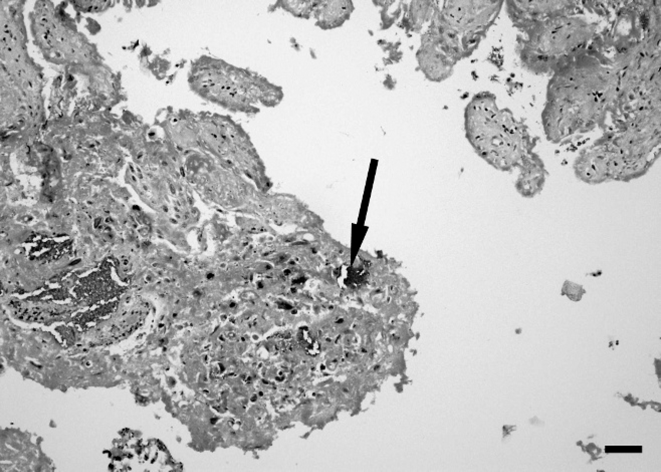
Placenta (chorioallantois). The affected chorionic surface reveals marked villous necrosis and fibrin deposition within which karyorrhectic debris and abundant bacterial colonies are embedded. Gram stain. Bar = 100 μm.
The isolates initially appeared to be catalase positive on blood agar; however, growth was insufficient to conduct additional biochemical tests. On subculture, it was observed that the isolates grew more readily in an anaerobic environment on Centers for Disease Control formula anaerobe agar a containing 5% sheep blood; however, subsequent anaerobic brain heart infusion agar subcultures were also catalase positive. The isolates were identified as P. acnes with a commercial biochemical test kit b designed for use with anaerobes. Biochemical reactions were identical for isolates from each of the 3 tissues, and the isolates were identified as P. acnes with 99.9% probability. Positive test results included a-glucosidase, N-acetyl-glucosaminidase, leucyl-glycine arylamidase, glycine arylamidase, proline arylamidase, arginine arylamidase, serine arylamidase, pyrrolidonyl arylamidase, and indole production. Negative test results were observed for urease, β-disaccharidase, α-arabinosidase, β-galactosidase, β-glucosidase, α-glucosidase, α-fucosidase, and alkaline phosphatase. An equivocal negative or weak positive reaction was observed for phenylalanine arylamidase.
Partial 16S ribosomal RNA (rRNA) gene sequencing, performed as previously described, 2 confirmed the identities of isolates from each of the 3 tissues as P. acnes. BLAST (http://www.ncbi.nlm.nih.gov/blast/Blast.cgi) searches of the GenBank database showed that DNA sequences of the clinical isolates (418 nt, within the 5′ end of the 16S rRNA gene) were identical to each other and to those of P. acnes (GenBank DQ672261). A longer composite sequence (618 nt) of overlapping and nonoverlapping sequences from the 3 isolates had 99% sequence identity to those of multiple P. acnes strains. No other named species were identified by BLAST search.
The Mycoplasma culture was discontinued because of overgrowth of eubacteria. A Geotrichum-like fungus was observed after 1 week on Sabouraud's agar but was not considered significant because fungal elements were not observed in histological examinations, and Gomori methenamine silver stains on sections of lung and placenta were negative. Formalin-fixed, paraffin-embedded lung and placental tissues that were sent to the California Animal Health and Food Safety Laboratory System for immunohistochemistry for Neospora antigen were negative. Likewise, real-time PCR for leptospirosis was negative on DNA extracted from fetal tissues with a commercial blood and tissue kit c following the manufacturer's instructions and published methodology. 17 An investigation implementing virus isolation with Madin-Darby bovine kidney (MDBK) cells was used as a standard assay for bovine viruses, 3 and there were no cytopathic effects after 2 passages from pooled fetal tissues, including placenta, lung, liver, kidney, and thymus. Direct fluorescent antibody assays with the use of antibody coupled with fluorescein isothiocyanate d [FITC] for detection of Infectious bovine rhinotracheitis virus (IBRV), Bluetongue virus, Parainfluenza virus-3, Bovine coronavirus, Bovine herpesvirus 2, Bovine herpesvirus 4, Bovine adenovirus 3, Bovine adenovirus 5, and Bovine viral diarrhea virus (BVDV) were performed on prepared cells, and all were negative. Because the samples had been frozen for an extended period before virus isolation, PCR for BVDV was carried out according to previous methodology and was negative. 5 Nested PCR for IBRV was carried out according to previous methodology after being validated to detect IBRV and was negative. 19
Although there were mixed isolates obtained from the placenta, pure cultures of P. acnes were obtained from the fetal lung and stomach contents, indicating that the other isolates from the placenta were likely environmental contaminants. Whether isolation from the bovine reproductive tract is a consistent feature of P. acnes awaits further confirmation, in that anaerobic culture in routine abortion screens is rarely requested. The source of P. acnes infection in this case is unknown but is suspected to be from an ascending uterine infection after exposure to an environmental contaminant. The dam was not examined, but according to history provided by the owner, the dam was producing milk and there was no suggestion of systemic illness. The farm history was also sparse, but it was noted that no other abortions, stillbirths, or neonatal deaths had been observed. The presence of P. acnes was confirmed in stomach contents, but significant lesions were not observed in other tissues of the calf. This suggests that the abortion may have been primarily the result of the significant placentitis. The Gram-positive, rod-shaped organisms isolated from placental tissue, and those observed in association with the placental lesions, were most likely P. acnes. This report, along with the previous association of propionibacteria with granulomatous lesions in cattle, suggests that propionibacteria could be a significant emerging bacterial pathogen of sporadic diseases of cattle and a cause of sporadic abortion. Although anaerobes are rarely incriminated as causative agents of abortions in livestock animals, mostly because of the omission of anaerobic culture in routine abortion diagnostics, few case reports document anaerobe involvement in bovine abortion including Fusobacterium necrophorum, 16 Fusobacterium nucleatum, 12 and Bacteroides fragilis. 13 This case adds P. acnes to the growing list of anaerobes involved in bovine abortion, and microbiologists should be alerted to the possible identification of P. acnes in clinical samples. Continued reporting of their occurrence will allow further analyses of host range and pathogenicity and will reemphasize the importance of anaerobic culture in bovine abortion diagnostics.
Acknowledgements. The authors thank Ms. Anik Vasington for graphic assistance, Ms. Rupal Brahmbhatt for technical assistance, and Ms. Misty Bailey for editorial support.
Footnotes
a.
BBL™, BD Diagnostic Systems, Sparks, MD.
b.
RapID™ ANA II System, Remel Inc., Lenexa, KS.
c.
QIAamp® DNeasy blood and tissue kit, Qiagen, Valencia, CA.
d.
VMRD Inc., Pullman, WA.