
Abstract
A multicentric basal cell carcinoma was diagnosed in a male multimammate mouse (Mastomys spp.) with widespread cutaneous alterations. Macroscopically, the skin was thickened and extremely wrinkled. Histopathological examination showed multicentric expanding cell-rich tumors composed of basaloid cells interpreted as basal cell carcinoma. Immunohistochemistry detected strong cytokeratin 14 positivity in the epidermal basal layer and in loosely arranged areas of these tumors but only a minimal positive reaction in densely packed areas of tumor cells. Furthermore, samples from the abdomen showed 3 nodular proliferations diagnosed as keratoacanthomas.
Introduction
Mastomys spp., belonging to the genus of rodent and to the family Muridae, is also called multimammate mouse or multi- mammate rat. In Mastomys spp., only very few spontaneously occurring skin tumors have been described. Most of the tumors are classified as keratoacanthomas, squamous cell papillomas, and squamous cell carcinomas (SCCs).23,25
Basal cell tumors, in general, are cutaneous neoplasms arising from the basal cell layer of the epidermis. They occur occasionally in rats and rarely in mice.2,3 In domestic animals, basal cell tumors can be found commonly in cats and uncommonly in dogs and horses, and they are rarely found or have not been described in other species. 5 The benign form of basal cell tumors, also called basal cell epitheliomas or basaliomas, in rodents has been reclassified as trichoepitheliomas in domestic animals.2,3,5,10 The malignant form, basal cell carcinoma (BCC), has a very low incidence of spontaneous occurrence in laboratory rats and mice (0.14% and 0.1%, respectively).13,17 In domestic animals, BCC is described in cats most frequently. In other domestic animals, BCCs are rare or have not been described. 5 In human beings, BCC is the most common malignant tumor, with increasing incidence.7,26,28
Basal cell carcinomas are predominantly solitary but can occasionally occur as multiple masses. In domestic animals, multicentric BCCs have been reported but occur in only 1% of the cases. Murine BCC can be multicentric underneath a hyperplastic epidermis.3,5,8 Histologically, tumor cells resemble basal cells, are intensely stained with hematoxylin, and have scant cytoplasm. Basal cell carcinomas do not metastasize but show extensive local invasion. In mice, mitotic figures are rare, according to the World Health Organization classification of rodent tumors, whereas in rats and domestic animals, they may be numerous.2,3,10 A solid and a basosquamous form can each be distinguished in rats. 2 In domestic animals, BCCs are subdivided into infiltrative and clear cell BCC. The latter is an uncommon variant that is characterized by tumor cells with a clear or finely granular cytoplasm. 10
Transgenic mice, which overexpress smoothened or sonic hedgehog proteins spontaneously, develop skin lesions that resemble human BCC. 16 Keratoacanthomas are benign cutaneous neoplasms arising from the hair follicle or from the epidermis in mice and rats, respectively.2,3 An experimental infection with Mastomys natalensis papillomavirus (MnPV) induces benign skin tumors in Mastomys spp., especially keratoacanthomas and papillomas. 15
A 1.5-year-old male multimammate mouse (Mastomys spp.) kept in a private household developed large-scaled skin lesions with thickening of the epidermis, formation of pustules, and alopecia on head, legs, back, and anogenital region. Additionally, the animal showed few small nodules with a retracted surface in the abdominal skin. Fungi and ectoparasites as well as antibodies against Ectromelia virus (ECTV) were not detected at clinical investigations. Nevertheless, treatment included antiparasitic (ivermectin) and antimycotic (griseofulvin), antibiotic (enrofloxacin a ), pain relief (meloxicam b ), and immunomodulatory drugs. c After 6 months without improvement, euthanasia of the animal was elected.
Materials and methods
Following necropsy, samples of the skin and other tissues (lung, heart, liver, kidneys, spleen, stomach, intestine, pancreas, and brain) were fixed in 10% nonbuffered formalin and embedded in paraffin wax. Sections (3–4 µm) were stained with hematoxylin and eosin (HE).
For immunohistochemical characterization of the tumor cells, antibodies against cytokeratin (CK)1, CK4, CK5, CK6, CK10 and CK14 as well as against cluster of differentiation (CD)34 and smooth muscle actin (SMA) were used (Table 1). Streptavidin–biotin complex method with Fast Red d as chromogen was applied for detection of antibody binding. 24 Paraffin-embedded skin samples were used for making ultrathin sections (approximately 0.9 µm thick) and contrasted with uranyl acetate and lead citrate.
Primary antibodies used to characterize basal cell carcinoma.*
CK = cytokeratin; CD = cluster of differentiation; SMA = smooth muscle actin. Sources: Covance Research Products Inc., Dedham, MA; Sigma-Aldrich, St. Louis, MO; BD Pharmingen™, BD, Franklin Lakes, NJ; Dako North America Inc., Carpinteria, CA.
Results
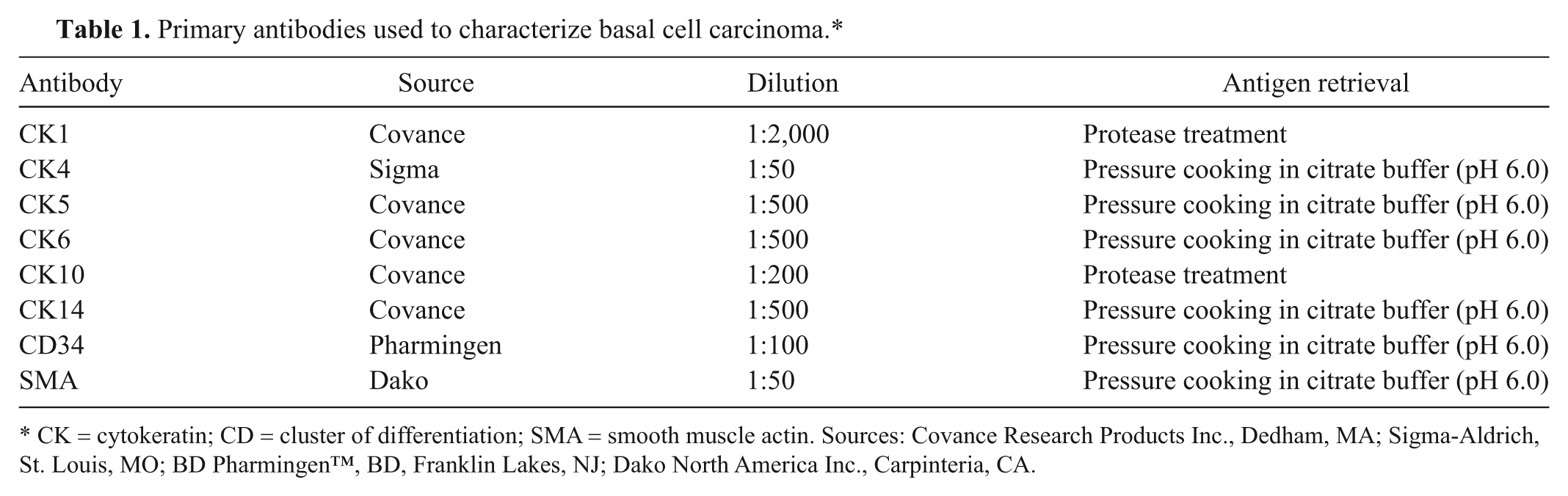
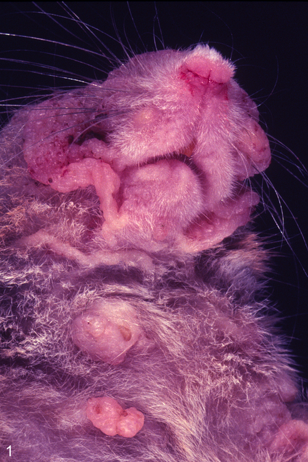
Postmortem examination revealed multiple extensive, poorly demarcated, soft, nonhaired masses in the oronasal region, cheeks, both ears, legs, abdomen, and anogenital region that diffusely infiltrated the epidermis and corium. The skin was markedly thickened, wrinkled, and multifocally covered by reddish-brownish crusts (Figs. 1, 2). The cut surface was homogenous and white, with a soft consistency (designated tumor no. 1). Additionally, 3 nodular masses with an indented surface, measuring approximately 0.5 cm in diameter, were found in the abdominal skin (tumor no. 2).
Multimammate mouse (Mastomys spp.). Skin on cheeks and chest with basal cell carcinoma. A prominent thickening and wrinkling of the skin in multiple locations is evident.
Multimammate mouse (Mastomys spp.). Skin of right hind limb with basal cell carcinoma. A prominent thickening and wrinkling of the skin in multiple locations is evident.
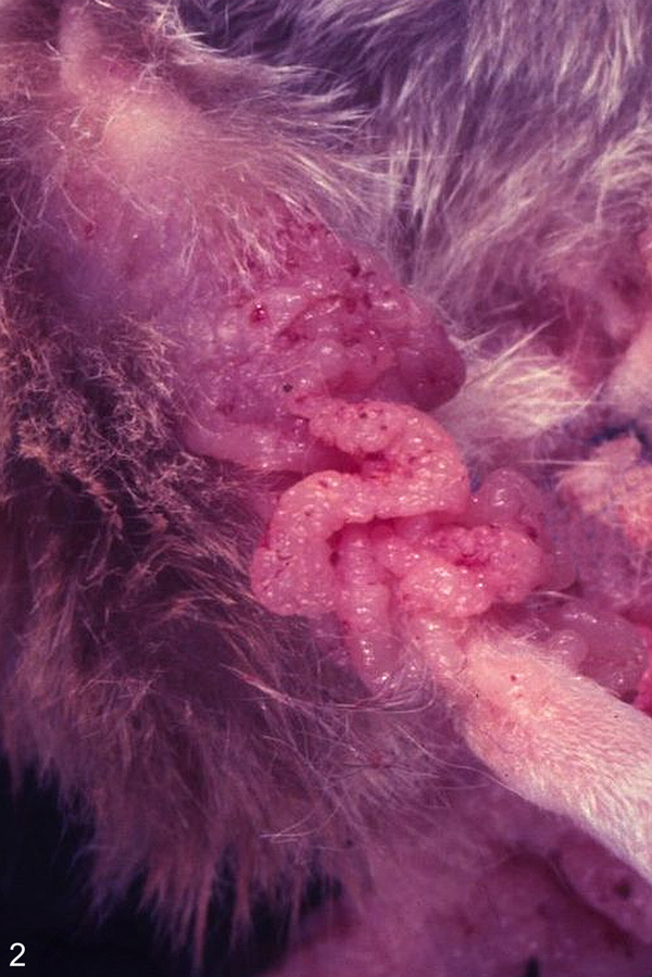
The histopathological examination of multiple masses (tumor no. 1) of the skin revealed poorly demarcated, cauliflower-like masses supported by a fibrous stalk (Fig. 3). The closely packed cells were arranged in sheets, divided by a delicate fibrous stroma, and expanded in the subcutis. Tumor masses consisted of polygonal cells, ranging from oval to elongated, which were mostly dark and basophilic but showed a varying staining intensity. These basaloid cells showed scant cytoplasm and a large, round to oval nucleus with a single nucleolus (Fig. 4). A mild anisokaryosis and a moderate mitotic rate with 0–2 mitotic figures per high-power field were present. The tumors were diagnosed as multicentric BCC.
Multimammate mouse (Mastomys spp.). Cutaneous basal cell carcinoma. The epidermis is expanded by tumor cells. Hematoxylin and eosin. Bar = 500 µm.
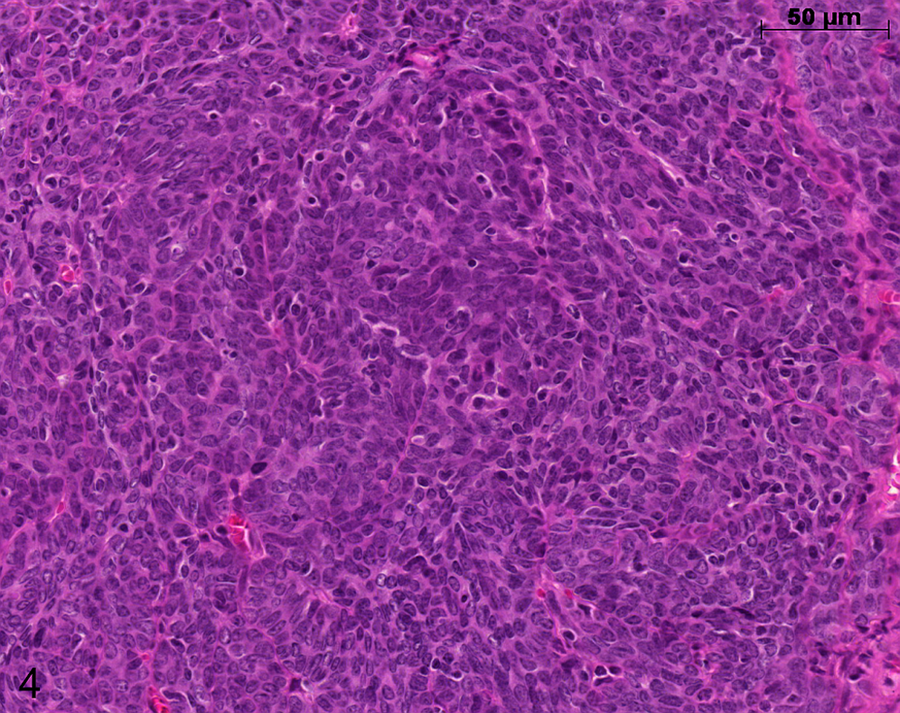
Multimammate mouse (Mastomys spp.). Cutaneous basal cell carcinoma. Tumor cells have a basaloid appearance and scant cytoplasm. Hematoxylin and eosin. Bar = 50 µm.
Additionally, the samples from the abdomen (tumor no. 2) consisted of approximately 3 mm in diameter, large, keratin-filled structures with a pore. The structures were well demarcated by a thin fibrous capsule and were lined by a multilayered, basophilic epithelium consisting of large, basophilic, oval cells with a moderate amount of basophilic cytoplasm. The structures contained a large, round, light basophilic nucleus with a single, prominent nucleolus. The mitotic rate was low (0–1 mitotic figures per high-power field). Intraluminal large masses of lamellated keratin were present. These nodular masses were diagnosed as keratoacanthoma. A mild, lymphohistiocytic dermatitis, partly with superficial serocellular crusts, and a moderate orthokeratotic hyperkeratosis were visible in nontumorous skin areas. The animal showed minimal lymphoplasmacytic infiltrates in stomach, intestine, and salivary gland as well as a focal cortical adenoma of 1 adrenal gland.
Immunohistochemically, a strong positivity of BCC tumor cells (tumor no. 1) for CK14 could be detected, although only in areas where tumor cells were loosely arranged (Fig. 5). Densely packed areas of the tumors showed minimal positive reaction for CK14. The tumor was partly covered by unaltered CK10- and CK1-positive suprabasal cells (Fig. 6). The tumor cells of the BCC stained negative for CK1, CK4, CK6, and CK10 as well as for CD34 and SMA. Connective tissue around the tumor exhibited a slight positivity for CD34. Expression of SMA was only detected in the vessel walls, not in basaloid tumor cells.
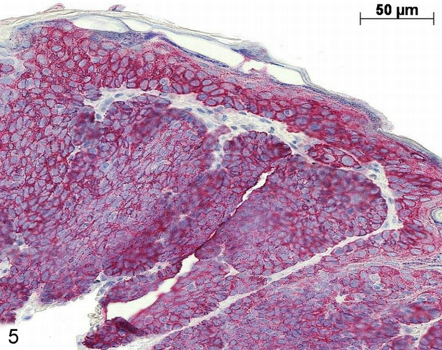
Multimammate mouse (Mastomys spp.). Cutaneous basal cell carcinoma (BCC). Cytokeratin (CK)14 antigen is strongly expressed by BCC tumor cells. Immunohistochemistry (CK14), Fast Red, counterstained with hematoxylin. Bar = 50 µm.
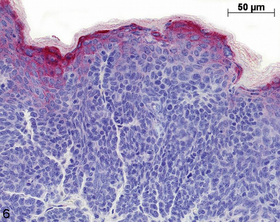
Multimammate mouse (Mastomys spp.). Cutaneous basal cell carcinoma (BCC). Cytokeratin (CK)10-negative BCC tumor cells covered by normal CK10-positive cells. Immunohistochemistry (CK10), Fast Red, counterstained with hematoxylin. Bar = 50 µm.
In control animals, epidermal basal layers were CK14 positive and suprabasal cells showed CK10 and CK1 expression. Keratoacanthomas (tumor no. 2) showed overall a similar CK expression pattern to control skin. Detailed information about immunohistochemical results is given in Table 2. In ultrathin sections of various localizations of tumorous and nontumorous skin, no viral particles could be detected by electron microscopy.
Primary antibodies, patterns of positivity in normal skin according to the literature, and immunohistochemical results of control skin, basal cell carcinoma (BCC), and keratoacanthoma.
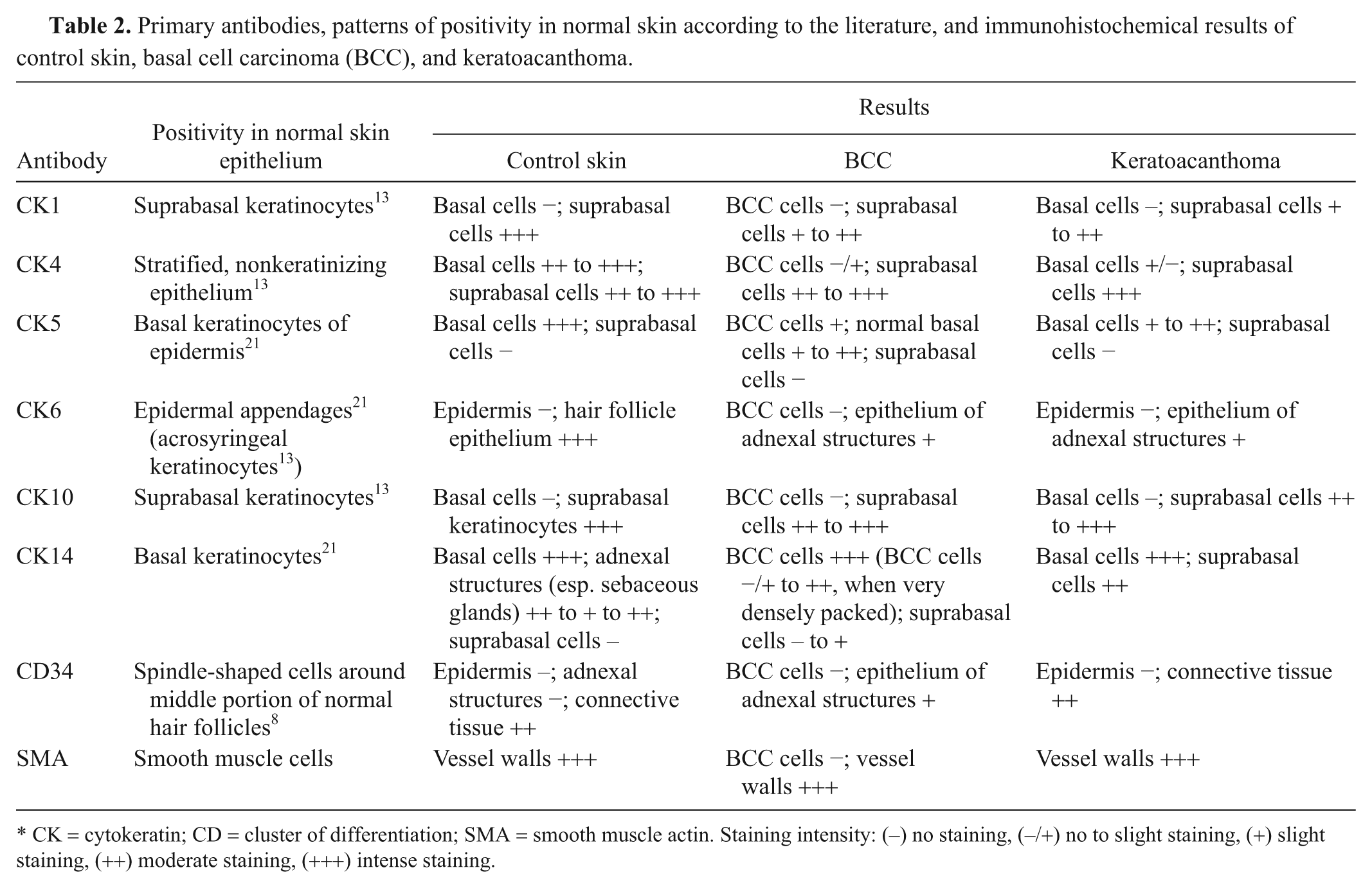
CK = cytokeratin; CD = cluster of differentiation; SMA = smooth muscle actin. Staining intensity: (–) no staining, (–/+) no to slight staining, (+) slight staining, (++) moderate staining, (+++) intense staining.
Discussion
The skin of a male Mastomys mouse showed an extreme thickening and wrinkling, which was due to a proliferation of basaloid, CK14-positive cells leading to the diagnosis of BCC. Additionally, 3 keratin-filled structures lined by an epithelium, located in the abdominal skin, were diagnosed as keratoacanthomas.
Macroscopically, murine BCC can appear as bulged nodules or show a granular surface. 8 In the present case, large folded skin areas were present. Typical histological features for malignant BCC are closely packed and intensely basophilic-stained cells that resemble basal cells of the epidermis. The cells are arranged in sheets or strands, often palisading, and are characterized by a scant cytoplasm and a dark blue nucleus. In poorly differentiated carcinomas, the cells might have an elongated or fusiform shape.2,3,10,17 Squamous, trichogenic, or sebaceous differentiation may occur.2,3 The amount of mitotic figures in murine BCC varies, depending on the reference, and may be numerous in rats and domestic animals.2,3,8,10 Major differential diagnoses for BCC include squamous cell carcinoma and benign basal cell tumor. The latter is mostly well demarcated in contrast to the invasive nature of the present tumor masses. Squamous cell carcinomas mostly show nuclear atypia as well as numerous, often bizarre mitotic figures and invasion of the dermis and striated muscle. Keratinization may be present to a varying extent. 2 In the present case, an invasion of the dermis was evident, but keratinization was lacking. Furthermore, an immunohistochemical-positive staining for CK6 is described in SCC, which could not be detected in the present case. 6 Additionally, malignant adenoacanthoma and adenocarcinoma of the mammary gland should be considered as differential diagnoses. However, adenocarcinomas would show adenomatous structures, which were not present in the current case. An undifferentiated mammary carcinoma can be taken into account as well because rodents, such as mouse and rat, show a widespread appearance of mammarian tissue, although an invasion of a mammary carcinoma to head and legs would be uncommon. Moreover, the incidence of mammary tumors in Mastomys spp. is very low. 25
Immunohistochemistry of normal skin shows an expression of CK5 and CK14 by basal keratinocytes and CK1 as well as CK10 by suprabasal keratinocytes. 1 Basal cell carcinomas stain immunohistochemically positive for CK14 and negative for CK1 and CK10. 29 A positive staining for CK14 is indicative for basal cell origin, which supports the diagnosis of BCC in the present case. 20 The lack of immunoreactivity of the densely packed areas could be due to undifferentiation of the tumor cells in these areas.
Cluster of differentiation 34 has been depicted as being strongly expressed by spindeloid cells around trichoepithelioma but not around BCC in human beings. 11 In the present case, only a slight positivity of the surrounding connective tissue could be detected. Smooth muscle actin is regularly expressed by smooth muscle fibers. Interestingly, a strong SMA reactivity could be seen in a number of human BCC, but in the current study, the rodent BCC tumor cells did not stain positive for SMA antigen (Miller RT: 2004, Immunohistochemistry in the differential diagnosis of basal cell carcinoma and squamous cell carcinoma. Available at: http://www.ihcworld.com/_newsletter/2004/2004-12_basal_cell_vs_squamous_v1.pdf. Accessed on March 12, 2012).
Keratoacanthomas arise from epidermal cells in rats and hair follicle epithelium in mice. Cavities filled with lamellated keratin masses, like in the present case, are frequently observed in rodent species.2,3 Keratoacanthomas are reported to be the most common spontaneous skin tumor in laboratory Mastomys spp. 25 In Mastomys natalensis, the incidence of skin tumors is very high because they are induced by an infection with MnPV.9,22 In addition, SCCs and papillomas are etiologically related to an MnPV etiology. 21 After experimental infection with MnPV virions, large amounts of virus particles can be seen in the nuclei of keratinized cells of the stratum granulosum and corneum by electron microscopy. 21 In the present case, however, no viral particles were observed ultrastructurally.
Hamsters develop benign skin tumors after infection with Hamster polyomavirus, which grossly resembles the keratoacanthomas of the animal in the current study and where viral particles are not visible by electron microscopy as well. Polyomavirus-induced tumors present themselves histologically as trichoepitheliomas in contrast to the keratoacanthomas in the present case. Nevertheless, the transition between the 2 skin tumors is fluent and depends on the presence of keratohyalin granules. 4
Large areas of BCC were found adjacent to the localizations diagnosed as keratoacanthoma in the animal in the current study. Therefore, a transition between both neoplasms can be assumed. In mice, a pronounced proliferation of basal cells in keratoacanthomas and, to a lesser extent, has been described. 3
Spontaneously occurring skin tumors in rats, mice, and Mastomys spp. are infrequent.18,23,25 Most of the tumors are classified as keratoacanthomas, squamous cell papillomas, and SCCs.23,25 In a histopathological survey of aged Mastomys spp., only squamous cell papillomas and SCCs were observed, but not BCC. 25 Intracutaneous cornifying epitheliomas arising from the epidermal epithelium have been described as well. 19
Human beings with BCC have a PTCH gene mutation or often have a history of arsenic or radiation exposure. 26 In mice, BCCs are naturally rarely occurring tumors.3,8 Such tumors can be experimentally induced in transgenic mice, which overexpress smoothened or sonic hedgehog proteins in the skin resulting in an overexpression of the transcription factor Gli2.6,12 Furthermore, an inactivation of the patched gene (PTCH ) and the p53 gene can lead to ultraviolet light–induced BCC. 12 In 2010, 13 a spontaneously occurring BCC was described in a young rat. The tumor was solitary and likewise showed a strong positivity for CK14 antigen. 13
Basal cell carcinomas have also been induced experimentally in M. natalensis and mice by application of 7,12-dimethylbenz(a)anthracene (DMBA) and 12-0-tetra-decanoyl-phorbol-13-acetate (TPA) and other mutagenic chemicals.14,27 Squamous cell carcinomas have been induced by the same 2-stage skin carcinogenesis (DMBA+TPA) in transgenic mice that carried an oncogene of MnPV. 9
An unknown toxic substance in the animal’s environment or a genetic defect (e.g., in the PTCH gene) could not be ruled out in the current case and has to be considered tumorigenically. Furthermore, an infection with papillomavirus must be taken into account as cause for the keratoacanthomas, although viral particles were not detected by electron microscopy.
Footnotes
a.
Baytril®, Bayer, Leverkusen, Germany.
b.
Metacam®, Boehringer Ingelheim, Ingelheim, Germany.
c.
Zylexis®, Pfizer, Berlin, Germany.
d.
Biogenex, San Ramon, CA.
Declaration of conflicting interests
The author(s) declared no potential conflicts of interest with respect to the research, authorship, and/or publication of this article.
Funding
The author(s) received no financial support for the research, authorship, and/or publication of this article.