
Abstract
Several plants that contain indolizidine alkaloids, including swainsonine, are toxic to livestock, causing dysfunctional lysosomes and storage disease. Swainsonine induces a neurovisceral disease, known as locoism, in sheep, goats, and cattle, which occurs in several parts of the world, including, but not limited to, the western United States, China, and parts of Australia. In South America, locoism has been described in the Andean region of Argentina affecting sheep, cattle, and llamas. Intoxication by consumption of Astragalus punae was suspected in 4 llamas in Jujuy Province, northwestern Argentina. The grazing area contained abundant specimens of A. punae. The clinical course was ~15 d, and included moderate ataxia, incoordination of hindlimbs, and progressive loss of body condition. Microscopically, fine cytoplasmic microvacuolation was observed in the proximal convoluted renal tubules. Ultrastructurally, these changes consisted of severely dilated lysosomes. Swainsonine was detected in stem and leaf samples of A. punae at a concentration of 0.06%. Based on clinical history and signs, histologic and ultrastructural changes, and plant analysis, a diagnosis of swainsonine toxicosis caused by consumption of A. punae was made, which has not been reported previously, to our knowledge.
Lysosomal storage diseases are acquired or inherited disorders characterized by dysfunctional lysosomes, which are intracytoplasmic accumulations of undegraded substrates that lead to cell death. 1 Acquired lysosomal storage diseases include locoism, which is caused by ingestion of plants containing indolizidine alkaloids, of which swainsonine and its N-oxide are the most critical given their ability to inhibit lysosomal α-mannosidase and Golgi α-mannosidase II. 20 Swainsonine acts as an α-mannosidase and mannosidase II inhibitor, altering glycoprotein processing and resulting in lysosomal storage disease. Swainsonine is present in several plant families including 6 genera (Ipomoea, Turbina, Astragalus, Oxytropis, Swainsona, Sida) of the Convolvulaceae, Fabaceae, and Malvaceae families. Several of these plants have been associated with cases of locoism in several animal species.9,12,17,19 Locoism is an acquired α-mannosidosis that is associated with consumption of plants of the genus Astragalus,12,17 Sida, 13 Oxytropis, 19 Swainsona, 3 and Ipomoea. 11 Approximately 70 species of the genus Astragalus are native to Argentina,6,7 where natural intoxication by Astragalus pehuenches in sheep 17 and cattle 10 in the Patagonia region, and Astragalus garbancillo var. garbancillo in sheep 12 and llamas 9 in the northwest region of the country have been described.
Astragalus punae (known locally as “mulato”) is a plant found only in the Susques Department (equivalent to a county) of Jujuy Province within the Puna region, 8 which is a grassland plateau in northwestern Argentina, at ~4,500 m above sea level (http://buscador.floraargentina.edu.ar, Spanish). Although for many years there was anecdotal evidence that A. punae is toxic (authors’ unpublished observation), this was never proven.
We describe here a cluster of cases of intoxication by A. punae in llamas. The cases occurred in Jujuy Province. Breeding llamas is the most relevant livestock activity for smallholding producers in the Puna region, and Jujuy Province has the largest number of llamas in Argentina. 14
One of the authors of this paper (R. Marin) visited a smallholding producer in a remote area near the town of El Toro, Susques Department, Jujuy Province, in the winter of 2019. The producer had a herd of 124 llamas grazing on a natural pasture with an abundance of A. punae (Fig. 1). At the time of the visit, 4 adult llamas had been observed for ~15 d losing nutritional condition and with moderate ataxia. The producer reported that similar clinical signs had been observed in ~3–5% of the herd in late winter and spring for the past several years. Eight producers from neighboring properties, in which A. punae was present, were interviewed and asked about histories of incoordination and loss of condition in their llamas. They all reported variable prevalence of similar clinical signs in grazing areas in which A. punae was present. The general consensus was that llamas that had consumed A. punae lost condition over several weeks and developed gait incoordination. Although spontaneous death was rarely observed, severe loss of condition prompted euthanasia in most cases. In addition, low reproductive indexes were also reported by most producers. All producers reported that their llamas appeared to develop a predilection for A. punae.
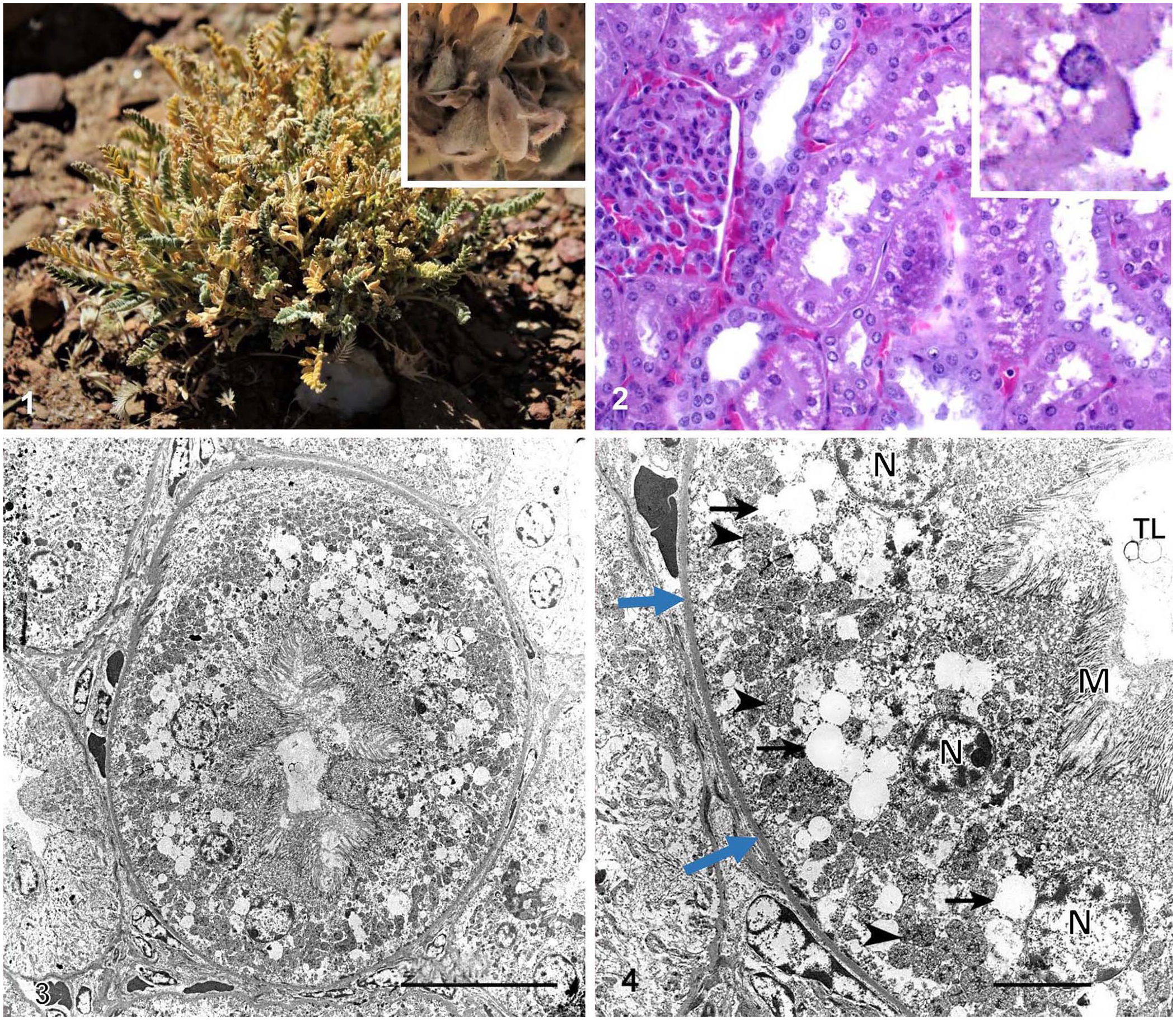
Astragalus punae and tissues from a llama intoxicated with this plant.
The 4 affected llamas were examined clinically. They were all standing and were able to walk but had ambulatory abnormalities, including moderate symmetric ataxia of all 4 legs, which was most marked in the hindlimbs, slight hypermetria, and slight head tremors. They all stumbled frequently when walking. In response to dorsiflexion of the head by the examining veterinarian, one of the animals fell to the ground, rising up shortly, and walking with marked hypermetria for a few minutes, after which the signs became milder. The other 3 animals did not fall to the ground after dorsiflexion of the head and neck. The lumbar and hindlimb muscles of the 4 affected llamas had moderate and diffuse atrophy.
Whole blood and serum were collected from the llama that collapsed after dorsiflexion of the neck. A CBC and serum chemistry revealed no abnormalities. No cytoplasmic vacuolation was identified in monocytes in peripheral blood smears. 20 This animal was euthanized with an overdose of sodium pentobarbital. Although the owner was asked for authorization to euthanize the 3 other affected animals, permission was not granted. A postmortem examination was performed on the euthanized llama. The carcass was in poor nutritional condition, with very small fat reserves and moderate but generalized muscle atrophy, which was most marked in the lumbar area and hindlimbs. No other significant gross changes were observed. In particular, no plant remains with exomorphologic characters compatible with Astragalus spp. were observed in the gastric compartments.
The whole brain, and samples of liver, kidney, spleen, lung, adrenal, pancreas, thyroid and pituitary glands, small and large intestine, heart, and skeletal muscle were collected and fixed in 10% neutral-buffered formalin, pH 7.2, for 24 h. The brain was then sliced at 1-cm intervals and fixed in fresh formalin for an additional 7 d before subsamples were taken from cerebral cortex, corpus striatum, thalamus, midbrain at the level of anterior colliculi, pons, cerebellar peduncles, cerebellum, and medulla oblongata at the level of the obex. Tissue samples were routinely processed for the production of 4-µm thick H&E sections. Selected sections of brain were also stained with Holmes, Luxol fast blue, and periodic acid–Schiff (PAS). In addition, formalin-fixed samples of kidney, brain, thyroid, and liver were processed for transmission electron microscopy (TEM). For TEM, ~2-mm3 tissue fragments from these organs were post-fixed in 2.5 mL of 0.1 M cacodylate–buffered glutaraldehyde (Electron Microscopy Sciences), followed by post-fixation in 1% 0.1 M cacodylate–buffered osmium tetroxide (Electron Microscopy Sciences). Using a graded series of ethanol, tissue fragments were dehydrated and embedded in resin and sectioned on an ultra-microtome (UC6; Leica). Sections (70–90-nm thick) were collected on 100-mesh nickel grids (Electron Microscopy Sciences) and stained with 5% uranyl acetate for 20 min and Sato lead citrate for 6 min. Stained sections were examined under a transmission electron microscope (1400 Plus; JEOL). Images were captured with a camera and software (Gatan).
Microscopically, fine cytoplasmic vacuolation was observed in the epithelium of most proximal convoluted renal tubules. These vacuoles were distributed more or less homogeneously throughout the cytoplasm (Fig. 2). No significant microscopic abnormalities were observed in any of the other tissues examined. Ultrastructurally, the cytoplasmic microvacuolar degeneration observed in the proximal convoluted renal tubules on light microscopy consisted of variably sized electron-lucent secondary lysosomes of up to 2.67 µm diameter (Figs. 3, 4). The fine structure of brain, thyroid, and liver was unremarkable, although mild postmortem artifacts were present.
Immunohistochemistry (IHC) for West Nile virus (WNV), rabies virus (Lyssavirus rabies), and Chlamydia spp. was performed on sections of cortex, brainstem, and cerebellum, as described previously.5,15,18 IHC for all infectious agents studied was negative in the test specimens and negative controls.
The area that the animals had grazed for the previous several weeks was examined the same day that the autopsy of the llama was performed. A. punae comprised ~30% of the vegetation in the grazing area. Many of these plants had indications of having been grazed. Other plants present included Tetraglochin cristatum, Junellia seriphioides, Ocyroe armata, Jarava ichu, Festuca dolichophylla, and Adesmia spp. No other species of Astragalus were found in this area. Specimens of A. punae in the fruiting stage were collected from the paddock in which the affected animals described here had been grazing, press-dried, and then submitted for identification to the Natural Resources Research Center, National Institute of Agricultural Technology, Argentina, where they were identified as A. punae and recorded as specimen BAB 97039 (Digitization BAB00010832). In addition, specimens of A. punae were collected in the fall of 2020 from the area in which the affected llamas described here had been grazing; the specimens were analyzed for swainsonine using methods described previously.2,4 The concentration of swainsonine on a composite sample of leaves and fine stems was 0.06% (dry weight basis).
A diagnosis of intoxication by A. punae was established based on clinical signs, microscopic and ultrastructural lesions, abundance of these plants in the grazing area, and determination of swainsonine in plant specimens.3,20 The microscopic and ultrastructural lesions observed in kidney are compatible with lysosomal storage disease.
The levels of swainsonine in the analyzed plant samples were higher than the minimum level established to produce neurologic damage (0.001%) when plants are consumed for a sufficient period of time. 4 The amount of swainsonine found in these specimens of A. punae (0.06 %) was higher than that previously found in A. garbancillo (0.03%). 9 This could explain the reports from several producers that A. punae is more toxic than A. garbancillo (authors’ unpublished observation). However, this observation should be analyzed cautiously because the swainsonine levels in A. punae were analyzed at the fruiting stage, whereas the analysis of A. garbancillo performed previously was done at an early vegetative stage and during a different time of a different year (summer of 2014). 9 Also, whereas the clinical examination of affected animals and autopsy of one of the llamas was performed in the winter of 2019, the collection of A. punae for swainsonine analysis was performed in the fall of 2020. It is possible that the concentration of swainsonine in the plants was different in these 2 periods.
A. punae appears to be restricted to a very limited geographic area. This may have some bearing upon why it is allegedly more toxic than other species of Astragalus. The presence of certain growing conditions in that area may arguably be unique to support the growth and toxicity of this plant. However, this is speculative, and further studies are needed to explain if the higher concentration of swainsonine in this plant is a result of the phenologic state, interaction of an endophyte fungus, type of soil where it grows, or other factors.
Considering the high concentrations of swainsonine in the plants collected and analyzed for our study, it is surprising that, in the llama described here, lesions were seen only in the kidneys, whereas in a llama intoxicated by A. garbancillo reported previously, lesions were found in brain, liver, and kidneys. 9 The reason for the organ specificity of the lesions affecting the kidney but not the liver or brain in the llama intoxicated with A. punae is unknown. It is possible, however, that because we collected the plant specimens months after the clinical event had occurred (see above), the level of swainsonine was different at that time and this animal had been subjected to lower doses of swainsonine than were present in the plants analyzed. If this was the case, a low concentration of swainsonine could have affected the kidney but not other organs, such as the liver, pancreas, and brain. In a study about the relationship between swainsonine concentration in tissues and lesions in various organs, the proximal convoluted renal epithelium exhibited vacuolar degeneration at very low doses of swainsonine, whereas lesions in several other tissues, including brain, liver, lymph nodes, spleen, and urinary bladder, occurred at higher concentrations of swainsonine. 21 The distribution of histologic changes was concluded to likely reflect tissue swainsonine concentrations. 21
No plant remains with morphology compatible with Astragalus spp. were found in the gastric compartments of our case. Because intoxication with this plant is chronic, it is possible that the animal had last consumed the plant several days before euthanasia, and this would explain the absence of the plant in the gastric compartments.
Intoxication by A. punae has been suspected in llamas in Jujuy Province for several years, but this had not been confirmed previously. The 4 llamas observed in this outbreak had moderate clinical signs, which allowed them to continue feeding more or less normally, and because they appeared to develop a predilection for A. punae, their condition worsened.
The microvacuolation observed in renal tubular epithelium is likely produced by swainsonine, given that similar changes have been described in swainsonine-intoxicated sheep.11,17,19 Renal changes appear to be early lesions associated with locoweed intoxication, and studies indicate that the proximal convoluted renal epithelium exhibits vacuolar degeneration at doses of ≥0.1 mg/kg; microvacuolation occurs with higher doses in other organs. 20
Reproductive problems have also been associated with swainsonine. These include abortion, long estrus cycles, hydrops, and weak neonates in ewes and cows. 16 The owners of the llamas in our study reported reproductive failure, which they considered to be swainsonine associated. There is, however, no scientific evidence that A. punae causes reproductive failure, and this remains speculative and anecdotal. Information about other causes of reproductive failure of camelids is not available for the area of study.