
Abstract
Sida carpinifolia is a small subshrub that is distributed throughout Brazil and is responsible for lysosomal storage disease and occasional reproductive problems in cattle, goats, equids, sheep, and deer. We describe herein the clinical, epidemiologic, and pathologic features of hydrallantois in 3 cows naturally poisoned by S. carpinifolia in Rio Grande do Sul State, Brazil. Clinically, all cows had marked abdominal distension and mild ataxia. After natural death or euthanasia, autopsies revealed that the abdominal distension in all 3 cases was caused by severe enlargement of the uterus, which contained 100–120 L of translucent fluid within the allantois, in addition to adventitial placentation. Microscopic evaluation of the placenta revealed marked diffuse edema, sometimes with a myxomatous appearance. Neurons in the cerebellum and obex were swollen, with mild-to-moderate cytoplasmic granular vacuolation. Histochemical examination with lectins ConA, WGA, and sWGA revealed mild-to-marked staining in the cytoplasm of neurons of the cerebellum and medulla at the level of the obex, indicating the occurrence of α-mannosidosis.
Sida carpinifolia (family Malvaceae, subfamily Malvaceae) is a small subshrub distributed throughout Brazil, mainly in humid and shaded areas in the south, southeast, and midwest regions. 11 S. carpinifolia poisoning has been reported in goats, 3 horses,1,9 sheep, 18 cattle, 4 and deer. 13 This plant contains an indolizidine alkaloid (swainsonine) that inhibits α-mannosidase, a lysosomal enzyme, and thus induces the accumulation of mannose-containing oligosaccharides in the lysosomes of several cell types. Swainsonine also inhibits Golgi α-mannosidase II, an enzyme that participates in the glycosylation of many glycoproteins, leading to mannose accumulation in lysosomes. 2
Transplacental poisoning and fetal damage leading to stillbirth associated with the ingestion of S. carpinifolia 3 has been described in goats and cattle. 15 Similarly, ingestion of Ipomea carnea, another swainsonine-containing plant, has been shown to reduce the survival of goats at birth and decrease the performance of survivors. 6 Other plants containing the same active principle, such as Astragalus spp. and Oxytropis spp., affect animal reproduction by interfering with fetal growth and development, reproductive maturity, and dam and offspring behavior. 12 Herein, we describe the epidemiologic and pathologic features of hydrallantois in 3 cows that were naturally poisoned with S. carpinifolia in Rio Grande do Sul State, Brazil.
From 2014 to 2016, autopsies were performed on 3 cows (cows 1–3), as well as their fetuses (fetuses 1–3), from different properties (properties A–C) in the municipality of Triunfo, Rio Grande do Sul, Brazil (29°93ʹ81ʺ S, 51°71ʹ53ʺ W). Epidemiologic and clinical data were obtained from the owners and the attending clinical veterinarian in the region. Samples of heart, lung, thyroid, liver, kidney, pancreas, uterus, spleen, and central nervous system (CNS) were collected at autopsy, fixed in 10% neutral-buffered formalin, processed routinely for histology, and stained with hematoxylin and eosin. Samples from the telencephalon, cerebellum, thalamus, and spinal cord of each cow were taken and subjected to the rabies virus direct fluorescent antibody test (DFAT). Sections of cerebellum and medulla (at the level of the obex) from the cows and fetuses were also processed for lectin histochemistry, as described previously, 15 using the following commercial lectins (Vector Laboratories, Burlingame, CA): ConA (concanavalin A from Canavalia ensiformis), SBA (soybean agglutinin from Glycine max), DBA (Dolichos biflorus agglutinin), UEA I (Ulex europaeus agglutinin I), WGA (wheat germ agglutinin from Triticum vulgaris), sWGA (succinyl-WGA), PNA (peanut agglutinin from Arachis hypogaea), RCA I (Ricinus communis agglutinin I), and BS I (Bandeiraea simplicifolia agglutinin I). The positive control consisted of cerebellar sections from cattle poisoned with S. carpinifolia as described previously 14 ; the negative control consisted of sections of CNS from cattle without any gross or microscopic lesions.
The cattle had access to a Eucalyptus spp. forest in which large numbers of S. carpinifolia plants were present (Fig. 1). On property A, a 2.5-y-old Girolando heifer (cow 1), 5-mo pregnant, had marked abdominal distension, ataxia, and recumbency. Given the unfavorable prognosis, the cow was euthanized. This cow was held in a paddock where 10 abortions had occurred in other heifers. The remaining cattle from this property were kept in another paddock and had no reproductive or neurologic abnormalities. On property B, a 6-y-old Braford cow (cow 2), 5-mo pregnant, had a distended abdomen, ataxia, and unsteadiness while in a standing position, which later evolved into sternal recumbency and death. On property C, a 3-y-old mixed-breed heifer (cow 3), 8-mo pregnant, had a 2-mo history of abdominal distension, and later underwent weight loss and death. In addition to these cows, 4 other cattle died after showing marked abdominal distension, dyspnea, ataxia, anorexia, and recumbency on other farms in this municipality, but autopsies were not performed. Grossly, all 3 cows had marked distension of the abdomen (Fig. 2), which was caused by a large increase in uterine volume (Fig. 3). Within the allantois, there was 100–120 L of yellow translucent fluid (Fig. 4). Adventitial placentation was also observed (Fig. 5). In addition, the lungs of all 3 cows were collapsed, dark red, and atelectatic. No significant gross anatomic changes were observed in fetuses 1 and 3; fetus 2 had moderate bilateral hydronephrosis.
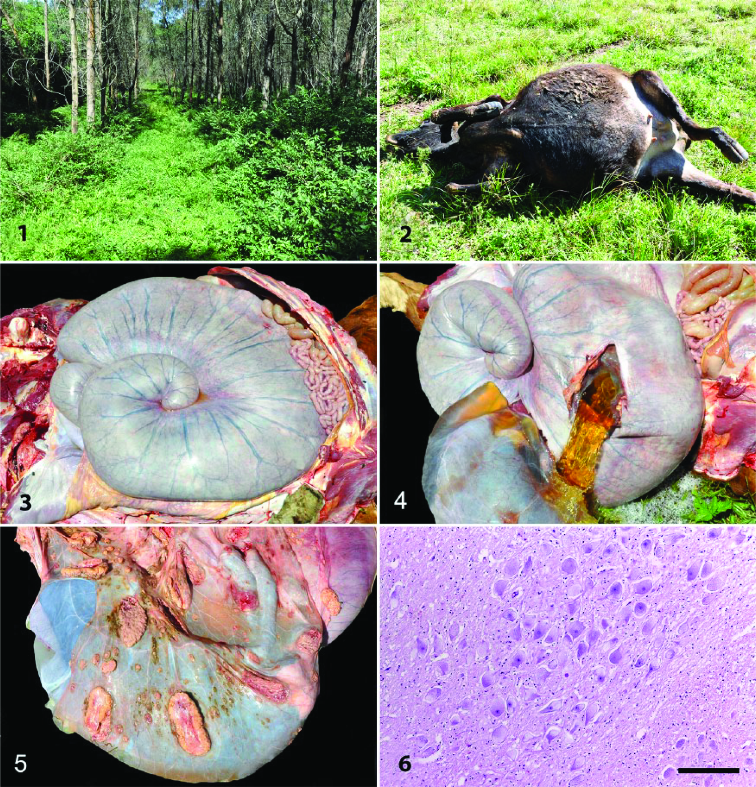
Hydrallantois in cows naturally poisoned by Sida carpinifolia (Malvaceae).
Microscopic evaluation of the placenta revealed marked diffuse edema, sometimes with a myxomatous appearance, in all 3 cattle. There was also thrombosis in the cotyledonary regions in cow 1. Swollen neurons with mild-to-moderate cytoplasmic granular vacuolation were observed in the CNS of all cows (Fig. 6), especially in the cerebellum and obex. Cows 1 and 2 also had hypereosinophilic neurons with cytoplasmic retraction, in addition to axonal spheroids in the white matter of the cerebellum. Other abnormalities observed included diffuse cytoplasmic vacuolation of the renal tubular epithelium (cow 1) and mild cytoplasmic vacuolation of follicular thyroid cells and pancreatic acinar cells (cow 3).
Microscopic evaluations of fetuses 1 and 2 revealed diffuse and marked swelling with cytoplasmic vacuolation in the acinar cells of the pancreas, follicular cells of the thyroid, hepatocytes, and renal tubular epithelial cells. Vacuolation and moderate swelling in the cytoplasm of the Purkinje neurons in the cerebellum and the telencephalic cortex, obex, thalamus, and hippocampus neurons were also observed. Hyperplasia of the Bergmann astrocytes in the Purkinje layer of the cerebellum, and axonal spheroids in sections of the cerebellum, obex, thalamus, and hippocampus, were also evident in fetuses 1 and 2. However, fetus 3 exhibited only mild cytoplasmic vacuolation and swelling of the pancreatic acinar cells and follicular thyroid cells.
Lectin histochemical examination of maternal and fetal samples revealed mild-to-marked positive cytoplasmic staining for ConA, WGA, and sWGA biomarkers in neurons of the cerebellum and obex. Staining for the remaining lectins tested (SBA, DBA, UEA I, PNA, RCA I, and BS I) was mild-to-absent, similar to that observed in the negative control. The DFAT was negative for rabies virus in all cases.
The diagnosis of hydrallantois in cows naturally poisoned by S. carpinifolia was made through our epidemiologic, clinical, and pathologic findings, and confirmed by lectin histochemistry. Hydrallantois is considered an infrequent condition in pregnancy, 16 and our findings suggested that the consumption of S. carpinifolia was responsible for this condition in the observed cases. This hypothesis was supported by the previously described high frequency of occurrence of this condition concomitantly with S. carpinifolia poisoning in this specific region (municipality of Triunfo). 14 Such a high frequency may also be explained by the production system employed in this region, in which integrated crop–livestock–forestry systems are common, with cattle and Eucalyptus spp. sharing the same environment. This shady environment creates suitable conditions for the proliferation of S. carpinifolia, which becomes readily accessible to the cattle. However, a survey of 6,706 cattle autopsies in the central region of Rio Grande do Sul State found no records of hydrallantois or poisoning by this plant. 10 This disagreement may be explained by the fact that different vegetation and lower production of Eucalyptus spp. occur at the studied sites than elsewhere in the region.
Fetal changes have been described in goats and cattle poisoned by S. carpinifolia, 15 and reproductive changes have been reported in animals poisoned by other plants containing swainsonine.5,12 An experiment conducted with pregnant sheep fed Astragalus lentiginosus, a toxic swainsonine-containing plant, revealed that the chorionic epithelium of the placenta initially had cytoplasmic vacuoles, but later became metaplasic. 19 In another experiment with sheep fed A. lentiginosus, smaller and irregularly shaped cotyledons were observed, in addition to an increase in the amount of uterine fluid in the poisoned sheep. 8 Such reproductive changes previously reported in ruminants may have occurred as a result of the transplacental passage of the active principle (swainsonine) of these plants, similar to that described in pregnant rats experimentally poisoned with I. carnea. 7
The 3 cases presented herein had adventitial placentation, which is a compensatory mechanism for the inadequate development of placentomes, and consists of an increase in the size of the remaining caruncles during gestation and the development of primitive placental villi. This condition may be congenital or acquired, as in cases in which there are inflammation and destruction of portions of the endometrium, and is often associated with hydrallantois in cattle. 16 In our cases, swainsonine intoxication was possibly associated with inadequate placentation and an increased volume of fluid in the allantois. However, the pathogenesis is still unknown, and further studies are needed to clarify the mechanisms underlying this condition.
An important differential diagnosis for cases of hydrallantois is hydramnios, a condition that involves excessive accumulation of fluid in the amniotic sac; these 2 conditions do not usually occur simultaneously. Hydramnios is associated with fetal malformations, mainly skeletal and muscular abnormalities in addition to gross macroscopic alterations, 16 none of which were observed in the 3 fetuses examined in our study. One fetus had hydronephrosis, a condition associated with excessive accumulation of fluid in the allantois, which obstructs the exit of fetal urine into the allantois and results in dilation of the renal pelvis. 17
Lectin histochemical tests are employed to identify the types of lectin residues accumulated within cells.3,15 S. carpinifolia poisoning causes α-mannosidosis, a lysosomal accumulation of oligosaccharides containing α-mannose and β-N-acetyl-glucosamine residues.
3
In the lectin histochemistry analyses of maternal and fetal tissues done is our study, sWGA and WGA staining were detected, indicating the expression of N-acetyl-
Footnotes
Acknowledgements
We thank Dr. Laura Barreto for her support.
Declaration of conflicting interests
The authors declared no potential conflicts of interest with respect to the research, authorship, and/or publication of this article.
Funding
This study was funded by the National Council of Scientific and Technological Development (CNPq) and the Brazilian Federal Agency for the Support and Evaluation of Graduate Education (CAPES).