
Abstract
A pedunculated exophytic mass developed on the rump of a dog. Fine-needle aspiration revealed keratin debris suggestive of a follicular tumor. However, histology revealed a pigmented viral plaque that contained numerous keratin-filled cystic cavities. Canine papillomavirus 18 DNA sequences were detected in the lesion. Viral plaques are typically multiple sessile lesions of dogs. A viral plaque appearing as a solitary exophytic keratin-filled mass has not been reported previously, to our knowledge. The novel clinical findings in this case expand the ways that viral plaques may appear in dogs. In addition, the histologic findings represent a novel pathologic entity of dogs. Given that canine viral plaques can be progressive, and dogs typically develop numerous plaques, it is important to differentiate between a viral plaque and a hair follicle tumor.
Canine pigmented viral plaques are typically multiple dark sessile masses that most commonly develop on the ventrum and the medial aspects of the limbs.2,5 They are well established to be caused by papillomaviruses (PVs) within the Chipapillomavirus genus, 5 which includes canine PV 18 (CPV18; Chipapillomavirus 1). However, disease caused by this PV appears to be rare, and CPV18 infection has only been reported in 2 dogs.3,7 We describe here a viral plaque that contained CPV18 DNA sequences and had novel clinical and histologic features.
An 8-y-old castrated male British Bulldog was presented at a veterinary clinic because of a 2 × 1-cm brown pedunculated mass on the right rump. The mass had first been observed 3 mo previously and had grown slowly since that time. Small amounts of dark opaque fluid had recently been observed discharging from the mass. Cytology of a fine-needle aspirate revealed keratin debris, melanin, and small numbers of large angular nucleated keratinized squames. A hair follicle neoplasm was suspected, and the mass was excised and submitted for histology.
Histology revealed a pedunculated mass that was attached to the underlying skin by a thin stalk (Fig. 1). The epithelium overlying the mass had a clear transition from normal epidermis to epidermis that was hyperplastic and had a “scalloped” appearance typical of a canine viral plaque.1,2 The epidermis overlying the mass was continuous with a central pore. Deep to the central pore were numerous cystic cavities lined by hyperplastic squamous epithelium. Cysts contained angular squames as well as keratinocytes that had retained their nuclei (Fig. 2). Visible within the epithelium lining the cysts were melanin, marked keratohyalin clumping, and occasional cells with shrunken nuclei and clear cytoplasm (consistent with koilocytes; Fig. 3).
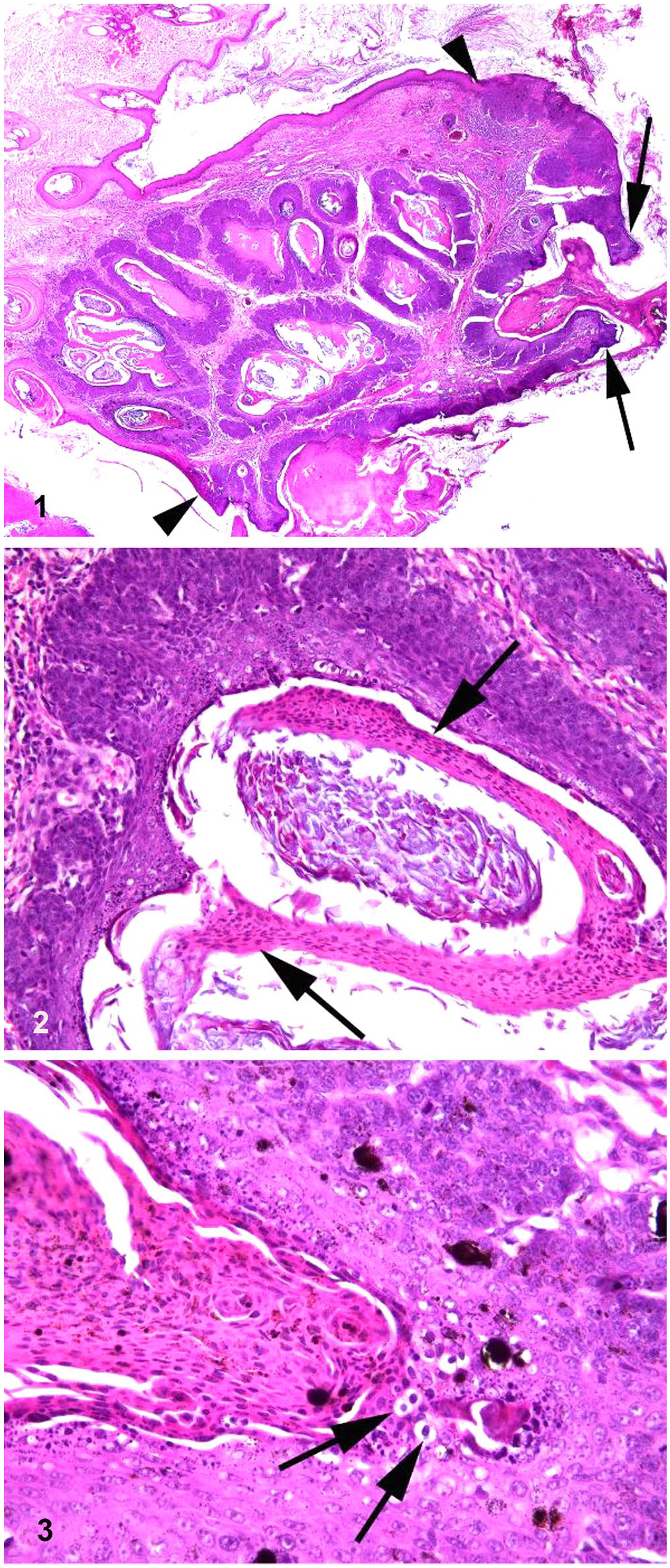
Cystic viral plaque associated with canine papillomavirus 18 in a British Bulldog.
DNA was extracted from a central part of the mass (NucleoSpin DNA FFPE XS kit; Macherey-Nagel) according to manufacturers’ instructions, and MY09/11 and CP4/5 consensus PCR primers were used to amplify PV DNA. 6 Amplified DNA was sequenced and found to be identical to CPV18 using a BLAST search (https://blast.ncbi.nlm.nih.gov/Blast.cgi).
The mass has not recurred 3 mo after surgical excision, and the dog has not developed any additional skin lesions. In contrast to the typical clinical presentation of canine viral plaques, 5 our case developed as a single large exophytic pedunculated mass on the rump. The mass contained large keratin-filled cysts and the keratin was detected by cytology, which prompted a presumptive diagnosis of a hair follicle cyst or neoplasm. Given that canine viral plaques can be progressive, and dogs typically develop numerous plaques,4,5 it is important to differentiate between a viral plaque and a hair follicle tumor.
This mass was diagnosed as a cystic viral plaque given the irregular epithelial hyperplasia both overlying the mass as well as within the epidermis lining the cysts. Other features consistent with a viral plaque included the presence of melanin, retention of nuclei in the keratin overlying the hyperplastic epidermis, and koilocytosis.1,2 The presence of a central pore opening onto the surface of the mass suggests continuity between the epidermis overlying the mass and the epidermis lining the cysts. The keratin-filled cysts within the mass probably developed as a result of folding and hyperplasia of the epidermis. It appears likely that the cysts communicated with the central pore, although this communication was not visible histologically. Although classification as a follicular neoplasm was considered, the histologic appearance of the mass was more consistent with proliferation and infolding of cells in the epidermis than proliferation of cells within a hair follicle.
This is the third report of canine skin disease caused by CPV18. CPV18 was reported in a pigmented plaque in 2016 and then in a trichoblastoma in 2019.3,7 Interestingly, the trichoblastoma also contained keratin-filled cysts, suggesting that infolding of affected epithelium could also have influenced the development of this neoplasm. Features of follicular differentiation, such as small basilar cells forming trabecular patterns, were reported in the earlier case. 7 In contrast, follicular differentiation was not visible in our case. Although additional cases are required, CPV18 may be more likely to stimulate cystic folding of the epidermis than other Chipapillomavirus types, possibly because CPV18 infects cells deeper in the epidermis resulting in more folding and a more basilar appearance to the hyperplastic cells.
The lesion in our case is a novel manifestation of PV infection in dogs. The lesion could easily be mistaken for a follicular cyst or neoplasm, although the characteristic irregular epidermal hyperplasia over the surface of the mass should allow recognition of this unusual viral-induced lesion of dogs. Although disease caused by CPV18 appears to be rare, infection by this virus may be more likely to cause masses with atypical clinical and histologic features.
Footnotes
Declaration of conflicting interests
The authors declared no potential conflicts of interest with respect to the research, authorship, and/or publication of this article.
Funding
The authors received no financial support for the research, authorship, and/or publication of this article.