
Abstract
A pig was in left lateral recumbency with limb spasticity, accentuated prostration, and strabismus, and was euthanized. During autopsy, yellowing of the leptomeninges at the ventral pons to medulla oblongata was noted. In the cerebellar peduncles, there was a focally extensive black-to-yellow area at the level of the vestibular nuclei. Histologic examination revealed a cross-section of a nematode larva, consistent with Stephanurus dentatus, bordered by edema and marked infiltration of mononuclear cells, plasma cells, and a few eosinophils. Vacuolation of the neuropil, with rare gitter cells and axonal spheroids, was also observed. We diagnosed parasitic encephalitis caused by S. dentatus migration based on the pathology findings and characterization of the parasite.
Stephanurus dentatus, the swine kidney worm, is widely distributed in tropical and subtropical countries,2,7 affecting mainly free-ranging and feral pigs.1,2,6,8,10 Adult worms encyst around the renal pelvis and ureter. Nematode eggs are eliminated in the urine.3,6,7 Pigs are infected by ingestion of infectious larvae free in the environment or in infected earthworms, or through cutaneous penetration by larvae. Larvae migrate to the liver through the portal or systemic circulation; some are trapped and encapsulated, others migrate to the retroperitoneal tissues.3,6,7 During migration, larvae and young adults cause necrosis, fibrosis, and abscess formation, mainly in the liver, urethral wall, renal pelvis, and renal capsule.2,3,5-7 Aberrant migration can cause atypical lesions and clinical signs. Migration of S. dentatus larvae in the spinal cord can cause paraplegia,6,7,13,15 but migration to other parts of the central nervous system has not been reported, to our knowledge.
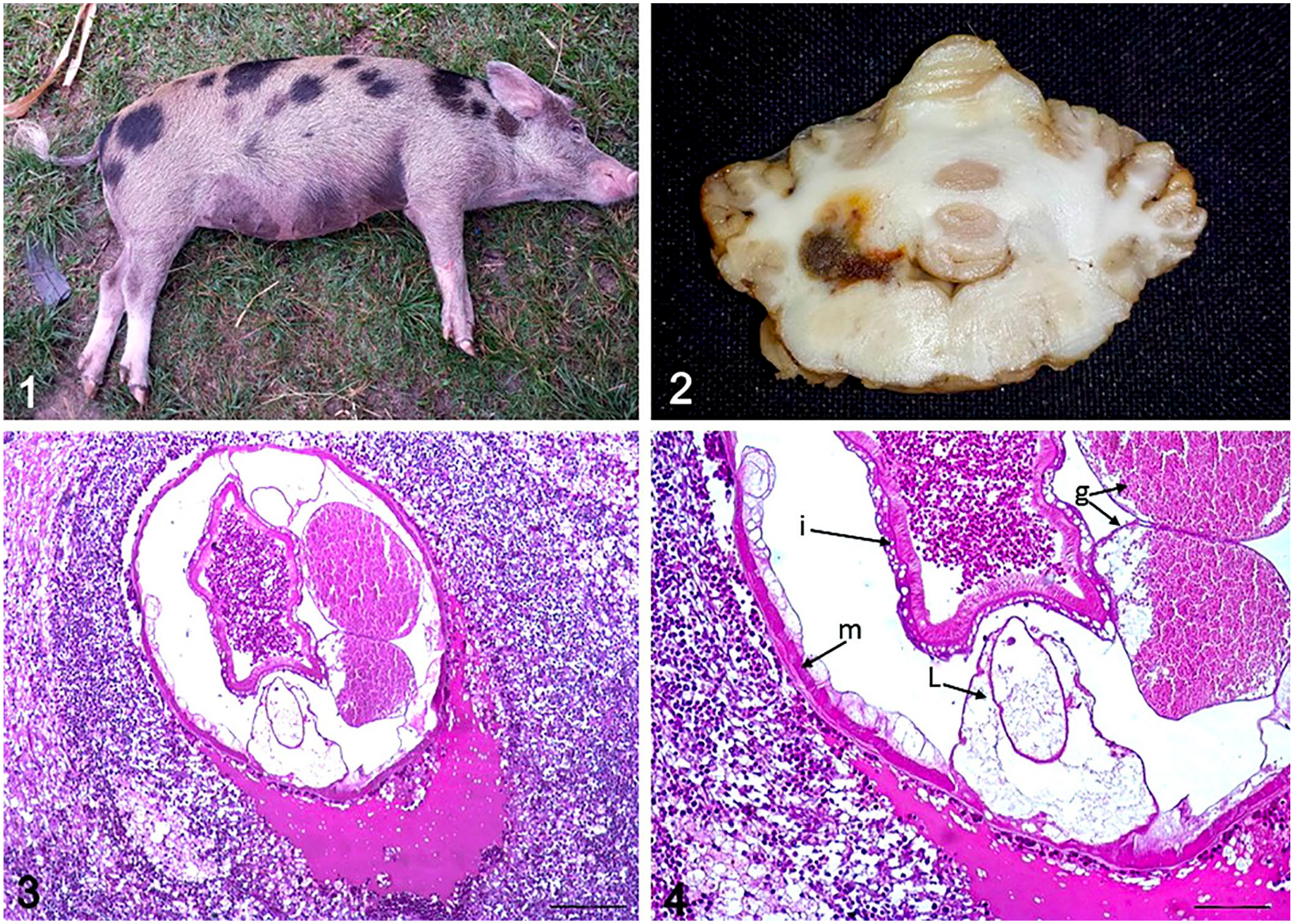
A 4-y-old, 130-kg, mixed-breed sow was admitted to the Veterinary Hospital of the Universidade Federal do Pará (UFPA; Brazil). The sow was in left lateral recumbency, with limb spasticity and strabismus (Fig. 1). When the animal was placed in right lateral recumbency, it moved back to left lateral recumbency. The sow was from a group of 6 pigs housed in stalls with brick walls and fed rice bran and common salt. It was the only clinically ill animal, and there was no history of recent trauma. After observation for 19 d and no clinical improvement, the animal was euthanized and submitted to the Animal Pathology Laboratory at UFPA for autopsy.
Parasitic encephalitis caused by Stephanurus dentatus in a pig.
At autopsy, the liver had an irregular surface, with many evenly distributed white areas. Yellowing of the leptomeninges of the brain was observed ventrally at the pons and medulla oblongata. On section of the cerebellum, there was a black-to-yellow area in the region of the cerebellar peduncles at the level of the vestibular nuclei (Fig. 2). Histologic examination of the cerebellum revealed a cross-section of a nematode larva bordered by edema and marked infiltration of mononuclear cells, plasma cells, and a few eosinophils. Vacuolation of the neuropil, with rare gitter cells and axonal spheroids, was observed (Fig. 3). The nematode was up to 110 µm in diameter. The cuticle was thin, and musculature was platymyarian-meromyarian. The intestine was large, lined by a few multinucleate cells with a prominent brush border. The lateral chords were vacuolated and often divided into 3 sublaterals (Fig. 4). These characteristics are those of the strongyle nematode S. dentatus. 9 The nematode was an immature female, identified through the presence of 2 developing genital tracts (Fig. 4). Adult parasites were not observed during the autopsy. We diagnosed parasitic encephalitis caused by S. dentatus migration based on the pathology findings and characterization of the parasite. 9
In Brazil, stephanuriasis is found mainly in free-range pig production1,2,11,12,14; the sow in our case was housed in an intensive breeding unit with no outside access. Clinical signs of disease are not usually seen, although moderate or massive infection can cause poor growth and reduced weight gain. S. dentatus is rarely a cause of mortality, but significant economic loss may occur as a result of decreased production and condemnation of portions of the carcass.2,3,6,7,17,18 Atypical clinical signs can occur when aberrant larval migration produces thrombi in the portal veins, hepatic artery, or caudal vena cava; peritonitis, pleuritis, and adhesions; and/or abscesses in the lungs, pancreas, and lymph nodes.4,6,7 Migration to the psoas muscles may cause pain and stiffness. 7
Paraplegia caused by aberrant migration of S. dentatus in the spinal cord has been described in pigs.3,6,7,13,15 Paralysis is usually associated with spinal cord compression from a granuloma. These cases usually occur with massive infections and abscesses in multiple organs, mainly the kidneys, the perirenal region, liver, abdominal wall, and lymph nodes. 13 S. dentatus has been found in the spinal canal of a heavily infected pig with no signs of paralysis. 16 In our case, the parasite migrated aberrantly to the brain with no visible lesions in other organs. The clinical signs observed suggested upper motor neuron syndrome associated with brain stem damage, with vestibular and cerebellar involvement. These signs were confirmed by the neuroanatomic location of the lesions. Our case demonstrates that aberrant migration of S. dentatus can occur in the brain of swine and that this disease must be considered in the differential diagnosis of neurologic syndromes in pigs in areas in which S. dentatus is endemic.
Footnotes
Declaration of conflicting interests
The authors declared no potential conflicts of interest concerning the research, authorship, and/or publication of this article.
Funding
The authors received no financial support for the research, authorship, and/or publication of this article.